
Explain upper limb of human anatomy
Reading File
Now I'll search the medical library for upper limb anatomy content in parallel.
Finding Sources
Finding Sources
Gray's Anatomy for Students is the primary source. Let me read several key sections in parallel - the overview, bones, muscles, and nerves of the upper limb.
Reading File
Reading File
Finding Sources
The block3 file has the full chapter. Let me read the detailed conceptual overview and regional anatomy sections.
Reading File
Reading File
Excellent content. Let me now read the detailed regional anatomy - bones/joints, muscles, nerves, and blood supply sections.
Reading File
Reading File
Now let me read the bones/joints and muscles sections in more detail.
Reading File
Reading File
I now have rich, comprehensive content from Gray's Anatomy for Students. Let me also fetch a couple of key anatomical images and read the muscles section to round out the response.
Reading File
Loading Image
Loading Image
Excellent - I have very rich material from Gray's Anatomy for Students, along with two key anatomical diagrams. Let me fetch two more useful images and read the brachial plexus section.
Reading File
Loading Image
I now have comprehensive information from Gray's Anatomy for Students. Let me compose a thorough, well-organized answer.
Anatomy of the Upper Limb
Source: Gray's Anatomy for Students
The upper limb is associated with the lateral aspect of the lower neck and the thoracic wall. It is suspended from the trunk by muscles and one small bony joint - the sternoclavicular joint (between clavicle and sternum). Based on its major joints and component bones, the upper limb is divided into four regions:
- Shoulder - area of attachment to the trunk
- Arm - shoulder to elbow joint
- Forearm - elbow joint to wrist joint
- Hand - distal to the wrist joint
1. Bones and Joints
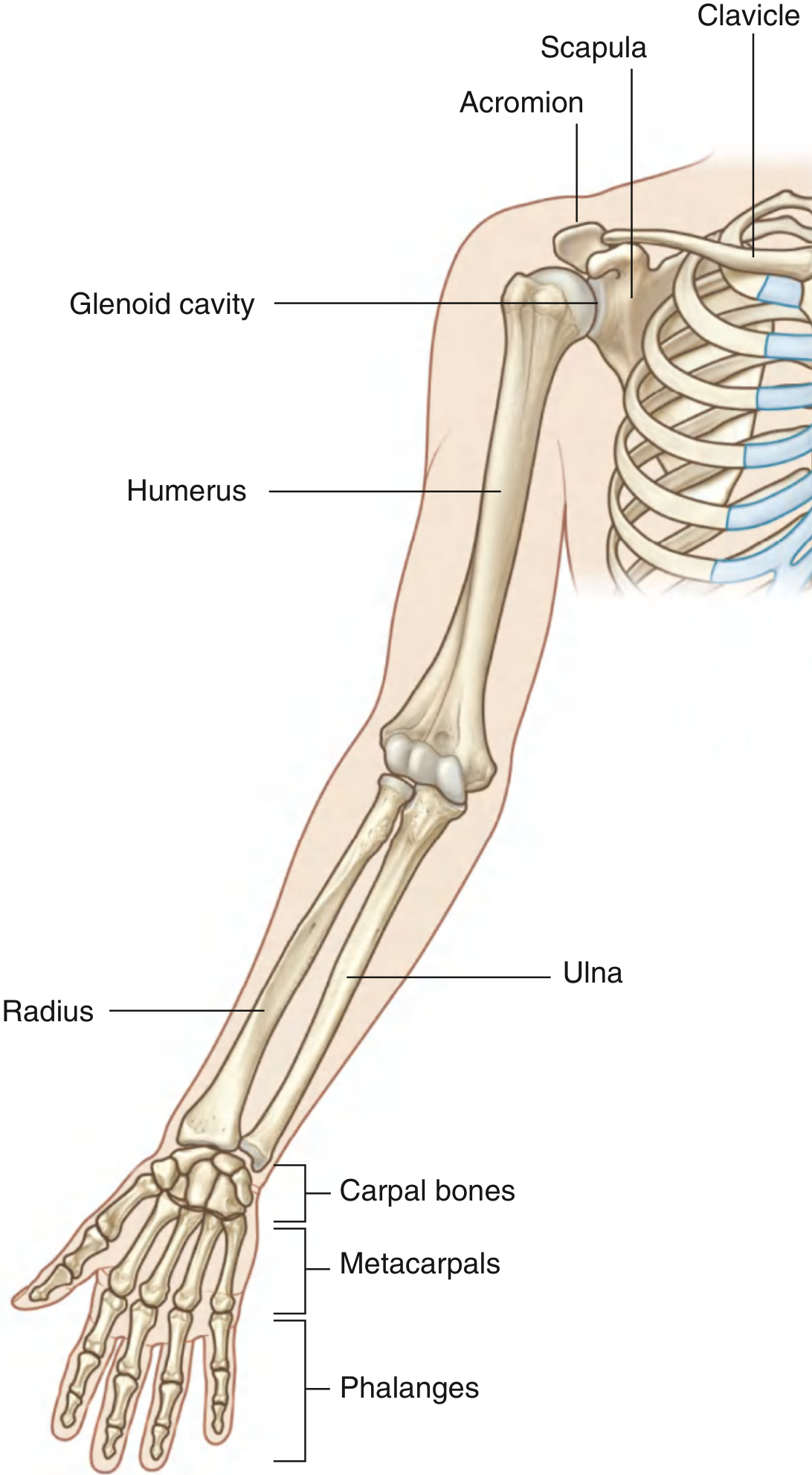
Fig. 7.7 - Bones of the upper limb (Gray's Anatomy for Students)
Shoulder Girdle
The pectoral (shoulder) girdle consists of the clavicle and scapula. The clavicle articulates medially with the sternum (sternoclavicular joint) and laterally with the acromion process of the scapula (acromioclavicular joint). The glenohumeral joint (shoulder joint) is a highly mobile ball-and-socket joint where the head of the humerus articulates with the shallow glenoid cavity of the scapula.
Arm
Contains the single bone: the humerus. The humerus articulates proximally at the glenohumeral joint and distally at the elbow.
Forearm
Contains two bones:
- Radius - the lateral bone; its distal end can flip over the ulna during pronation/supination
- Ulna - the medial bone
At the elbow joint, both bones articulate with the humerus, and also with each other at the proximal and distal radioulnar joints - allowing pronation (palm posterior) and supination (palm anterior).
Hand
- 8 carpal bones (wrist) - arranged in two rows; work together as a unit with limited individual movement
- 5 metacarpals - form the skeleton of the palm
- Phalanges - thumb has 2; other fingers have 3 each (proximal, middle, distal)
The wrist (radiocarpal) joint is formed between the radius, an articular disc distal to the ulna, and the carpal bones; it allows flexion, extension, abduction (radial deviation), adduction (ulnar deviation), and circumduction.
The metacarpophalangeal (MCP) joints are biaxial condylar (ellipsoid) joints. The interphalangeal (IP) joints are hinge joints permitting flexion and extension only.
2. Movements
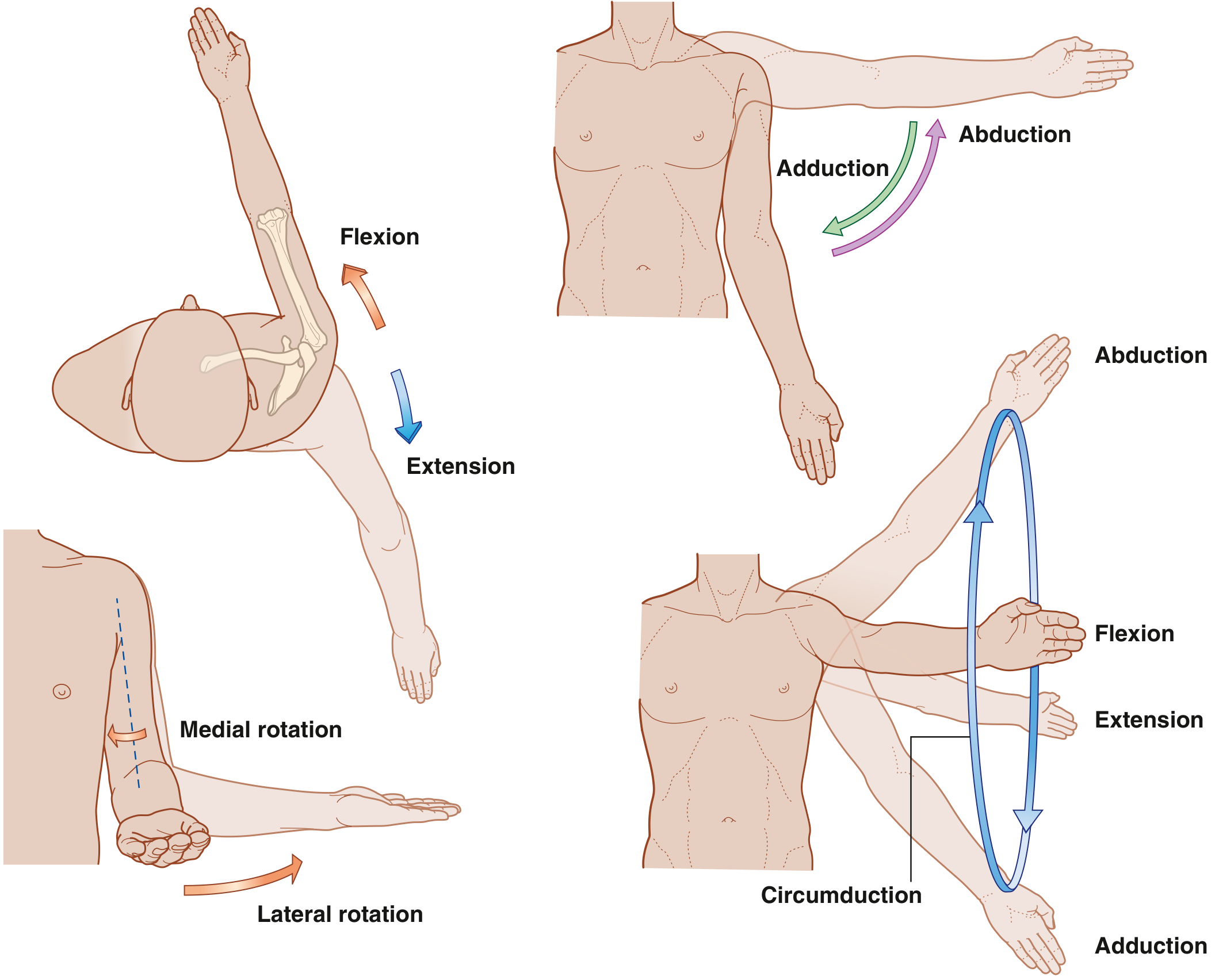
Fig. 7.4 - Movements of the arm at the glenohumeral joint (Gray's Anatomy for Students)
| Joint | Movements |
|---|---|
| Glenohumeral | Flexion, extension, abduction, adduction, medial rotation, lateral rotation, circumduction |
| Elbow | Flexion, extension |
| Radioulnar (proximal + distal) | Pronation, supination |
| Wrist | Flexion, extension, abduction (radial deviation), adduction (ulnar deviation), circumduction |
| MCP | Flexion, extension, abduction, adduction, circumduction |
| IP | Flexion, extension only |
3. Muscles
Muscles of the arm and forearm are separated into anterior (flexor) compartments and posterior (extensor) compartments by fascial septa, bones, and ligaments.
Shoulder Muscles
- Deltoid - major abductor of the arm at the glenohumeral joint
- Rotator cuff (4 muscles - SITS): Supraspinatus (abduction), Infraspinatus (lateral rotation), Teres minor (lateral rotation), Subscapularis (medial rotation). These provide dynamic stability to the glenohumeral joint.
Arm (Anterior Compartment - Flexors)
- Biceps brachii - flexes forearm, powerful supinator
- Brachialis - primary flexor of the forearm
- Coracobrachialis - flexes and adducts the arm
Arm (Posterior Compartment - Extensors)
- Triceps brachii - only extensor of the elbow
Forearm
- Anterior compartment (flexors, pronators): flexor carpi radialis, flexor carpi ulnaris, flexor digitorum superficialis, flexor digitorum profundus, flexor pollicis longus, pronator teres, pronator quadratus
- Posterior compartment (extensors, supinator): extensor carpi radialis longus/brevis, extensor digitorum, extensor carpi ulnaris, supinator, extensor pollicis longus/brevis, abductor pollicis longus
Hand - Intrinsic Muscles
These generate delicate digital movements and modify forces produced by the long flexor/extensor tendons from the forearm:
- Thenar muscles (3 muscles) - form the thenar eminence over metacarpal I; give the thumb its freedom of movement and enable opposition
- Hypothenar muscles - act on the little finger
- Lumbricals and interossei - fine control of finger movements; flex MCP joints while extending IP joints
4. Nerves - Brachial Plexus
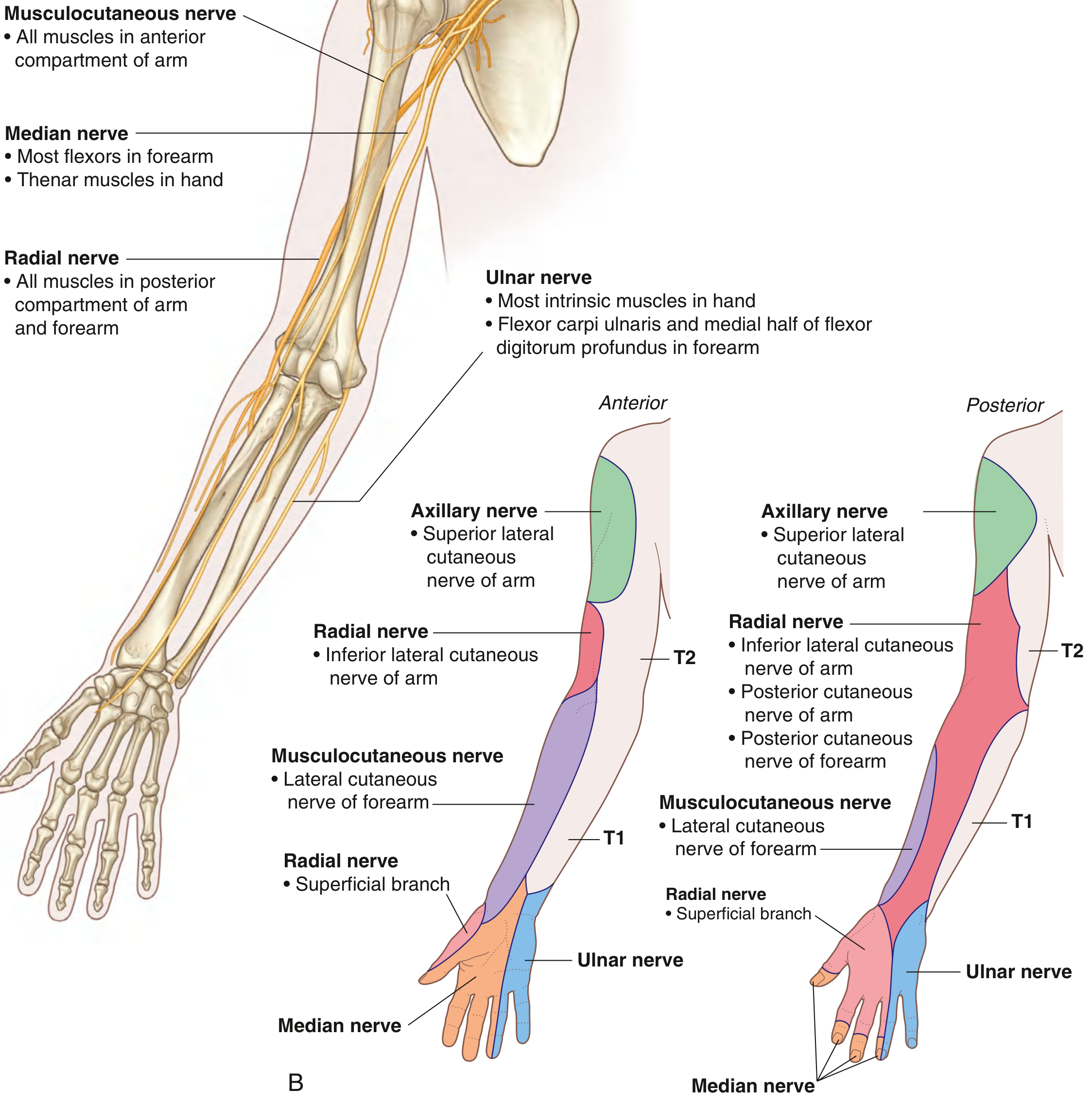
Fig. 7.16 - Nerves of the upper limb showing motor supply and cutaneous territories (Gray's Anatomy for Students)
The entire upper limb is innervated by the brachial plexus (spinal roots C5-T1), formed in the neck and continuing through the axillary inlet into the axilla. The five major terminal nerves are:
| Nerve | Motor Supply | Cutaneous Supply |
|---|---|---|
| Musculocutaneous | All muscles in anterior compartment of arm (biceps, brachialis, coracobrachialis) | Anterolateral forearm |
| Median | Most flexors in anterior forearm (except FCU and medial FDP); thenar muscles; lateral 2 lumbricals | Palmar surface of lateral 3½ digits |
| Ulnar | Flexor carpi ulnaris, medial half of FDP; most intrinsic hand muscles | Medial 1½ digits |
| Radial | ALL muscles in posterior compartments of arm and forearm (extensors) | Posterior forearm; dorsolateral hand |
| Axillary | Deltoid, teres minor | Superior lateral arm (regimental badge area) |
Nerves Related to Bone - Clinically Important
Three nerves are directly related to parts of the humerus:
- Axillary nerve - wraps around the surgical neck of humerus (vulnerable in shoulder dislocation and proximal humerus fractures)
- Radial nerve - passes diagonally in the radial groove on the posterior shaft of the humerus (vulnerable in mid-shaft fractures - causes "wrist drop")
- Ulnar nerve - passes posterior to the medial epicondyle (vulnerable in elbow injuries - causes "claw hand")
Dermatomes (Key Landmarks)
- C5 - upper lateral arm
- C6 - palmar pad of thumb
- C7 - pad of index finger
- C8 - pad of little finger
- T1 - medial aspect of elbow
Myotomes (Key Movements)
- C5 - arm abduction at glenohumeral joint
- C6 - forearm flexion at elbow; biceps reflex
- C7 - forearm extension at elbow; triceps reflex
- C8 - finger flexion
- T1 - finger abduction/adduction
5. Blood Supply
Arterial
- The subclavian artery becomes the axillary artery at the lateral border of rib I
- The axillary artery becomes the brachial artery at the lower border of teres major
- The brachial artery divides at the cubital fossa into the radial artery (lateral) and ulnar artery (medial)
- In the hand, these form the superficial palmar arch (mainly ulnar) and the deep palmar arch (mainly radial)
Superficial Veins (Clinically Important)
The two main superficial veins - commonly used for venepuncture and IV access:
- Cephalic vein - originates over the anatomical snuffbox at the base of the thumb, runs up the anterolateral surface of the limb, and drains into the axillary vein via the clavipectoral triangle
- Basilic vein - originates from the medial side of the dorsal venous network of the hand, passes up the posteromedial forearm, and pierces the deep fascia midway up the arm
- Median cubital vein - connects cephalic and basilic veins at the elbow (common site for venepuncture)
6. The Axilla
The axilla is the pyramidal gateway between the neck and the arm. It has four walls (anterior, posterior, medial, lateral), an apex (axillary inlet), and a floor (base). All major vessels and nerves passing into and out of the upper limb pass through the axilla - including the brachial plexus, axillary artery, and axillary vein.
7. The Hand - Special Features
The hand serves two primary roles:
Mechanical tool - gripping and manipulating objects. This involves flexing the fingers against the thumb. The intrinsic hand muscles modify the forces of the long tendons and produce combinations of joint movements (e.g., simultaneous MCP flexion with IP extension) that cannot be achieved by forearm tendons alone.
Sensory tool - fine tactile discrimination. The fingertip pads have the highest density of sensory receptors in the body.
The thumb is positioned at right angles to the other fingers. This unique orientation means:
- Flexion sweeps the thumb across the palm
- Abduction moves it away at right angles to the palm
- Opposition (rotation of metacarpal I on the wrist) brings the pad of the thumb to face the pads of the other fingers - essential for normal hand function
All anatomical content sourced from Gray's Anatomy for Students (ISBN 9780323934237), Chapter 7: Upper Limb.
This is a shared conversation. Sign in to Orris to start your own chat.