from Guyton and Hall textbook of medical physiology, 4th South Asia edition, write a 10-mark long answer on Deglutition , Achalasia Cardia

Reading File
Finding Sources
Finding Sources
Reading File
Finding Sources
Reading File
Finding Sources
Reading File
Reading File
Loading Image
Searching Images

achalasia cardia megaesophagus lower esophageal sphincter
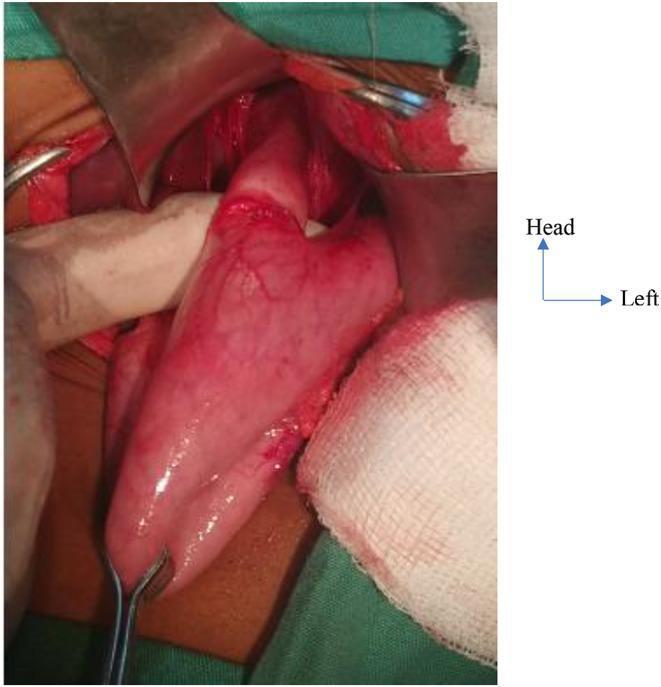
This clinical photograph captures an intraoperative view during a laparotomy for Achalasia Cardia in a pediatric patient. The image provides surgical exposure of the hiatal region, demonstrating the hallmark anatomical findings of the disease. Centrally, a severely dilated esophagus (megaesophagus) is visible, presenting with a widened lumen and a glistening, reddish-pink serosal surface. Distal to this dilation, there is a distinct circumferential stricture at the cardia of the stomach, representing the hypertensive lower esophageal sphincter that fails to relax. Surgical retractors are positioned to maintain visualization of the gastroesophageal junction, and sterile white gauze is used for tissue protection and fluid management. The orientation is indicated by an superimposed axis showing the cranial ('Head') and 'Left' directions. This visual serves as a primary educational example of the gross pathological changes in end-stage achalasia, emphasizing the transition from upstream esophageal stasis and dilation to the narrowed, non-relaxing cardia.
This clinical photograph captures an intraoperative view during a laparotomy for Achalasia Cardia in a pediatric patient. The image provides surgical exposure of the hiatal region, demonstrating the hallmark anatomical findings of the disease. Centrally, a severely dilated esophagus (megaesophagus) is visible, presenting with a widened lumen and a glistening, reddish-pink serosal surface. Distal to this dilation, there is a distinct circumferential stricture at the cardia of the stomach, representing the hypertensive lower esophageal sphincter that fails to relax. Surgical retractors are positioned to maintain visualization of the gastroesophageal junction, and sterile white gauze is used for tissue protection and fluid management. The orientation is indicated by an superimposed axis showing the cranial ('Head') and 'Left' directions. This visual serves as a primary educational example of the gross pathological changes in end-stage achalasia, emphasizing the transition from upstream esophageal stasis and dilation to the narrowed, non-relaxing cardia.

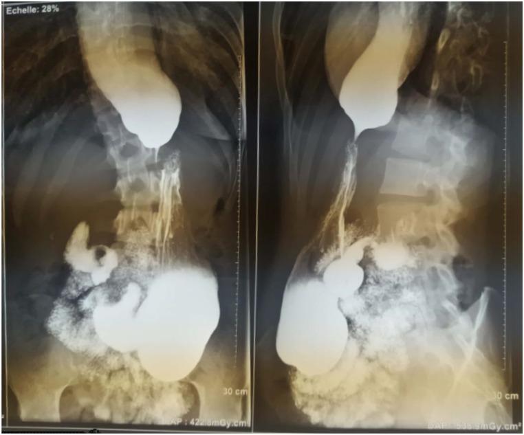
This diagnostic image consists of two views of a barium swallow x-ray radiography, showing the upper gastrointestinal tract in an anteroposterior and lateral perspective. The esophagus is markedly dilated (megaesophagus) with significant retention of the radiopaque barium contrast medium. At the gastroesophageal junction, there is a smooth, tapered narrowing of the distal esophagus, creating the classic 'bird's beak' appearance characteristic of achalasia. This narrowing represents the failure of the lower esophageal sphincter (LES) to relax. In the stomach, the barium presents a 'snowflake-falling' pattern, indicating irregular distribution and altered gastric filling. These findings are primary indicators of esophageal motility disorders, specifically achalasia, where the primary educational focus is on identifying the combination of proximal dilation and distal muscular obstruction at the cardia. This clinical imaging is essential for students and residents to recognize stages of Achalasia Cardia (AC) based on esophageal diameter and morphology.
This diagnostic image consists of two views of a barium swallow x-ray radiography, showing the upper gastrointestinal tract in an anteroposterior and lateral perspective. The esophagus is markedly dilated (megaesophagus) with significant retention of the radiopaque barium contrast medium. At the gastroesophageal junction, there is a smooth, tapered narrowing of the distal esophagus, creating the classic 'bird's beak' appearance characteristic of achalasia. This narrowing represents the failure of the lower esophageal sphincter (LES) to relax. In the stomach, the barium presents a 'snowflake-falling' pattern, indicating irregular distribution and altered gastric filling. These findings are primary indicators of esophageal motility disorders, specifically achalasia, where the primary educational focus is on identifying the combination of proximal dilation and distal muscular obstruction at the cardia. This clinical imaging is essential for students and residents to recognize stages of Achalasia Cardia (AC) based on esophageal diameter and morphology.

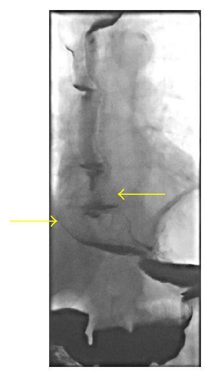
This diagnostic image is a barium swallow fluoroscopy showing the lower esophagus and gastroesophageal junction. The primary finding is a significantly dilated and tortuous esophagus, which appears atonic and uniformly filled with radiopaque barium contrast. Two yellow arrows highlight the lateral margins of the widened esophageal lumen. Despite the severe proximal dilation, the lower esophageal sphincter (LES) region demonstrates patency with a relatively smooth transition into the stomach. Notably, the classic 'bird's beak' tapering associated with achalasia is absent, as the contrast flows freely into the gastric cardia without evidence of a distal stricture or mechanical obstruction. This imaging is clinically significant for evaluating esophageal dysmotility disorders and differentiating megaesophagus from structural or functional obstructions at the gastroesophageal junction.

This diagnostic image is a barium swallow fluoroscopy showing the lower esophagus and gastroesophageal junction. The primary finding is a significantly dilated and tortuous esophagus, which appears atonic and uniformly filled with radiopaque barium contrast. Two yellow arrows highlight the lateral margins of the widened esophageal lumen. Despite the severe proximal dilation, the lower esophageal sphincter (LES) region demonstrates patency with a relatively smooth transition into the stomach. Notably, the classic 'bird's beak' tapering associated with achalasia is absent, as the contrast flows freely into the gastric cardia without evidence of a distal stricture or mechanical obstruction. This imaging is clinically significant for evaluating esophageal dysmotility disorders and differentiating megaesophagus from structural or functional obstructions at the gastroesophageal junction.

I now have all the content needed from Guyton and Hall to write a complete, detailed 10-mark long answer. Let me compose the answer:

Deglutition and Achalasia Cardia

(Guyton and Hall Textbook of Medical Physiology, 4th South Asia Edition - Chapters 64 & 67)

DEGLUTITION (SWALLOWING)

Swallowing (deglutition) is a complex coordinated mechanism by which food is propelled from the mouth to the stomach. Because the pharynx serves both respiration and swallowing, it is converted into a digestive conduit for only a few seconds at a time during each swallow.
Swallowing is divided into three stages:
  1. Voluntary stage
  2. Involuntary pharyngeal stage
  3. Involuntary esophageal stage

1. Voluntary Stage

When the food bolus is ready for swallowing, it is voluntarily squeezed or rolled posteriorly into the pharynx by pressure of the tongue upward and backward against the palate. Once the bolus enters the posterior pharynx, swallowing becomes entirely automatic and cannot be stopped voluntarily.

2. Involuntary Pharyngeal Stage

As the bolus enters the posterior mouth and pharynx, it stimulates epithelial swallowing receptor areas all around the pharyngeal opening - especially on the tonsillar pillars. The following automatic sequence is triggered:
  1. Soft palate pulled upward - closes the posterior nares, preventing reflux of food into the nasal cavities.
  2. Palatopharyngeal folds drawn medially - they approximate each other to form a sagittal slit, allowing only adequately masticated food to pass into the posterior pharynx. This selective action filters large, poorly chewed food particles.
  3. Laryngeal protection - the vocal cords are strongly approximated, and the larynx is pulled upward and anteriorly by the neck muscles. This causes the epiglottis to swing backward over the laryngeal opening, preventing passage of food into the trachea. The tight approximation of vocal cords is the most essential protective mechanism.
  4. Upper esophageal sphincter (pharyngoesophageal sphincter) relaxes - the upper 3-4 cm of esophageal muscular wall, which normally remains strongly contracted between swallows (preventing air entry during respiration), relaxes to allow food to pass from the posterior pharynx into the upper esophagus.
  5. Pharyngeal peristalsis - the muscular wall of the pharynx contracts from superior to inferior, propelling the bolus by peristalsis into the upper esophagus. The entire pharyngeal stage is completed in less than 2 seconds.
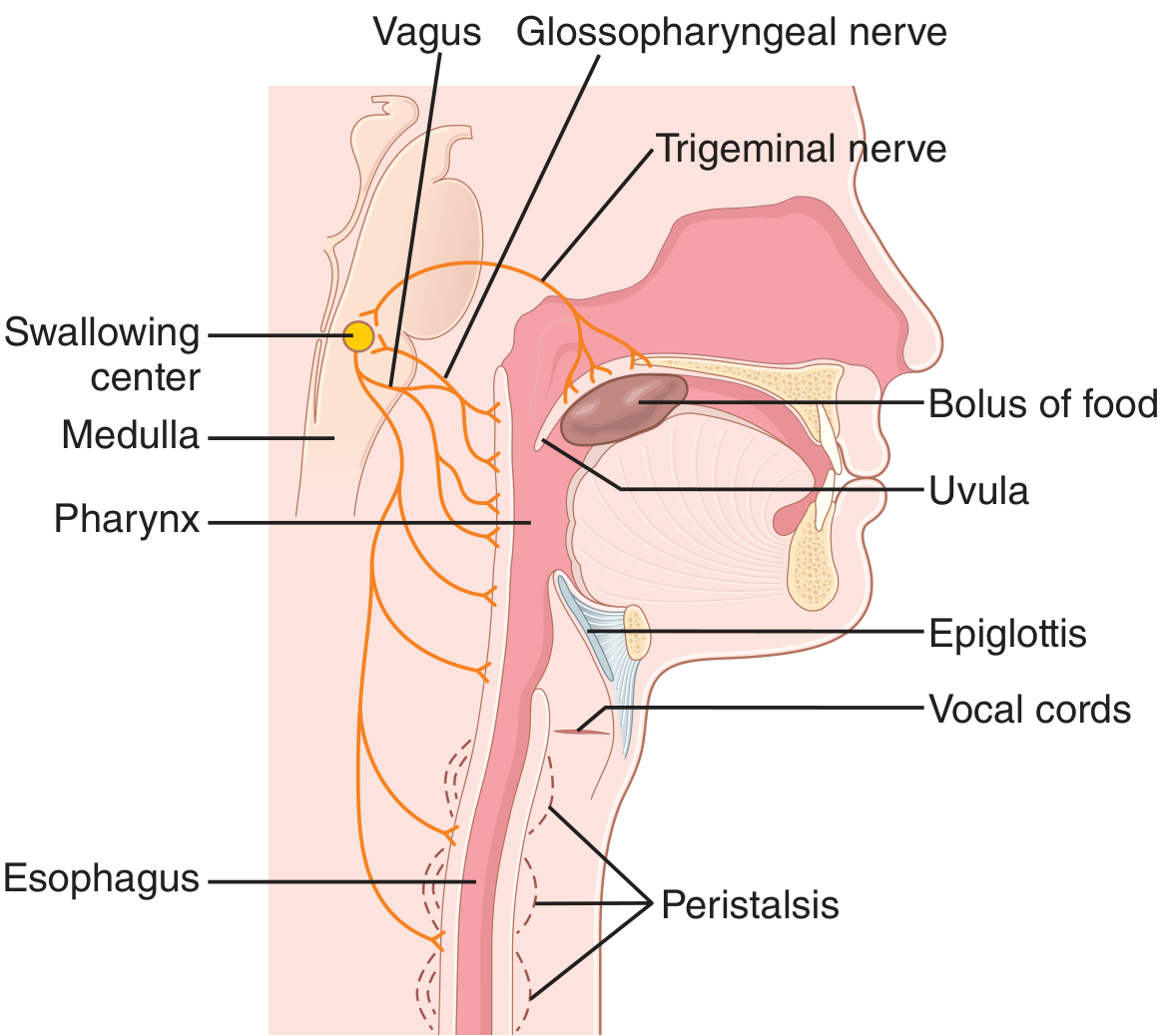
Figure 64.1 Swallowing mechanism - Guyton & Hall
Nervous Control of the Pharyngeal Stage:
Sensory impulses from the tonsillar pillars and surrounding pharyngeal mucosa pass via the trigeminal (V) and glossopharyngeal (IX) nerves into the medulla oblongata - specifically into or near the tractus solitarius. The swallowing sequence is then automatically coordinated by neural areas in the reticular substance of the medulla and lower pons - collectively called the deglutition center (swallowing center). Motor impulses from this center are transmitted via the 5th, 9th, 10th, and 12th cranial nerves and some superior cervical nerves to the pharynx and upper esophagus.
Interruption of Respiration: The pharyngeal stage occurs in under 6 seconds. The swallowing center specifically inhibits the respiratory center of the medulla during this interval, halting respiration at any point in its cycle to allow safe swallowing.

3. Esophageal Stage

The esophagus functions primarily to rapidly conduct food from the pharynx to the stomach. Two types of peristaltic movements accomplish this:
a) Primary Peristalsis: This is a continuation of the peristaltic wave that began in the pharynx. It travels the entire length of the esophagus to the stomach in about 8-10 seconds. In an upright person, gravity assists the bolus, reducing transit time to 5-8 seconds.
b) Secondary Peristalsis: If the primary wave fails to clear all food from the esophagus, distension of the esophagus by retained food triggers secondary peristaltic waves. These arise from the myenteric nerve plexus of the esophagus itself and continue until all retained food is propelled into the stomach.
Muscular Control:
  • Upper one-third of esophagus: striated muscle - controlled by skeletal nerve impulses via the glossopharyngeal and vagus nerves.
  • Lower two-thirds: smooth muscle - controlled by the vagus nerve via the myenteric nervous system. Even after vagal section, the myenteric plexus becomes sufficiently excitable after a few days to sustain secondary peristalsis independently.
Receptive Relaxation of the Stomach: As the peristaltic wave approaches the stomach, a wave of relaxation transmitted through myenteric inhibitory neurons precedes it, relaxing the stomach (and to a lesser extent the duodenum) to receive the incoming food.
Function of the Lower Esophageal Sphincter (LES / Gastroesophageal Sphincter): At the lower end of the esophagus, the circular muscle over approximately 3 cm functions as the LES. It normally remains tonically constricted with an intraluminal pressure of about 30 mmHg (compared to the relaxed mid-esophagus). As a peristaltic wave approaches, receptive relaxation of the LES allows smooth propulsion of food into the stomach. The LES also prevents gastroesophageal reflux of acidic stomach contents, which would digest the esophageal mucosa.
An additional anti-reflux mechanism is a valvelike closure: a short portion of the esophagus extends into the stomach, and increased intraabdominal pressure caves this segment inward, sealing the lumen and preventing reflux during coughing, walking, and straining.

ACHALASIA CARDIA

Definition

Achalasia is a condition in which the lower esophageal sphincter fails to relax during swallowing. As a result, food swallowed into the esophagus cannot pass into the stomach.
  • Guyton and Hall, Chapter 67: "Achalasia is a condition in which the lower esophageal sphincter fails to relax during swallowing."

Pathophysiology

Pathophysiological studies in patients with achalasia have demonstrated damage to the neural network of the myenteric plexus in the lower two-thirds of the esophagus. As a result:
  • The musculature of the lower esophagus remains spastically contracted.
  • The myenteric plexus loses its ability to transmit the signal that causes "receptive relaxation" of the gastroesophageal sphincter as food approaches it during swallowing.
  • Consequently, peristaltic waves arriving at the LES cannot overcome its persistent tonic contraction, and food accumulates in the esophagus above.

Consequences - Megaesophagus

When achalasia becomes severe:
  • The esophagus cannot empty swallowed food into the stomach for many hours (normal transit is a few seconds).
  • Over months to years, the esophagus becomes tremendously enlarged - a condition called megaesophagus - capable of holding up to 1 liter of food.
  • Retained food undergoes putrid infection due to prolonged esophageal stasis.
  • Infection leads to ulceration of the esophageal mucosa, sometimes causing severe substernal pain or even rupture and death.
Intraoperative view showing megaesophagus with hypertensive LES stricture in achalasia cardia
Intraoperative photograph showing the severely dilated esophagus (megaesophagus) and circumferential stricture at the cardia representing the non-relaxing LES in achalasia.
Barium swallow showing megaesophagus with bird's beak sign at the gastroesophageal junction in achalasia
Barium swallow X-ray showing the classical "bird's beak" appearance: a markedly dilated esophagus with smooth, tapered narrowing at the gastroesophageal junction - the hallmark radiological sign of achalasia.

Treatment

Guyton and Hall describes two approaches:
  1. Balloon dilation (pneumatic dilatation): Stretching the lower end of the esophagus with a balloon inflated on the end of a swallowed esophageal tube. This forcibly dilates the spastic LES and provides considerable symptomatic benefit.
  2. Antispasmodic drugs: Drugs that relax smooth muscle can be helpful by reducing the tonic contraction of the LES.
(Contemporary management also includes surgical myotomy - Heller's myotomy - and per-oral endoscopic myotomy (POEM), though these are discussed in detail in surgical/gastroenterology texts.)

SUMMARY TABLE

FeatureNormal DeglutitionAchalasia Cardia
LES behaviorReceptive relaxation during swallowingFails to relax - remains spastically contracted
Myenteric plexusIntactDamaged in lower 2/3 of esophagus
Esophageal transit5-10 secondsHours - food retained
Esophageal sizeNormalMegaesophagus (up to 1 litre)
ComplicationsNoneInfection, ulceration, rupture
TreatmentNot applicableBalloon dilation, antispasmodics

Sources:
  • Guyton and Hall Textbook of Medical Physiology, 4th South Asia Ed. - Chapter 64 (Propulsion and Mixing of Food in the Alimentary Tract), pp. 788-790 - covering all three stages of deglutition, nervous control, LES function.
  • Guyton and Hall Textbook of Medical Physiology, 4th South Asia Ed. - Chapter 67 (Physiology of Gastrointestinal Disorders), p. 823 - covering Achalasia and Megaesophagus.
This is a shared conversation. Sign in to Orris to start your own chat.