
Amyloidosis
amyloidosis diagnosis treatment
amyloidosis Congo red apple green birefringence histology
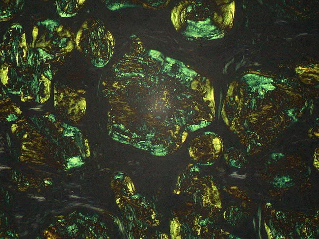
Light microscopy image of a lymph node biopsy stained with Congo Red and examined under polarized light. The tissue reveals extracellular, amorphous eosinophilic deposits distributed throughout nodal architecture consistent with amyloid. The deposits assume a dense, granular to plaque-like appearance on routine brightfield, and demonstrate distinctive apple-green birefringence when illuminated with polarized light, confirming the beta-pleated sheet rich configuration of amyloid fibrils. Congo Red positivity with birefringence distinguishes amyloid from other PAS-positive glycoprotein material and helps differentiate from non-amyloid nodal deposits. The beta-pleated sheet conformation accounts for the characteristic staining behavior and optical properties used in diagnosis. Clinically, identification of nodal amyloid supports systemic or localized amyloidosis and prompts workup for light-chain (AL) or serum amyloid A (AA) subtypes, including serum free light chains, immunofixation, and mass spectrometry-based typing. This image is relevant for educational illustrations of amyloid histology, pathophysiology, and diagnostic pathology workflows, including differential diagnosis with glycoprotein-rich PAS-positive deposits that are Congo Red negative. Potential clinical scenarios include workup of lymphadenopathy in patients with chronic inflammatory states, plasma cell dyscrasias, and lymphoproliferative disorders. High quality images like this support interdepartmental communication, teaching, and reproducible reporting of amyloid histology for junior pathologists and medical trainees in pathology education programs.
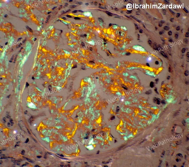
Renal biopsy histology of a patient with multiple myeloma showing AL-type amyloid deposition within the kidney. Congo red–stained sections reveal extracellular, eosinophilic, acellular deposits in the glomerular mesangium and throughout the tubulointerstitium; these deposits are salmon-pink on routine light microscopy. When viewed under polarized light, the Congo red positive material displays the diagnostic apple-green birefringence, confirming amyloid. The deposits disrupt normal glomerular architecture, with mesangial expansion, thickened capillary loops, and variable involvement of the tubules and interstitium; vascular involvement may be appreciated as deposition in arterioles. The lamina appears to be produced by a monoclonal immunoglobulin light chain (AL type) associated with underlying plasma cell dyscrasia; in the setting of myeloma, AL amyloidosis accounts for this pattern. Clinically, such renal amyloid deposition commonly correlates with nephrotic-range proteinuria, hypoalbuminemia, edema, and progressive renal insufficiency leading to chronic kidney disease if untreated. The image illustrates Congo red positivity and apple-green birefringence, the gold-standard histologic features used to diagnose systemic AL amyloidosis. Confirmatory typing by immunohistochemistry or mass spectrometry is often pursued. This histology is critical for prognosis and guides therapy targeting the plasma cell clone rather than only symptomatic management. This pattern supports systemic evaluation for underlying plasma cell dyscrasia.
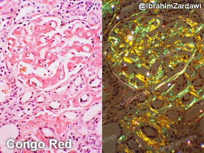
This renal cortical histology specimen demonstrates AL-type amyloidosis with kidney involvement in a patient with multiple myeloma. The left panel shows hematoxylin and eosin–stained tissue with amorphous, eosinophilic extracellular deposits within the glomerular mesangium and extending into the tubulointerstitium, causing mesangial expansion and variable basement membrane thickening. The right panel uses Congo Red staining to highlight the amyloid deposits, which appear salmon-pink on bright-field examination. Under polarized light, the same deposits display the diagnostic apple-green birefringence, a hallmark of Congo Red–positive amyloid. The deposits are diffuse, involving multiple glomeruli and extending into the tubulointerstitium, sometimes outlining capillary walls. In AL amyloidosis associated with plasma cell dyscrasia, the deposited light-chain–derived proteins disrupt normal renal architecture and contribute to proteinuria and progressive renal impairment. Differential considerations include AA (secondary) amyloidosis and other renal amyloidoses, but the combination of a known plasma cell disorder, Congo Red positivity, and apple-green birefringence strongly supports AL-type amyloidosis. This image serves as a valuable teaching reference for renal pathology, nephrology, and hematology-oncology, illustrating classic Congo Red histology, amyloid distribution patterns within glomeruli and interstitium, and the polarizing light signature. Correlates with proteinuria and renal dysfunction in many myeloma patients, guiding targeted therapy and monitoring of clonal plasma cell burden.
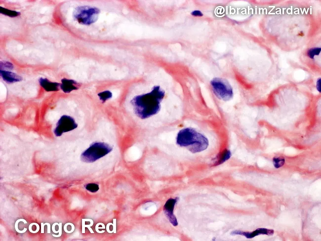
This histology image depicts human cardiac tissue (myocardium) stained with Congo Red and examined under bright-field microscopy. The image highlights extracellular, eosinophilic amyloid deposits that encircle and encroach upon individual cardiomyocytes (perimyocytic/interstitial deposition). The deposits appear as amorphous, orange to salmon-pink material on Congo Red, consistent with amyloid. When the same tissue is examined under polarized light, Congo Red–positive material exhibits characteristic apple-green birefringence, a pathognomonic feature of amyloidosis. The prevailing pattern is perimyocytic, with amyloid accumulating in the interstitium around myocytes and near small vessels, potentially contributing to myocardial stiffness and diastolic impairment. Histologic architecture shows preserved cellular morphology of myocytes with basophilic nuclei contrasted against the pink-red amyloid matrix. This image is diagnostic for cardiac amyloidosis on biopsy and would typically prompt typing (AL vs ATTR) by immunohistochemistry or mass spectrometry, as well as correlation with serum free light chains or transthyretin testing. Clinically, such infiltration predisposes to restrictive cardiomyopathy, diastolic dysfunction, arrhythmias, and heart failure symptoms. This slide underscores the utility of Congo Red staining in identifying myocardial amyloid deposits and the diagnostic value of polarized light birefringence for definitive confirmation. Useful for teaching, differential diagnosis, and research into infiltrative cardiomyopathies and treatment monitoring.
Amyloidosis
Definition
Structure of Amyloid
- Beta-pleated sheet conformation on X-ray diffraction
- Congo red positivity - salmon/orange on light microscopy, with pathognomonic apple-green birefringence under polarized light
- Thioflavin T fluorescence
- Ultrastructure: straight, non-branching, non-anastomosing fibrils, 8-12 nm in diameter
- Presence of serum amyloid P (SAP), a 25-kDa pentraxin glycoprotein, in all subtypes
- Association with sulfated glycosaminoglycans and proteoglycans
Pathogenesis
- Normal proteins with an inherent tendency to misfold and self-associate into fibrils, particularly when overproduced or when degradation is impaired
- Variant (mutant) proteins that are intrinsically prone to misfolding due to genetic mutations
Classification
| Type | Fibril Protein | Precursor | Clinical Setting |
|---|---|---|---|
| AL | Immunoglobulin light chains (chiefly λ) | Monoclonal plasma cells | Multiple myeloma, plasma cell dyscrasias |
| AA | Serum amyloid A (SAA) protein | SAA (acute-phase reactant) | Chronic inflammation (RA, Crohn, TB, FMF) |
| ATTRwt | Wild-type transthyretin | TTR (liver) | Aging males >70 years; cardiac predominant |
| ATTRv | Mutant transthyretin | TTR variant | Hereditary; cardiomyopathy + neuropathy |
| Aβ2M | β2-microglobulin | MHC class I component | Long-term hemodialysis; periarticular |
| Aβ | Amyloid β protein | Amyloid precursor protein (APP) | Alzheimer disease (localized cerebral) |
| ACal | Calcitonin | Calcitonin | Medullary thyroid carcinoma |
| AIAPP | Islet amyloid polypeptide | IAPP | Type 2 diabetes mellitus |
- Robbins, Cotran & Kumar Pathologic Basis of Disease, Table 6.17
Key subtypes in detail
Clinical Features
AL Amyloidosis
- Kidney (~60%): Proteinuria, often nephrotic-range; progressive renal insufficiency. Most common clinical presentation is nephrotic syndrome.
- Heart (~50-75%): Restrictive cardiomyopathy, heart failure with preserved EF (HFpEF), conduction abnormalities, arrhythmias
- Peripheral/autonomic nervous system: Sensorimotor neuropathy, orthostatic hypotension, GI dysmotility (early satiety, diarrhea, constipation), impotence, neurogenic bladder, dry eyes/mouth
- Soft tissue: Macroglossia - a pathognomonic sign of AL amyloidosis; carpal tunnel syndrome; "shoulder pad sign" (periarticular soft tissue infiltration)
- GI tract/liver: Hepatomegaly, malabsorption, bleeding
- Periorbital ecchymosis ("raccoon eyes"): from capillary fragility - considered a pathognomonic sign of AL amyloidosis
- Skin: Waxy papules/plaques, petechiae, purpura
Cardiac Amyloidosis (AL and ATTR)
- Restrictive filling pattern on echocardiography - "granular sparkling" myocardial texture
- Conduction system disease (AV block, requiring pacemaker)
- Atrial fibrillation (extremely common in ATTR; very high stroke risk due to atrial amyloid deposition even in sinus rhythm - TEE before cardioversion is mandatory)
- Sudden cardiac death less common than pulseless electrical activity
AA Amyloidosis
Diagnosis
Biopsy and Histology
- Abdominal fat pad aspirate: Most accessible first-line biopsy; positive in >80% of patients with systemic amyloidosis. Fat aspirated with a 16-gauge needle, stained with Congo red.
- Rectal mucosa biopsy: Historically common
- Organ biopsy (kidney, heart, liver, tongue, GI): For higher-yield confirmation when fat aspirate is negative
- Congo red staining + polarized light: Apple-green birefringence = gold standard for amyloid diagnosis
- Additional stains: Thioflavin T (fluorescence), crystal violet (metachromasia), PAS-positive/diastase-resistant
- Note on subtypes: Secondary AA amyloid loses birefringence after potassium permanganate treatment; AL and localized forms do not
Typing the Amyloid
- Immunohistochemistry (IHC): Antibodies against specific proteins (AL λ/κ chains, AA, transthyretin); limited sensitivity (difficulties in 14-35% of AL cases)
- Mass spectrometry (laser microdissection + LC-MS/MS): Current gold standard for protein identification; sensitivity 88%, specificity 96% - superior to IHC. Required for rare subtypes (ALECT2, etc.)
- Serum/urine protein electrophoresis + immunofixation: To detect monoclonal protein in AL
- Serum free light chains: Essential in AL workup
- Genetic sequencing: For hereditary ATTR and other familial forms
- Radiolabeled SAP scintigraphy: Whole-body imaging to assess amyloid burden and monitor regression (not universally available)
- Bone scan (99mTc-DPD/PYP scintigraphy): Highly specific for ATTR cardiac amyloidosis; positive scan + absence of monoclonal protein = non-invasive diagnosis of ATTRwt cardiac amyloid
Treatment
AL Amyloidosis
- Chemotherapy targeting the plasma cell clone to reduce light-chain production:
- Bortezomib + melphalan + dexamethasone (BMDex): Significantly improves progression-free and overall survival
- Daratumumab (anti-CD38 monoclonal antibody) + CyBorD: FDA-approved; the ANDROMEDA trial demonstrated superior hematologic response with daratumumab-based regimens - Goldman-Cecil Medicine
- High-dose melphalan + autologous stem cell transplant: For eligible patients (cardiac function must be adequate)
- Supportive: Diuretics for fluid overload (cautiously), compression stockings for orthostasis, anticoagulation if AF
ATTR Amyloidosis
- TTR stabilizers:
- Tafamidis: FDA-approved for wild-type or hereditary ATTR cardiomyopathy. Binds selectively to transthyretin, slowing monomer formation, misfolding, and amyloidogenesis
- Diflunisal (NSAID): Repurposed for ATTR amyloidosis treatment
- TTR silencers (reduce TTR production by liver):
- Patisiran (siRNA): FDA-approved for hereditary ATTR amyloidosis with polyneuropathy
- Inotersen (antisense oligonucleotide): Also approved for ATTRv polyneuropathy
- Additional novel agents in clinical trials
- Liver transplantation: Removes the source of mutant TTR (95% of TTR produced by liver); used for ATTRv prior to pharmacotherapy era; now combined with TTR-specific drugs post-transplant
- Heart transplantation: Selected patients with advanced cardiac amyloidosis; outcomes comparable to non-amyloid indications for ATTRwt and cardiac-restricted ATTRv
AA Amyloidosis
- Treat the underlying inflammatory disease to reduce SAA protein levels:
- Anti-TNF agents, IL-1 blockers (e.g., anakinra/canakinumab for FMF and Still's disease)
- Colchicine for Familial Mediterranean Fever
- Aggressive treatment of chronic infections (TB, etc.)
- SAA reduction leads to regression of amyloid deposits (demonstrated by SAP scintigraphy)
Dialysis-related (Aβ2M)
- High-flux dialysis membranes reduce β2-microglobulin accumulation
- Renal transplantation is definitive
Key Points Summary
| Feature | AL | AA | ATTRwt | ATTRv |
|---|---|---|---|---|
| Fibril | Light chain | SAA protein | Wild-type TTR | Mutant TTR |
| Cause | Plasma cell dyscrasia | Chronic inflammation | Aging | Inherited mutation |
| Age | Any, usually >50 | Any | Males >70 | Variable |
| Heart | +++++ | + | +++++ | ++ to +++++ |
| Kidney | +++++ | +++++ | + | Variable |
| Nerve | + | - | - | +++++ |
| Treatment | Daratumumab/bortezomib ± SCT | Treat underlying disease | Tafamidis | Patisiran/inotersen/tafamidis |