
What is meant by musctropy ular dys
Muscular dystrophy (MD) is a group of inherited genetic diseases that cause progressive weakening and degeneration of skeletal muscles. The underlying defect is a mutation in genes responsible for producing proteins that maintain muscle fiber structure and function.
Searching sources...
Reading File
Searching the Web
muscular dystrophy types mechanisms overview
Finding Sources
Searching Images
muscular dystrophy muscle biopsy histology
Reading File
Finding Sources
Reading File
Reading File
Loading Image
Here is a thorough explanation of Muscular Dystrophy (MD):
What is Muscular Dystrophy?
Muscular dystrophy refers to a group of hereditary disorders of skeletal muscle that cause progressive degeneration of skeletal muscle and associated weakness. The word itself comes from Greek: mys (muscle) + dys (faulty/bad) + trophe (nourishment). While the genetic mutations differ between types, all forms share the same core feature: progressive loss of muscle mass and strength.
- Campbell's Operative Orthopaedics 15th Ed
How It Happens - The Core Mechanism
Most forms involve defects in a protein complex called the dystrophin-glycoprotein complex, which:
- Spans the muscle cell membrane
- Links the internal cytoskeleton (inside the fiber) to the basement membrane (outside the cell)
- Provides mechanical stability to the myofiber during contraction
When this complex is defective, small tears develop in the membrane during contraction, allowing calcium to flood in, triggering a cascade that ends in myofiber degeneration and replacement by fibrofatty tissue.
- Robbins & Kumar Basic Pathology
Major Types
| Type | Inheritance | Key Defect | Onset | Severity |
|---|---|---|---|---|
| Duchenne (DMD) | X-linked recessive | Dystrophin completely absent | 3-5 years | Most severe; wheelchair by ~12, fatal by early adulthood |
| Becker (BMD) | X-linked recessive | Truncated/partial dystrophin | ~12 years | Milder; ambulation lost avg age 25-30 |
| Emery-Dreifuss | X-linked recessive | Nuclear envelope proteins | Childhood | Cardiac involvement prominent |
| Limb-girdle (LGMD) | Autosomal recessive (mostly) | Sarcoglycans or other proteins | Variable | Proximal limb weakness |
| Facioscapulohumeral (FSHD) | Autosomal dominant | DUX4 gene derepression | Teens-young adult | Face, shoulder, arm muscles |
| Myotonic | Autosomal dominant | DMPK gene (CTG repeat) | Adults | Most common adult form; muscle stiffness + weakness |
| Congenital (CMD) | Autosomal recessive | Merosin (laminin-a2) and others | Birth | Severe, early onset |
| Oculopharyngeal | Autosomal dominant | PABPN1 gene | >50 years | Eyelid drooping, swallowing problems |
Duchenne MD - The Most Common & Severe Form
- Incidence: 1 in 3,500 live male births
- Caused by deletions or frameshift mutations in the dystrophin gene (Xp21 locus), one of the largest human genes (2.3 million base pairs, 79 exons)
- Results in total absence of dystrophin
- Boys typically present with toe-walking and delayed walking milestones
- Progression: proximal weakness → Gowers' sign (using hands to "climb up" the legs when rising) → loss of ambulation → respiratory failure
- Cardiomyopathy develops in many patients as cardiac muscle is also affected
- No cure currently exists; treatment is supportive (corticosteroids, physiotherapy, respiratory support)
Microscopic Appearance (DMD)
The image below shows immunostaining for dystrophin in a DMD patient's muscle. Compare to normal muscle where a strong, uniform brown outline is seen at every cell membrane - in DMD, many fibers show absent or severely reduced dystrophin staining:
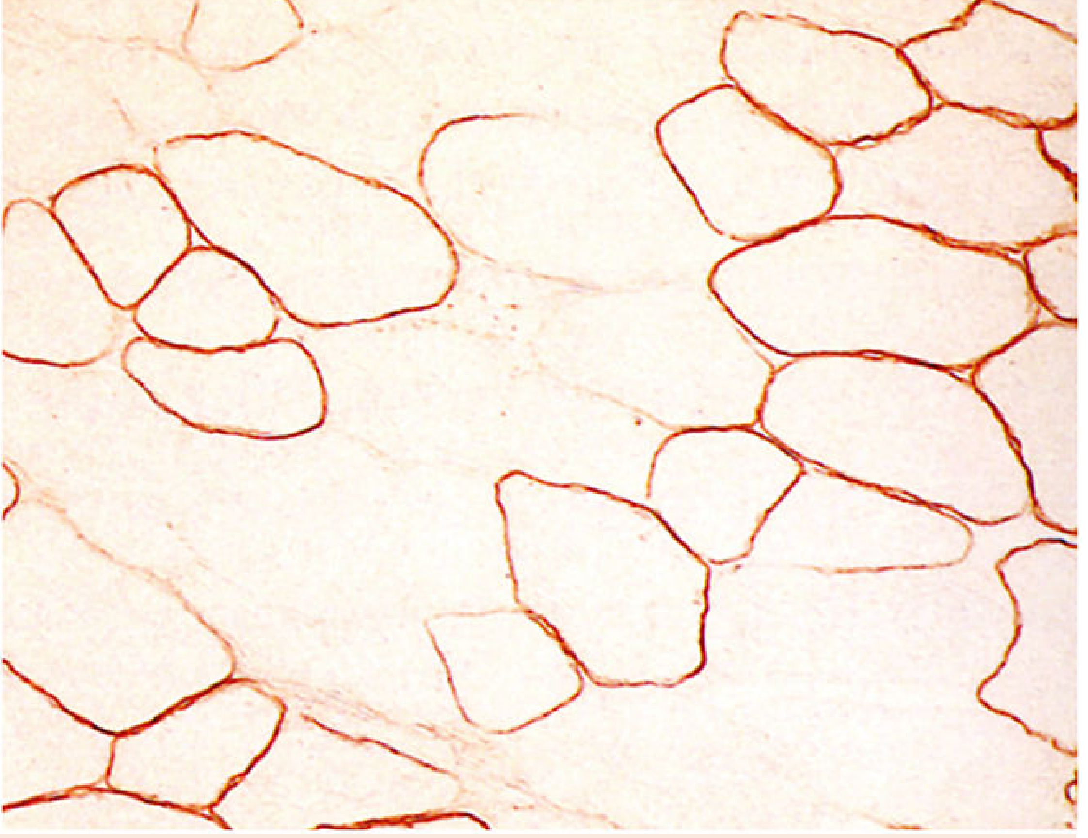
Skeletal muscle cross-section from a DMD patient. Many fibers show absent or variable dystrophin expression (compare to normal where every cell membrane is uniformly outlined). x480. - Histology: A Text and Atlas
Key Points to Remember
- Genetic - all forms are hereditary, caused by mutations in various genes
- Progressive - muscle weakness worsens over time
- Proximal-to-distal pattern in most types (hip/shoulder girdle muscles affected first)
- Pseudohypertrophy of calves is classic in DMD (muscle replaced by fat and connective tissue, but appears bulky)
- Elevated CK (creatine kinase) in blood is an early marker
- Diagnosis confirmed by genetic testing, muscle biopsy, and protein analysis (immunostaining for dystrophin)
- Treatment is largely supportive; gene therapy and exon-skipping strategies (e.g., eteplirsen for DMD) are active research areas
This is a shared conversation. Sign in to Orris to start your own chat.