Doc for chicken pox ?

Reading File
Finding Sources
Searching Images

chickenpox varicella rash vesicles skin

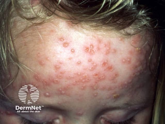
Clinical photography of active varicella (chickenpox) rash on the frontal scalp and forehead of a pediatric patient. Modality: digital color photograph taken under ambient lighting; standard clinical technique without dermoscopy or fluorescence. The image shows numerous small vesicles on an erythematous, slightly raised base distributed across the forehead and extending toward the temple. Lesions are in multiple evolutionary stages, with some vesicles capturing clear serous content, others showing crusted surfaces, and crusts around the margins. The pattern is characteristic of varicella: centripetal distribution with trunk and face involvement, crops of lesions appearing sequentially, and a tendency for vesicles to be more numerous on exposed skin areas. The epidermis and superficial dermis contain superficial fluid-filled vesicles, each about 2-4 mm in diameter, some conforming to the classic dew drop on a rose petal description. Facial involvement may accompany fever and malaise in pediatric patients. Although lesions may be pruritic, Nikolsky sign is typically absent; scarring is uncommon in children. Diagnosis is clinical, supported by history of varicella exposure or vaccination status; differential includes disseminated herpes zoster, pityriasis rosea, and vesicular dermatitis. Recognizing multistage lesions in a contagious exanthem is clinically significant for isolation, supportive therapy, and public health reporting as Varicella-zoster virus infection.

Clinical photography of active varicella (chickenpox) rash on the frontal scalp and forehead of a pediatric patient. Modality: digital color photograph taken under ambient lighting; standard clinical technique without dermoscopy or fluorescence. The image shows numerous small vesicles on an erythematous, slightly raised base distributed across the forehead and extending toward the temple. Lesions are in multiple evolutionary stages, with some vesicles capturing clear serous content, others showing crusted surfaces, and crusts around the margins. The pattern is characteristic of varicella: centripetal distribution with trunk and face involvement, crops of lesions appearing sequentially, and a tendency for vesicles to be more numerous on exposed skin areas. The epidermis and superficial dermis contain superficial fluid-filled vesicles, each about 2-4 mm in diameter, some conforming to the classic dew drop on a rose petal description. Facial involvement may accompany fever and malaise in pediatric patients. Although lesions may be pruritic, Nikolsky sign is typically absent; scarring is uncommon in children. Diagnosis is clinical, supported by history of varicella exposure or vaccination status; differential includes disseminated herpes zoster, pityriasis rosea, and vesicular dermatitis. Recognizing multistage lesions in a contagious exanthem is clinically significant for isolation, supportive therapy, and public health reporting as Varicella-zoster virus infection.

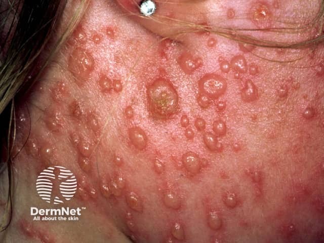
Clinical photograph of facial skin demonstrates an acute varicella-zoster infection (chickenpox) with a vesiculopapular eruption at varying stages. The image captures a close-up view of the left facial region (cheek/temporal area) showing numerous small translucent vesicles atop an erythematous, inflamed surface. Some lesions appear as dew drop vesicles; others are crusted, eroded, or coalesced into shallow plaques. The lesions are distributed in crops, with evidence of ongoing new vesicle formation alongside older crusted lesions, reflecting the characteristic centripetal pattern of varicella. The surrounding skin is mildly edematous and inflamed, with focal tenderness and pruritus typically reported in clinical practice. This presentation is diagnostic in the appropriate clinical context when accompanied by fever, malaise, and a diffuse vesiculopapular rash, though varicella can resemble herpes simplex virus infections or disseminated contact dermatitis. The image emphasizes cutaneous involvement as the primary visual clue for clinicians, educators, and researchers assessing varicella morphology, staging, and differential diagnosis. Clinically, recognition supports prompt isolation to reduce transmission and informs management decisions, including antiviral therapy considerations in adults or immunocompromised patients. This photograph serves as a visual educational resource for dermatology, pediatrics, infectious disease, and medical training exams.

Clinical photograph of facial skin demonstrates an acute varicella-zoster infection (chickenpox) with a vesiculopapular eruption at varying stages. The image captures a close-up view of the left facial region (cheek/temporal area) showing numerous small translucent vesicles atop an erythematous, inflamed surface. Some lesions appear as dew drop vesicles; others are crusted, eroded, or coalesced into shallow plaques. The lesions are distributed in crops, with evidence of ongoing new vesicle formation alongside older crusted lesions, reflecting the characteristic centripetal pattern of varicella. The surrounding skin is mildly edematous and inflamed, with focal tenderness and pruritus typically reported in clinical practice. This presentation is diagnostic in the appropriate clinical context when accompanied by fever, malaise, and a diffuse vesiculopapular rash, though varicella can resemble herpes simplex virus infections or disseminated contact dermatitis. The image emphasizes cutaneous involvement as the primary visual clue for clinicians, educators, and researchers assessing varicella morphology, staging, and differential diagnosis. Clinically, recognition supports prompt isolation to reduce transmission and informs management decisions, including antiviral therapy considerations in adults or immunocompromised patients. This photograph serves as a visual educational resource for dermatology, pediatrics, infectious disease, and medical training exams.

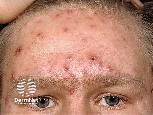
Clinical photograph of facial skin during varicella (chickenpox) infection, frontal view of the forehead. Imaging modality: Clinical photography using standard color photography under visible light; single exposure. The photograph documents vesicular lesions at multiple stages, including dew-drop vesicles on an erythematous base, small papules, and crusted plaques. The forehead demonstrates a cluster of shallow vesicles with surrounding dermal erythema and occasional crusts, while adjacent skin shows mild edema and pruritus-related rubbing. Lesions are predominantly superficial, with translucent fluid-filled vesicles and some crusted centers, consistent with secondary infection risk being low in this image. The distribution appears facial and possibly centripetal with more lesions on the face than on surrounding regions, a common pattern in pediatric varicella. No mucosal involvement is evident in this view. Differential considerations include disseminated herpes zoster in older adults or immunocompromised patients, pityriasis rosea, hand-foot-and-mouth disease, and impetiginized lesions; however, the vesicular morphology and clinical context favor varicella. Diagnostic significance: characteristic varicella rash pattern supports clinical diagnosis when combined with fever and malaise; useful for educational, triage, and telemedicine documentation. Potential clinical use cases include teaching dermatology residents, documenting eruption stage for treatment planning, and monitoring response to antiviral therapy.

Clinical photograph of facial skin during varicella (chickenpox) infection, frontal view of the forehead. Imaging modality: Clinical photography using standard color photography under visible light; single exposure. The photograph documents vesicular lesions at multiple stages, including dew-drop vesicles on an erythematous base, small papules, and crusted plaques. The forehead demonstrates a cluster of shallow vesicles with surrounding dermal erythema and occasional crusts, while adjacent skin shows mild edema and pruritus-related rubbing. Lesions are predominantly superficial, with translucent fluid-filled vesicles and some crusted centers, consistent with secondary infection risk being low in this image. The distribution appears facial and possibly centripetal with more lesions on the face than on surrounding regions, a common pattern in pediatric varicella. No mucosal involvement is evident in this view. Differential considerations include disseminated herpes zoster in older adults or immunocompromised patients, pityriasis rosea, hand-foot-and-mouth disease, and impetiginized lesions; however, the vesicular morphology and clinical context favor varicella. Diagnostic significance: characteristic varicella rash pattern supports clinical diagnosis when combined with fever and malaise; useful for educational, triage, and telemedicine documentation. Potential clinical use cases include teaching dermatology residents, documenting eruption stage for treatment planning, and monitoring response to antiviral therapy.

Searching PubMed

varicella chickenpox treatment management

Reading File
Reading File
Finding Sources
Reading File
Reading File
Reading File
Reading File
Reading File
I now have comprehensive information from multiple authoritative textbooks. Let me compile the full document on Chickenpox.

Chickenpox (Varicella) - Clinical Overview

Definition

Varicella (chickenpox) is an acute, highly infectious disease caused by Varicella-Zoster Virus (VZV), also called Human (alpha) herpesvirus 3. It is characterized by a generalized vesicular rash accompanied by fever and malaise. Chickenpox represents the primary infection with VZV; the same virus establishes latency in dorsal root ganglia and can reactivate decades later as herpes zoster (shingles).
  • Goldman-Cecil Medicine, p. 3608
  • Harrison's Principles of Internal Medicine 22E, p. 1548

Pathogen

VZV is a member of the Herpesviridae family with the following features:
  • Structure: Lipid envelope + icosahedral nucleocapsid; ~180-200 nm diameter; double-stranded DNA (~125,000 bp)
  • Glycoproteins on the viral envelope are targets for neutralizing antibodies
  • Encodes a thymidine kinase that phosphorylates acyclovir → inhibits viral DNA polymerase (basis of antiviral therapy)
  • Harrison's Principles of Internal Medicine 22E, p. 1548

Epidemiology

ParameterDetail
Global disease burden~4.2 million severe complications/year; ~4,200 deaths/year
Pre-vaccine case fatality rate~3 per 100,000 cases (high-income countries)
TransmissionRespiratory droplets + direct contact with skin lesions
Attack rate≥90% in susceptible (seronegative) contacts
ReservoirHumans only
Incubation period14-16 days (range: 10-21 days)
Period of communicability1-2 days before rash to 6 days after onset
Peak ageChildren (most cases <10 years); adults are more severely affected
  • Park's Textbook of Preventive and Social Medicine, p. 163

Clinical Features

Prodrome

  • Children: Usually absent or mild (low-grade fever, malaise 1-2 days before rash)
  • Adults: More pronounced prodrome - fever, headache, malaise, myalgia, arthralgia precede the rash by 2-4 days

Rash - The Hallmark

The rash appears in successive crops (new crops every 3-4 days), giving lesions at different stages simultaneously:
  1. MaculePapuleVesicle ("dew drop on a rose petal") → PustuleCrust/Scab
  2. Distribution: Centripetal - starts on trunk/face/scalp, then spreads to extremities (face, trunk > limbs)
  3. Mucous membrane involvement: Oral mucosa, conjunctiva, genitalia
  4. Pruritus is intense
  5. Lesions at different stages simultaneously is pathognomonic
Varicella rash - vesicular lesions at multiple stages on forehead
Facial chickenpox lesions showing dew-drop vesicles with erythematous base
  • Harrison's Principles of Internal Medicine 22E, p. 1548

Pathogenesis

  1. Respiratory droplets inhaled → replicate in nasopharynx → seed lymphatics/reticuloendothelial system → viremia (primary)
  2. Virus seeds skin → vesicle formation: ballooning degeneration, multinucleated giant cells, eosinophilic intranuclear inclusions
  3. Vesicular fluid becomes cloudy with PMNs → ruptures or reabsorbs
  4. After primary infection: VZV travels via sensory nerves to dorsal root ganglia → establishes lifelong latency
  5. Reactivation → Herpes zoster (shingles)
  • Harrison's Principles of Internal Medicine 22E, p. 1548

Complications

In Healthy Children

  • Usually mild and self-limiting

Serious Complications (especially adults, immunocompromised, neonates, pregnant women)

ComplicationNotes
Varicella pneumoniaMost common life-threatening complication in adults; rare in healthy children; interstitial pneumonitis
EncephalitisPerivascular cuffing in brain; less common than in measles
Acute cerebellar ataxiaMore common in children
Reye's syndromeAcute encephalopathy + fatty liver degeneration (associated with aspirin use - NEVER give aspirin)
Secondary bacterial infectionsGroup A beta-hemolytic Streptococcus, Staphylococcus aureus → cellulitis, erysipelas, epiglottitis, osteomyelitis
Hemorrhagic varicellaRare, life-threatening
DICIn immunocompromised; rapidly fatal
Congenital varicella syndromeMaternal infection → cutaneous scars, limb atrophy, microcephaly, cataracts, chorioretinitis, deafness
Neonatal varicellaMother develops rash within 5 days before/48 hours after delivery → severe disseminated neonatal disease
Acute retinal necrosisMajor VZV manifestation, especially in AIDS patients
  • Park's Textbook of Preventive and Social Medicine, p. 163

Laboratory Diagnosis

TestNotes
PCR (vesicular fluid, crusts, saliva, CSF)Gold standard - most sensitive and specific
Cell cultureVZV isolation; slower
Direct immunofluorescence (DIF)Rapid but lower sensitivity than PCR
IgM antibodyLess sensitive; not method of choice for acute varicella
IgG serologyUsed to assess immunity in unvaccinated individuals (e.g., healthcare workers)
Clinical diagnosis is usually sufficient; labs reserved for atypical or complicated cases, or post-eradication era differentiation from smallpox.
  • Park's Textbook of Preventive and Social Medicine, p. 163

Treatment

Supportive (All Cases)

  • Antipyretics: Paracetamol (acetaminophen) - NEVER aspirin (risk of Reye's syndrome)
  • Antihistamines and calamine lotion for pruritus
  • Keep nails trimmed; avoid scratching (prevents secondary infection and scarring)
  • Maintain hydration

Antiviral Therapy

DrugDoseDurationIndication
Acyclovir (oral)800 mg 5x/day (adults); 20 mg/kg/dose 4x/day (children >2 yrs, max 800 mg)5-7 daysHealthy adolescents >13 yrs, adults, secondary household cases
Valacyclovir1 g PO 3x/day5-7 daysPreferred over acyclovir (better bioavailability, less frequent dosing)
Famciclovir500 mg PO 3x/day7 daysAlternative; at least as effective as acyclovir
IV Acyclovir10 mg/kg every 8 hours7 daysSeverely immunocompromised, varicella pneumonia, encephalitis
Start antivirals within 24 hours of rash onset for maximum benefit.
  • Harrison's Principles of Internal Medicine 22E, p. 1548

Prevention

1. Live Attenuated Varicella Vaccine (Oka strain - VAR)

  • Children 12-15 months: First dose
  • 4-6 years: Second dose (booster)
  • Seronegative persons >13 years: Two doses at least 1 month apart
  • Contraindicated if CD4 count <200 cells/mm³ (immunocompromised)
  • Results in significant decline in chickenpox incidence in vaccinated communities

2. Varicella-Zoster Immunoglobulin (VZIG) - Post-Exposure Prophylaxis

Given within 96 hours of exposure to high-risk individuals:
  • Immunocompromised susceptible children (no prior varicella or vaccination)
  • Susceptible pregnant women
  • Newborn whose mother had chickenpox within 5 days before or 48 hours after delivery
  • Hospitalized premature infants (≥28 weeks) whose mother is seronegative
  • Premature infants <28 weeks (regardless of maternal history)

3. Control Measures

  • Isolation of cases for ~6 days after onset of rash
  • Notification to public health authorities
  • Disinfection of articles soiled with discharge
  • Harrison's Principles of Internal Medicine 22E, p. 1548
  • Park's Textbook of Preventive and Social Medicine, p. 163

Special Situations

SituationManagement
PregnancyOral acyclovir for mild disease; IV acyclovir for severe/pneumonia; VZIG for susceptible women exposed; delivery-associated varicella requires neonatal VZIG
ImmunocompromisedIV acyclovir initially; reduce immunosuppression if possible
Neonatal varicellaVZIG + IV acyclovir
Varicella pneumoniaIV acyclovir + ventilatory support if needed

Key Differentials

ConditionDistinguishing features
SmallpoxAll lesions in same stage; centrifugal distribution; deeper lesions; ERADICATED
Herpes simplex (disseminated)Localized clusters; history of HSV
Hand-foot-mouth diseaseLesions on palms/soles/oral mucosa
ImpetigoHoney-crusted; no systemic features
Insect bitesGrouped, no multiple stages simultaneously

Summary Table

FeatureDetail
Causative agentVZV (Human herpesvirus 3)
TransmissionRespiratory droplets, direct contact
Incubation10-21 days (mean 14-16 days)
Infectivity1-2 days before rash to 6 days after
RashCentripetal, polymorphic, crops of vesicles
Key diagnostic signLesions at multiple stages simultaneously
Complication to fearVaricella pneumonia (adults), Reye's (aspirin), bacterial superinfection
TreatmentAcyclovir/Valacyclovir; NO aspirin
PreventionLive attenuated vaccine (Oka); VZIG post-exposure

Sources: Harrison's Principles of Internal Medicine 22E (2025) | Goldman-Cecil Medicine | Park's Textbook of Preventive and Social Medicine | Andrews' Diseases of the Skin
This is a shared conversation. Sign in to Orris to start your own chat.