
adnenoida size variation with age quick data easy to remember
adenoid size variation with age normal growth involution
https://pmc.ncbi.nlm.nih.gov/articles/PMC9499359
adenoid size age chart growth involution nasopharynx
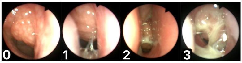
A series of four endoscopic clinical photographs of the nasopharynx demonstrating the Mucus of Adenoid Scale by Nasopharyngoscopy Assessment (MASNA). The images are labeled 0 through 3, illustrating a progressive increase in mucus volume and viscosity on the adenoid tissue. Scale 0 shows a normal, pink adenoid with no visible mucus. Scale 1 illustrates a mild residue of clear, watery, and transparent mucus. Scale 2 shows a moderate amount of dense, semi-opaque mucus accumulating on the surface. Scale 3 depicts copious, thick, dense, and opaque white-to-yellow mucus largely occluding the field of view and covering the adenoid. This comparison chart is used in Otolaryngology to standardize the evaluation of postnasal drip, rhinosinusitis, and adenoiditis. The educational focus is on distinguishing mucus characteristics—transparency, color, and thickness—to objectively score inflammatory status during diagnostic nasopharyngoscopy.
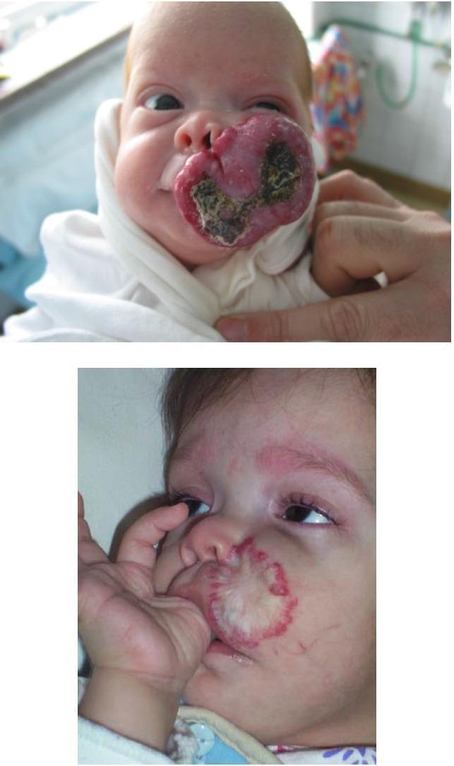
This comparison chart consists of two clinical photographs showing the progression and involution of an infantile hemangioma (IH) on the upper lip and left cheek of a pediatric patient. The first image depicts the patient at 1 month of age with a large, exophytic, bright red vascular tumor characterized by a lobulated, raised surface and central areas of dark crusting/necrosis, suggesting ulceration. The lesion involves the philtrum and extends onto the left malar region. The second image shows the same patient at age 2 years, demonstrating significant involution of the hemangioma. The previously large mass has regressed into a flatter, depressed scar-like area with central pallor/fibrofatty replacement and a peripheral telangiectatic red rim. This timeline illustrates the natural history of proliferative infantile hemangiomas, transitioning from an active growth phase with potential complications like ulceration to the late involution stage. The images are highly relevant for dermatology and pediatrics regarding vascular birthmark management and clinical observation.
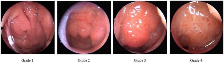
This clinical comparison chart presents four nasoendoscopic images illustrating the grading of adenoid hypertrophy according to the Parikh grading standard. The images provide a sequential view of the nasopharynx, focusing on the relationship between adenoid tissue and adjacent anatomical landmarks. Grade 1 shows a small adenoid mass with a clearly patent airway and visible surface folds. Grade 2 demonstrates moderate enlargement, reducing the airway space while maintaining visible landmarks such as the vomer bone. Grade 3 exhibits significant hypertrophy where the adenoid tissue occupies more than two-thirds of the choanal space, partially obscuring the Eustachian tube pillow. Grade 4 represents severe obstruction, with the adenoid tissue making contact with the soft palate and completely or near-completely obstructing the posterior nasal choana. Visually, the progression shows the tissue shifting from a small, pink, textured mass to a large, bulbous, and sometimes erythematous structure with prominent vascularity. This diagnostic series is essential for Otolaryngology (ENT) students to understand the endoscopic evaluation of pediatric airway obstruction and sleep-disordered breathing.
Adenoid Size Variation with Age
The Core Pattern: Born → Grow → Peak → Shrink
| Age | What Happens | Memory Hook |
|---|---|---|
| Birth | Present but tiny | "Born with it" |
| 0-6 months | Usually invisible / undetectable | Too small to see |
| 6 months - 6 years | Rapid growth, occupies up to 50% of nasopharynx by age 2 | "Grows fast, fills the space" |
| 7-10 years | PEAK size (~14-15 mm on MRI) | "7-10 = Maximum" |
| After 10-12 years | Gradual involution begins | "Teen years = shrinking" |
| Adult (60 yrs) | Shrinks to ~5 mm | "Nearly gone" |
Quick Memory Anchor: "2-7-10-14-gone"
- 2 yrs - fills ~50% of nasopharynx
- 7-10 yrs - PEAK (largest)
- 14-15 mm - peak MRI measurement
- Teens onward - involutes, nearly "gone" by adulthood (~5 mm by age 60)
Direction of Changes (MRI data)
- Growth goes: anterior + inferior direction
- Involution goes: posterior + superior direction (retreats the way it came)
What Peaks When (fine detail)
| Dimension | Peaks at |
|---|---|
| Adenoid depth | ~4 years |
| Adenoid area | ~6-7 years |
| Adenoid height & thickness | ~8-9 years |
Clinical Relevance
- Adenoid hypertrophy is most symptomatic at ages 3-8 yrs (nasal obstruction, otitis media with effusion, OSA, "adenoid facies")
- Size alone is NOT an indication for removal - it is the consequences (OSA, nasal block, recurrent AOM) that matter
- By teenage years, physiological involution usually resolves most obstruction without surgery
- The Parikh grading (1-4) is used endoscopically to assess obstruction relative to the choanal space
Waldeyer's Ring Context