
This is his cbc and esr
python3 -c " import pdfplumber with pdfplumber.open('/tmp/workspace/attachments/912a6712-0c19-48d8-88a7-c803e53a56ca/VAIBHAV_20RAJ-Male18_20years-375128.pdf') as pdf: for i, page in enumerate(pdf.pages): print(f'--- PAGE {i+1} ---') text = page.extract_text() print(text) print() tables = page.extract_tables() for j, table in enumerate(tables): print(f'TABLE {j+1}:') for row in table: print(row) print() "
CBC & ESR Report - Vaibhav Raj | 18M
Red Cell Parameters
| Parameter | Value | Reference | Status |
|---|---|---|---|
| Haemoglobin | 15.0 g/dL | 13.0-18.0 | ✅ Normal |
| RBC Count | 4.61 mil/cu.mm | 4.5-6.0 | ✅ Normal |
| PCV (Haematocrit) | 48.6% | 30-50% | ✅ Normal |
| MCV | 105.42 fL | 78-100 fL | ⚠️ HIGH - Macrocytosis |
| MCH | 32.54 pg | 27-31 pg | ⚠️ HIGH |
| MCHC | 30.86 g/dL | 32-36 g/dL | ⚠️ LOW - Hypochromia |
| RDW | 13.6% | 11.5-15.0% | ✅ Normal |
White Cell Parameters
| Parameter | Value | Reference | Status |
|---|---|---|---|
| Total WBC | 9,600 /cu.mm | 4,000-11,000 | ✅ Normal |
| Neutrophils | 57% | 40-70% | ✅ Normal |
| Lymphocytes | 36% | 20-50% | ✅ Normal |
| ALC | 3,456 | 1,000-3,000 | ⚠️ Mildly high |
| ANC | 5,472 | 2,000-7,000 | ✅ Normal |
Platelets
| Parameter | Value | Reference | Status |
|---|---|---|---|
| Platelet Count | 2,26,000 /cmm | 1,50,000-4,50,000 | ✅ Normal |
| MPV | 11.4 fL | 7.2-11.7 fL | ✅ Normal |
ESR
| Parameter | Value | Reference | Status |
|---|---|---|---|
| ESR (Wintrobe's) | 10 mm/1st hr | 0-20 | ✅ Normal |
Key Findings & Interpretation
1. Macrocytosis with Relative Hypochromia (the main abnormality)
- MCV 105.42 fL - clearly elevated, indicating macrocytic red cells
- MCH 32.54 pg - high (larger cells carry more haemoglobin in absolute terms)
- MCHC 30.86 g/dL - mildly low, suggesting the cells are not densely packed with haemoglobin despite being large (relative hypochromia)
- The peripheral smear confirms: "Macrocytic hyperchromia" (this is the lab's morphology descriptor - the large cells appear darker due to their size, but the MCHC tells us the concentration is actually slightly low)
2. Overall Haemoglobin is Normal
- Despite macrocytosis, Hb is 15 g/dL - he is not anaemic. This is an early or compensated state.
3. Absolute Lymphocyte Count mildly elevated (3,456 vs upper limit 3,000)
- Mild, likely reactive (viral illness, recent infection). Not clinically significant in isolation.
4. ESR is normal (10 mm/hr)
- No significant systemic inflammation at this time.
Differential Diagnosis for Macrocytosis in an 18-year-old male
| Cause | Notes |
|---|---|
| Vitamin B12 deficiency | Most common at this age - dietary (vegetarian/vegan diet), malabsorption |
| Folate deficiency | Poor diet, increased demand, malabsorption |
| Hypothyroidism | Can cause macrocytosis; check TSH |
| Liver disease / alcohol | Less likely at 18, but worth asking |
| Medications | Methotrexate, hydroxyurea, antiretrovirals |
| Reticulocytosis | Reticulocytes are larger - check if haemolysis/blood loss present |
Suggested Next Steps
- Serum Vitamin B12 level - most likely cause at this age
- Serum Folate (RBC folate) - rule out folate deficiency
- Peripheral smear review - look for hypersegmented neutrophils (classic for B12/folate deficiency)
- TSH - screen for hypothyroidism
- LFTs - if any history of alcohol/liver issues
- Reticulocyte count - to rule out haemolysis as a cause
18 yr old pt presented with eczema type appearance around nose on scalp , chest and behind ears After stretching and rubbing it showed whitish flakes what is diagnosis and treatment This is his symptoms and above shown are lab reports
seborrhoeic dermatitis scalp face nose behind ears greasy scales
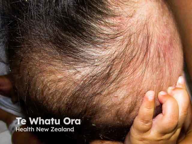
This is a high-resolution clinical photograph of an infant scalp demonstrating seborrhoeic dermatitis with yellow greasy scales preferentially located on the frontal/temporoparietal scalp and sparing the face. The image captures the characteristic matting and flaking scales adherent to hair shafts, with mild erythema and diffuse scale accumulation at the crown and temple regions. The lesion is non-vesicular, non-exudative, and appears symmetrical across the scanned scalp area. The findings are consistent with cradle cap, a common, benign dermatologic condition of infancy believed to reflect sebaceous gland activity and infant skin immaturity. Differential considerations include tinea capitis if alopecia or perifollicular scaling is prominent, atopic dermatitis with more diffuse erythema, or psoriasis with well-demarcated plaques though these are less typical in early life. Clinically correlated, this pattern typically responds to routine infant scalp hygiene and gentle, non-irritating cleansing; medicated soaps or antifungal shampoos may be used under pediatric guidance if scales persist. This image is valuable for educational reference in pediatric dermatology, dermoscopy correlation not shown, and radiology cross-disciplinary discussions about differential diagnoses of scalp scaling. Potential applications: medical education, image-based differential diagnosis training, clinical case discussions, and telemedicine triage for infants presenting with cradle cap.
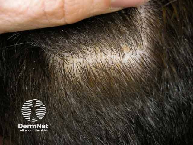
Clinical photograph of the scalp demonstrates seborrhoeic dermatitis characterized by honey-coloured, greasy scales adherent to hair shafts with mild surrounding erythema. Involvement is patchy to diffuse over hair-bearing skin, most pronounced at the vertex and temple regions, with scales accumulating along follicular openings. The yellowish scales are waxy and easily removable, yet can be persistent; pruritus is commonly reported in patients. Hair shafts appear preserved and no frank alopecia is evident in this image. This presentation aligns with the typical clinical phenotype of seborrhoeic dermatitis, a common inflammatory scalp dermatosis linked to sebaceous gland activity and colonization by Malassezia species. The visual features help distinguish it from psoriasis (silvery scales, well-demarcated plaques) and tinea capitis (hair loss, broken hairs, erythematous patches). Diagnostic significance lies in recognizing the greasy, yellow scales on the scalp as a hallmark, guiding topical therapies such as selenium sulfide, zinc pyrithione, ketoconazole antifungal shampoos, and low‑potency corticosteroids when appropriate. Clinically, the image is relevant for dermatology education, differential diagnosis exercises, and treatment response documentation in adult and pediatric patients. The image supports hands-on recognition of seborrhoeic dermatitis patterns in scalp dermatology curricula and serves as a reference for teledermatology consultations.
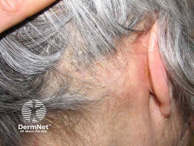
Clinical photography of the scalp demonstrating seborrhoeic dermatitis with thick adherent scales. The image presents a macroscopic view of the left temporal scalp and retroauricular region. Specimen type: human scalp skin. Imaging perspective: close-up, lateral scalp with ear visible. The scales are thick, greasy, yellowish, lamellate, coating the scalp surface and obscuring underlying hair shafts. Surrounding skin shows mild erythema and minimal crusting; hair density is preserved; no ulceration. This pattern is characteristic of seborrhoeic dermatitis: chronic relapsing inflammatory dermatosis associated with Malassezia yeast, sebaceous gland activity, and host inflammatory response. The lesion displays confluent plaques with adherent scale, diffuse but more prominent in chronically affected areas such as the temporal and retroauricular scalp. Diagnostic significance: photographic documentation aids clinical diagnosis, guides topical antifungal or anti-seborrheic therapy, and serves as baseline for response assessment. Differential considerations include psoriasis capitis (more silvery scale and well-demarcated plaques), tinea capitis (alopecia, hair shedding, broken hairs), and contact dermatitis. Clinical correlation includes pruritus, oiliness, and seasonal flares. This image is useful for education, dermatology training, and image-based search queries for seborrheic dermatitis of the scalp. Images like this support differential diagnosis, treatment planning, and educational demonstrations for medical students, residents, and clinicians in clinics.
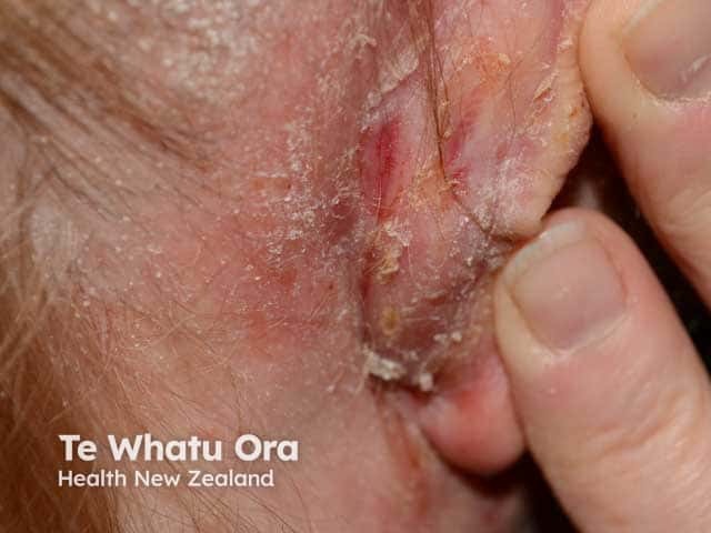
Comprehensive Description: A high-resolution clinical photograph captures the retroauricular skin behind the ear on an adult patient. The image demonstrates erythema and scale with crusted, greasy plaques localized to the posterior auricular region. The skin shows focal desquamation with lamellar white scales adherent to the lesion edges; surrounding skin appears mildly dry with faint erythema. There is no visible purulence, ulceration, or vesiculation. The distribution is localized to the retroauricular fold, consistent with seborrhoeic dermatitis involving sun-exposed or sebaceous-rich areas; reflectivity of the skin appears slightly oily, with fissuring minimized by moisture. The appearance is characteristic of chronic inflammatory dermatosis in adults, often presenting bilaterally on the scalp, eyebrows, nasolabial folds, and behind-ears. In this image, the retroauricular involvement aligns with typical patterns described for seborrheic dermatitis, as opposed to psoriasis or contact dermatitis, which may show more scaling, well-defined plaques, or vesiculation. The clinical context suggests a benign chronic inflammatory process related to Malassezia yeast overgrowth and sebaceous gland activity; the lesion responds to antifungal or anti-inflammatory therapy. This image is useful for dermatology education, differential diagnosis practice, and documentation of retroauricular SD for clinician reference and patient education.
Diagnosis: Seborrhoeic Dermatitis (SD)
- Eczema-like erythematous plaques in sebaceous-gland-rich zones - around the nose (nasolabial folds), scalp, chest (sternal area), and behind the ears (postauricular region)
- Whitish/yellowish greasy scales on rubbing/stretching - the hallmark sign
- Young male (males are more commonly affected)
- Normal ESR rules out significant systemic inflammation
Pathogenesis
- Malassezia yeast (lipophilic fungus, formerly Pityrosporum) - overgrowth on sebaceous skin
- Sebaceous gland activity - adolescents have high sebum production (explains why it's active at 18)
- Host inflammatory response - individual susceptibility to Malassezia triggers inflammation
Treatment
Scalp
| Step | Drug | Formulation | How to use |
|---|---|---|---|
| 1st line | Ketoconazole 2% | Shampoo | Apply 3x/week, leave 5 min, rinse |
| 1st line | Selenium sulfide 2.5% | Shampoo | 2-3x/week |
| 1st line | Zinc pyrithione 1-2% | Shampoo | Daily or alternate days |
| Add-on | Mild topical corticosteroid | Solution/lotion (1% hydrocortisone or 0.05% fluocinolone) | Short term for flares |
| Alternative | Coal tar shampoo | Shampoo | 2-3x/week |
Face (nasolabial folds, nose, ears)
| Step | Drug | Notes |
|---|---|---|
| 1st line | Ketoconazole 2% cream | Apply thinly BD for 2-4 weeks |
| 1st line | Mild corticosteroid (1% hydrocortisone) | Short-term only (1-2 weeks max) - avoid strong steroids on face |
| Alternative | Tacrolimus 0.1% or Pimecrolimus 1% cream (calcineurin inhibitors) | Preferred for long-term maintenance on face - no risk of skin atrophy |
| Adjunct | Emollients (non-comedogenic moisturiser) | Helps scale management |
Chest
- Ketoconazole 2% cream or shampoo used as a body wash
General Measures
- Gentle cleansing with a mild non-soap cleanser
- Avoid harsh scrubbing (worsens inflammation)
- Sun exposure helps - SD worsens in winter/low sunlight
- Avoid greasy/oily hair products
For Severe or Refractory Cases
- Oral itraconazole 200 mg/day x 7 days/month for a few months
- Oral fluconazole - alternative
- Narrowband UVB phototherapy - safe and effective option
- Low-dose isotretinoin 0.1-0.5 mg/kg/day for very severe cases
Addressing the CBC Findings in Context
- Serum B12 and Folate levels
- Peripheral smear for hypersegmented neutrophils