
Hemodynamic disorders. Embolism. Etiopathogenesis morphology
"Embolism"[MeSH Terms]
pulmonary embolus saddle embolus lung artery
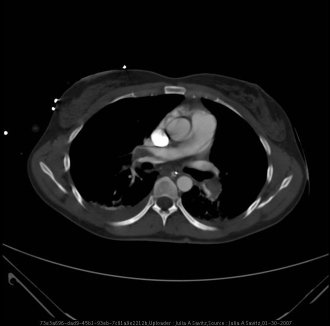
**Imaging Modality:** Contrast-enhanced Computed Tomography (CT) of the chest, axial view (soft tissue window).
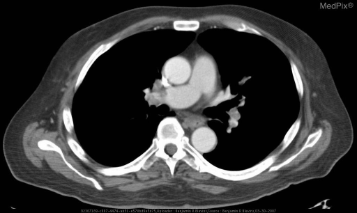
**Imaging Modality:** Contrast-enhanced Computed Tomography (CT) of the chest, axial view (CT Pulmonary Angiogram protocol).
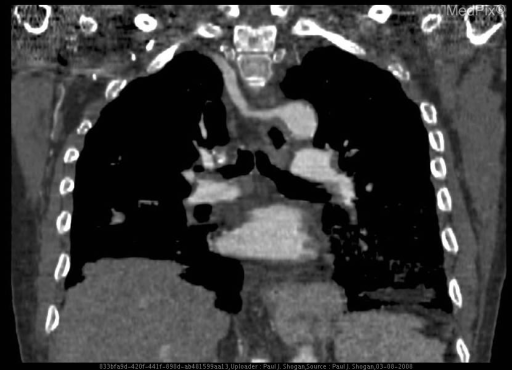
**Imaging Modality:** Computed Tomography (CT) scan, coronal reformatted view.
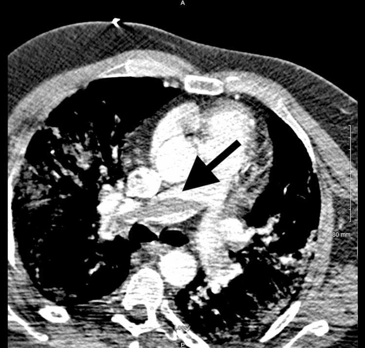
This diagnostic image is an axial contrast-enhanced computed tomography (CT) scan of the chest at the level of the pulmonary artery bifurcation. The central mediastinal structures, including the heart and ascending aorta, are visible. A prominent black arrow points to a significant filling defect within the main pulmonary artery that straddles the bifurcation into the right and left pulmonary arteries. This hypodense (darker) area within the bright, contrast-opacified vessel is characteristic of a 'saddle embolus,' a high-risk form of pulmonary embolism. The surrounding lung parenchyma is visible, showing peripheral lung markings and aeration. The image serves as a critical educational tool for identifying acute vascular emergencies in radiology and pulmonology, demonstrating the classic radiographic appearance of massive pulmonary thromboembolism.
Embolism: etiopathogenesis and morphology
Definition
- fat droplets
- air or nitrogen bubbles
- atherosclerotic debris (cholesterol emboli)
- tumor fragments
- bone marrow
- foreign bodies
- amniotic fluid
Classification
| Type | Main sources |
|---|---|
| Thromboembolism: pulmonary (venous) | Deep leg veins |
| Thromboembolism: systemic (arterial) | Left heart mural thrombi, valves, aorta |
| Paradoxical embolism | Venous thrombus crossing an ASD, VSD or PFO |
| Fat and marrow embolism | Long-bone fractures |
| Air and gas embolism | Surgery, procedures, decompression |
| Amniotic fluid embolism | Labor and the postpartum period |
| Others | Atheroembolism, tumor, foreign body |
1. Pulmonary thromboembolism
- In 70-80% of cases the source is a leg deep vein thrombosis (DVT), usually in the popliteal, femoral or iliac veins. The risk factors are therefore those of DVT.
- These are Virchow's triad (stasis, endothelial injury, hypercoagulability). Clinical examples include immobilization, surgery, trauma, malignancy, pregnancy and inherited thrombophilias.
- Incidence is about 60-120 per 100,000. PE causes about 60,000-100,000 deaths a year in the US. About 20% of patients die before or soon after diagnosis.
- Fragments travel through progressively larger veins and the right heart, then lodge in the pulmonary arterial tree.
- A large embolus may block the main pulmonary artery or straddle the bifurcation (saddle embolus). Smaller ones pass into branching arteries.
- Emboli are often multiple, sequential or as a shower. A patient who has had one PE is at high risk for more.
- Grossly, the embolus is a coiled, laminated thrombus cast of the vein, lodged at a vessel branch point. Lines of Zahn show it formed in flowing blood.
- Most (60-80%) are small and silent. They organize and are incorporated into the vessel wall, sometimes leaving a delicate fibrous web.
- Massive PE blocks 60% or more of the pulmonary circulation. It causes sudden death, acute cor pulmonale or cardiovascular collapse.
- Medium-sized arteries: vascular rupture can cause pulmonary hemorrhage, but usually not infarction. The lung has a dual supply, and the bronchial arteries usually maintain perfusion. Infarction occurs if bronchial flow is compromised, as in left heart failure.
- Small end-arteriolar branches: these often cause hemorrhage or infarction. A pulmonary infarct is typically wedge-shaped, peripheral, pleural-based and red (hemorrhagic).
- Recurrent multiple emboli cause pulmonary hypertension and right ventricular failure (chronic cor pulmonale).
2. Systemic thromboembolism
- About 80% come from intracardiac mural thrombi. Two-thirds are linked to left ventricular wall infarcts, and about a quarter to left atrial dilation with atrial fibrillation.
- The rest come from aortic aneurysms, atherosclerotic plaques, valvular vegetations, or venous thrombi (paradoxical emboli). 10-15% have an unknown source.
- Unlike venous emboli, arterial emboli can lodge in many sites. The site depends on the source and on how much blood flow the downstream tissues receive.
- The lower limbs are the commonest site (about 75%), followed by the brain (about 10%). The intestines, kidneys and spleen are also affected. These figures are from my recall of Robbins, because the page text was cut off in my retrieval.
- Whether infarction follows depends on the site, the caliber of the vessel, and collateral supply. The brain, heart, kidney and spleen have end-arterial or poor collateral supply, so infarcts are common there. Organs with dual supply, such as the lung, liver and hand, are protected.
- The result may be a pale (anemic) infarct, or gangrene in a limb.
3. Fat and bone marrow embolism
- Microscopic fat globules, sometimes with hematopoietic marrow, enter the circulation after long-bone fractures. Rarely they follow soft tissue trauma or burns.
- Marrow sinusoids or venules rupture, and marrow and fat herniate into the vascular space and travel to the lung.
- Fat embolism is found in about 90% of severe skeletal injuries. Only a minority develop fat embolism syndrome, which is fatal in 5-15%.
- Mechanisms: mechanical obstruction of pulmonary and cerebral microvessels by fat with red cell and platelet aggregates, plus biochemical injury. Free fatty acids damage endothelium, activate platelets and recruit granulocytes, which release free radicals, proteases and eicosanoids.
- 1-3 days after injury, the patient develops tachypnea, dyspnea, tachycardia, irritability and restlessness. This can progress to delirium or coma.
- Thrombocytopenia (platelets adhere to fat globules, aggregate, or are sequestered in the spleen) and anemia (red cell aggregation or hemolysis) are typical.
- A diffuse petechial rash occurs in 20-50% and is a useful clue.
- Histology shows marrow elements (hematopoietic cells and fat vacuoles) in pulmonary vessels. Fat alone dissolves out during routine paraffin processing. Demonstrating it needs frozen sections with fat stains (Oil red O, Sudan).
4. Air and gas embolism
- Gas bubbles coalesce into frothy masses that obstruct flow and cause distal ischemia.
- Air entry needs a communication between the vasculature and outside air, plus a negative pressure gradient. Examples are neurosurgery in the sitting position, endovascular procedures, mechanical ventilation, obstetric or laparoscopic procedures, and chest wall injury.
- More than 100 mL is needed for a clinical effect in the pulmonary circulation. 300-500 mL over a few seconds may be fatal.
- In the lung, air blocks perfusion and also triggers a cytokine-mediated inflammatory injury of the alveoli. In the CNS, bubbles can cause memory loss, ataxia, visual disturbance and coma.
- This follows a rapid fall in atmospheric pressure, for example after a fast ascent by scuba or deep-sea divers, in underwater construction workers, or in unpressurized aircraft.
- At high pressure, extra nitrogen dissolves in blood and tissues. On rapid ascent it comes out of solution as bubbles.
- The bends: bubbles in muscle and periarticular tissues cause pain.
- The chokes: bubbles in the lung cause edema, hemorrhage, and focal atelectasis or emphysema, leading to respiratory distress.
- Caisson disease is the chronic form. Persistent gas emboli in bone cause multifocal ischemic necrosis, most often in the femoral heads, tibia and humeri.
- Treatment is a hyperbaric chamber to force bubbles back into solution, followed by slow decompression.
5. Amniotic fluid embolism
- Amniotic fluid or fetal tissue enters the maternal circulation through a tear in the placental membranes or through ruptured uterine veins, during labor or the immediate postpartum period.
- It is rare (2-6 per 100,000 deliveries) but has up to 60% mortality. About half of survivors have permanent neurologic deficits. It accounts for about 10% of maternal deaths in the US.
- Much of the harm is not mechanical obstruction. Amniotic fluid contains procoagulants (factors II, VII, X and tissue factor). These activate coagulation and innate immunity and release vasoactive substances. The result is acute pulmonary hypertension and right heart failure, then hypoxia, left heart failure, pulmonary edema and diffuse alveolar damage.
- Clinically: sudden dyspnea, cyanosis and shock, then headache, seizures or coma, and DIC.
- At autopsy, fetal squamous cells, lanugo hair, fat from vernix caseosa, and mucin from the fetal respiratory or GI tract are found in the maternal pulmonary microvasculature. Small organizing thrombi from DIC are also present.
- Histology shows pulmonary arterioles packed with laminated swirls of fetal squames, with edema and congestion. (Robbins, Cotran & Kumar Pathologic Basis of Disease, Fig. 4.17)
6. Other emboli (brief)
- Paradoxical embolism: a venous embolus crosses an ASD, VSD or PFO into the systemic circulation. It can cause stroke or limb ischemia.
- Atheroembolism (cholesterol emboli): plaque debris, often after aortic instrumentation, lodges in small arterioles. The needle-shaped cholesterol clefts seen on histology are a hallmark. This is from my own knowledge, not the retrieved text.
- Tumor emboli: these underlie hematogenous metastasis.
Key points for revision
- Most emboli are thromboemboli. Pulmonary emboli come from leg DVT. Systemic emboli come from left heart mural thrombi.
- PE is mostly silent, but an embolus blocking 60% or more of the pulmonary circulation causes sudden death or cor pulmonale. Infarction is uncommon because of the dual (bronchial) blood supply.
- Fat embolism syndrome: pulmonary, neurologic and petechial features, with thrombocytopenia, 1-3 days after fracture.
- Air embolism needs more than 100 mL in the lung. Decompression sickness (the bends, the chokes, caisson disease) is caused by nitrogen bubbles.
- Amniotic fluid embolism is mainly a coagulation and immune-mediated reaction. The finding to remember is fetal squames and lanugo in the maternal lung vessels.