Briefly discuss salient pelvic floor muscles and nerves with respect to ano-rectum and measures taken to prevent their injury during low anterior resection for carcinoma rectum. (Bailey 28th edn: chap. 80 Pg no: 1417)
Reading File
Finding Sources
Finding Sources
Finding Sources
Finding Sources
Finding Sources
Reading File
Reading File
Finding Sources
Reading File
Reading File
Finding Sources
Finding Sources
Reading File
Finding Sources
Finding Sources
Reading File
Reading File
Reading File
Reading File
Reading File
Reading File
Reading File
Reading File
Reading File
I now have all the necessary content from Bailey's. Let me compile the comprehensive answer:
Pelvic Floor Muscles and Nerves in Relation to the Ano-rectum, and Their Preservation during Low Anterior Resection
(Bailey and Love's Short Practice of Surgery, 28th ed., Ch. 79 & 80)
Part I: Salient Pelvic Floor Muscles
1. Levator Ani Complex
The levator ani is the most important pelvic floor muscle group. It comprises three components:
| Component | Origin | Insertion / Course | Function |
|---|---|---|---|
| Puborectalis | Posterior surface of pubis | Passes posteriorly around the anorectal junction as a U-shaped sling | Creates and maintains the anorectal angle (~120°); its sustained contraction is the principal mechanism of faecal continence |
| Pubococcygeus | Pubis | Coccyx and anococcygeal raphe | Elevates the pelvic floor; supports the rectum |
| Iliococcygeus | Ischial spine and arcus tendineus | Coccyx and anococcygeal raphe | Closes the pelvic outlet |
The puborectalis encircles the posterior and lateral aspects of the anorectal junction, and its tonic contraction produces the anorectal angle. Relaxation during defecation straightens this angle to allow evacuation. Levator ani muscle forms the lateral relation of the lower rectum and is in close proximity during TME dissection.
2. External Anal Sphincter (EAS)
A striated voluntary muscle surrounding the anal canal, composed of subcutaneous, superficial, and deep portions. It acts synergistically with puborectalis for continence. It receives somatic innervation from the inferior rectal branch of the pudendal nerve (S2, S3, S4) and the perineal branch of S4 directly.
3. Internal Anal Sphincter (IAS)
A thickened downward continuation of the circular smooth muscle of the rectum. It is under involuntary (autonomic) control, generating ~80% of resting anal canal tone (normal resting pressure ~60 cmH₂O). It relaxes in response to rectal distension - the rectoanal inhibitory reflex (RAIR), mediated via the myenteric plexus.
4. Obturator Internus
Though not a sphincter, this forms the lateral pelvic wall in close proximity to the lateral dissection plane during rectal mobilisation.
Part II: Salient Pelvic Nerves
A. Sympathetic Innervation
1. Superior Hypogastric Plexus (Presacral Nerve)
- Formed by convergence of the right and left lumbar sympathetic trunks and the aortic plexus, at the level of the aortic bifurcation and anterior to the sacral promontory (L5/S1)
- Divides into the right and left hypogastric nerves which descend into the pelvis along either side of the rectum (posterior to its fascia)
- Injury here causes loss of ejaculation (failure of seminal emission) in males and altered bladder neck function
2. Hypogastric Nerves (Right and Left)
- Descend from the superior hypogastric plexus along the posterolateral aspects of the rectum
- Lie on the posterior surface of the mesorectum - at risk during posterior rectal mobilisation if dissection strays into an incorrect plane
- Join the pelvic plexuses laterally
B. Parasympathetic Innervation
Nervi Erigentes (Pelvic Splanchnic Nerves)
- Arise from S2, S3, and S4 sacral nerve roots
- Exit the sacral foramina and travel anterolaterally to join the pelvic (inferior hypogastric) plexus
- They are the erection nerves - injury causes erectile dysfunction in males
C. The Pelvic (Inferior Hypogastric) Plexus
- A paired autonomic plexus located on the lateral pelvic sidewall, at the level of the lateral stalks/lateral ligaments of the rectum
- Receives both sympathetic (from hypogastric nerves) and parasympathetic (from nervi erigentes) inputs
- Sends branches to bladder, prostate/seminal vesicles, urethra, uterus/vagina, and rectum
- Contains the neurovascular bundle of Walsh (the cavernous nerves) which run alongside Denonvilliers' fascia anterolaterally
- Injury leads to bladder dysfunction (retention or incontinence) and sexual dysfunction
The anatomy of the pelvic plexus and its relation to Denonvilliers' fascia is illustrated in Bailey's Figure 79.25 - the "holy plane" of TME dissection is between the mesorectal fascia and the presacral fascia/autonomic nerves posteriorly.
D. Pudendal Nerve (S2, S3, S4)
- Exits the pelvis through the greater sciatic foramen, wraps around the ischial spine, and re-enters through the lesser sciatic foramen into Alcock's canal
- Gives the inferior rectal nerve (to external anal sphincter and perianal skin), perineal nerve, and dorsal nerve of the penis/clitoris
- Injury causes faecal incontinence, urinary incontinence, and impaired perineal sensation
Part III: Measures to Prevent Nerve Injury during Low Anterior Resection (LAR)
The key principle is Total Mesorectal Excision (TME) in the correct embryological avascular plane, combined with deliberate nerve identification.
1. High Ligation of the Inferior Mesenteric Artery - Identifying the Superior Hypogastric Plexus
- During sigmoid/left colon mobilisation, the superior hypogastric plexus lies just inferior to the aortic bifurcation and the sacral promontory, slightly to the left of the midline
- The IMA is ligated below the left colic artery origin where possible, or just at its origin from the aorta
- Dissection is kept in the correct retroperitoneal plane to avoid sweeping the plexus anteriorly; the plexus should be left adherent to the posterior presacral fascia
2. TME in the "Holy Plane" - Preserving Hypogastric Nerves
- Posterior rectal dissection is performed in the avascular plane between the mesorectal fascia (visceral layer) and the presacral fascia (parietal layer)
- This plane passes anterior to the hypogastric nerves, keeping them against the presacral fascia/sacrum
- Waldeyer's recto-sacral fascia is divided at the level of S4 as the dissection reaches the pelvic floor, taking care to remain anterior to the sacral fascia
- Sharp dissection rather than blunt avulsion prevents nerve traction injury
3. Lateral Dissection - Sparing the Pelvic Plexus
- The lateral ligaments (which contain the middle rectal artery and part of the pelvic plexus) are divided close to the rectal wall to minimise plexus injury
- The neurovascular bundles course on the lateral surface of the mesorectal fascia; the dissection stays medial to them
- Avoiding excessive traction or diathermy near the lateral stalks is important
4. Anterior Dissection - Preserving Nerves of Denonvilliers
- Anteriorly, the dissection enters the plane between the anterior mesorectal fascia and Denonvilliers' fascia (for posterior tumours) or between Denonvilliers' fascia and the prostate/seminal vesicles (for anterior tumours)
- The cavernous nerves (from pelvic plexus) run posterolaterally to the prostate in the neurovascular bundle - staying in the correct plane of Denonvilliers' fascia prevents erectile nerve injury
- In females, the rectovaginal septum is entered with care to protect the pelvic plexus branches coursing along the vaginal wall
5. Avoiding Excessive Lateral Traction
- Medialisation of the rectum with excessive tension can stretch and avulse the pelvic plexuses and nervi erigentes - gentle retraction is essential
6. Nerve Stimulation (Intraoperative Neuromonitoring)
- Intraoperative nerve stimulation with recording of bladder/urethral sphincter responses can help identify the pelvic plexus and nervi erigentes, especially in difficult re-operative or post-radiotherapy cases
7. Correct Patient Positioning and Lighting
- The steep Trendelenburg position and adequate lighting/laparoscopic magnification (particularly with robotic/laparoscopic approaches) enhance visualisation of the nerve structures; robotic surgery may reduce nerve injury by virtue of improved pelvic visibility
8. Avoiding Diathermy Near Nerves
- The nervi erigentes are at greatest risk near the sacral foramina; diathermy or clips here can cause thermal injury even when the anatomical plane seems correct
Summary
| Structure at Risk | Surgical Step at Risk | Consequence of Injury |
|---|---|---|
| Superior hypogastric plexus | High IMA ligation / sacral promontory dissection | Ejaculatory failure, bladder dysfunction |
| Hypogastric nerves | Posterior rectal mobilisation | Ejaculatory failure |
| Nervi erigentes (S2-S4) | Lateral rectal dissection at sacrum | Erectile dysfunction, bladder dysfunction |
| Pelvic (inferior hypogastric) plexus | Lateral ligament division | Mixed bladder + sexual dysfunction |
| Pudendal nerve | Deep pelvic floor dissection, Alcock's canal | Faecal/urinary incontinence, perineal anaesthesia |
| Neurovascular bundle (cavernous) | Anterior dissection at Denonvilliers' | Erectile dysfunction |
The golden rule is dissection in the correct embryological TME plane, with identification and preservation of pelvic autonomic structures at each step - from the sacral promontory (sympathetics) to the lateral ligaments (pelvic plexus) to the anterior Denonvilliers' plane (cavernous nerves).
- Bailey and Love's Short Practice of Surgery, 28th Ed., Chapter 79 (The Rectum), pp. 1415-1417 & 1433-1435
Create a visual diagram showing the nerve preservation planes in TME and LAR
Generating Image
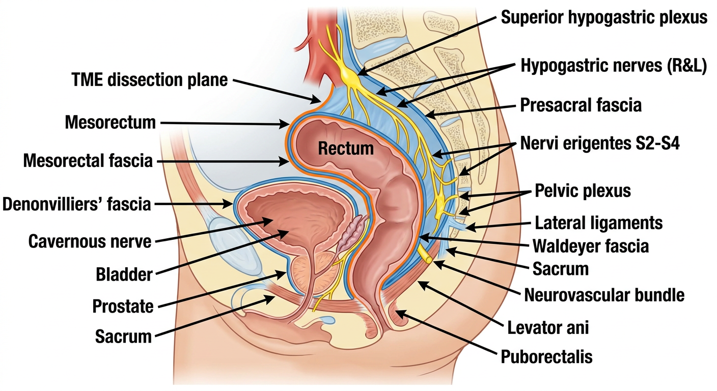
Here is a summary of what the diagram depicts across the three critical nerve preservation planes:
Posterior Plane - The "Holy Plane"
The TME dissection runs between the mesorectal fascia (visceral) and the presacral fascia (parietal). The hypogastric nerves run posteriorly along the sacrum, protected by staying anterior to the presacral fascia. Waldeyer's fascia is divided sharply at S4.
Lateral Plane - The Pelvic Plexus
The pelvic (inferior hypogastric) plexus sits on the lateral pelvic sidewall. The nervi erigentes (S2, S3, S4) enter from the sacral foramina laterally. The lateral ligaments are divided close to the rectal wall to preserve this plexus.
Anterior Plane - Denonvilliers' Fascia
Denonvilliers' fascia separates the anterior mesorectum from the prostate/seminal vesicles. The neurovascular bundle of Walsh (cavernous nerves) runs posterolateral to the prostate - staying in the correct Denonvilliers' plane protects erectile function.
Superior Level - Presacral Nerve
The superior hypogastric plexus at the aortic bifurcation/sacral promontory (L5-S1) is at risk during IMA ligation - it must be swept posteriorly, not anteriorly.
This is a shared conversation. Sign in to Orris to start your own chat.