
Describe the principal of training
Describe the principal of transdermal drug delivery, describe advantage of transdermal fentanyl patch
transdermal drug delivery fentanyl patch
transdermal drug delivery patch skin layers diagram
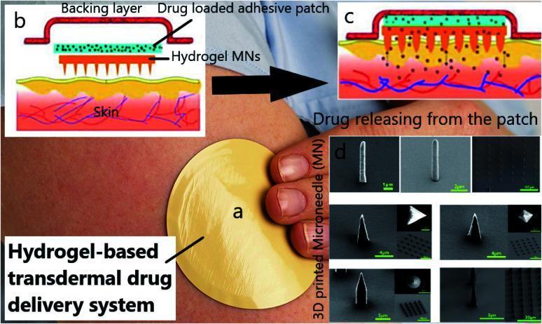
This composite educational graphic illustrates a hydrogel-based transdermal drug delivery system (TDDS). (a) A clinical photograph showing the application of a circular, tan adhesive patch to human skin. (b) A cross-sectional anatomical diagram detailing the patch's structural layers: an outer 'Backing layer,' an internal 'Drug loaded adhesive patch' containing therapeutic molecules (green dots), and a bottom layer of 'Hydrogel MNs' (microneedles). (c) A pathophysiology diagram demonstrating the clinical application, where the microneedles penetrate the stratum corneum to facilitate drug diffusion into the dermal capillary system (depicted by blue and red vascular structures). (d) High-magnification Scanning Electron Microscopy (SEM) images displaying various 3D-printed microneedle morphologies, including cylindrical, conical, and pyramidal shapes with scale bars ranging from 1μm to 20μm. The visual educational focus is on the mechanism of minimally invasive drug administration and the importance of microneedle architecture in enhancing skin permeability for systemic or localized therapy.
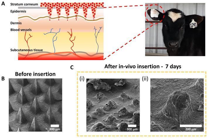
This educational graphic illustrates the in-vivo degradation and transdermal drug delivery mechanism of a chitosan-based microneedle patch. Panel A presents a cross-sectional pathophysiology diagram of bovine skin layers, showing microneedles penetrating the stratum corneum to reach the epidermis and dermis. Red markers indicate drug release into the systemic circulation via dermal blood vessels. A clinical photograph identifies the application site on a calf's ear. Panel B displays a scanning electron microscopy (SEM) image of the microneedle array 'Before insertion,' showing sharp, cone-shaped structures with smooth surfaces and defined geometry (scale bar 300 μm). Panel C provides SEM images 'After in-vivo insertion - 7 days' at two magnifications. Image (i) shows a wide view where the needles appear blunted and partially dissolved (scale bar 300 μm), while image (ii) provides a high-magnification view of a single, significantly degraded microneedle with a rough, 'melted' surface texture (scale bar 200 μm). The visual data demonstrates the biodegradable nature of the chitosan matrix for sustained drug delivery.
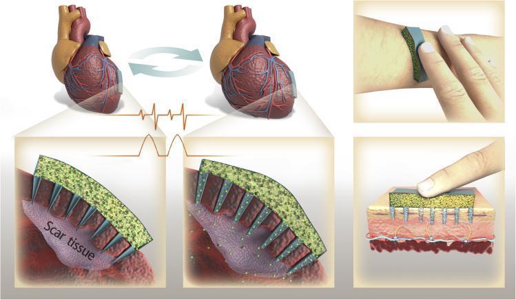
Medical infographic and schematic diagram illustrating a pulsatile drug delivery microneedle patch for cardiac and transdermal applications. The diagram depicts a device consisting of a porous sponge-like drug reservoir (container) integrated with an array of hollow microneedles. In the cardiac repair scenario, the patch is shown applied to the left ventricle of a human heart. Magnified views demonstrate the microneedles penetrating dense, purple-toned 'Scar tissue' (myocardial infarction site). Synchronized with an electrocardiogram (ECG) waveform, the diagram illustrates how mechanical compression of the sponge container during cardiac contraction forces the release of drug particles (green spheres) into the infarcted tissue. In the transdermal scenario, the device is applied to the forearm skin. A cross-sectional view shows a finger applying pressure to the patch, illustrating 'on-demand' delivery. The microneedles are seen penetrating the epidermal and dermal layers, reaching toward subcutaneous vascular structures. The graphic demonstrates two distinct clinical uses: targeted regenerative therapy for acute myocardial infarction and localized anesthesia via transdermal on-demand administration.
Principles of Transdermal Drug Delivery
What Is Transdermal Drug Delivery?
The Skin as a Barrier
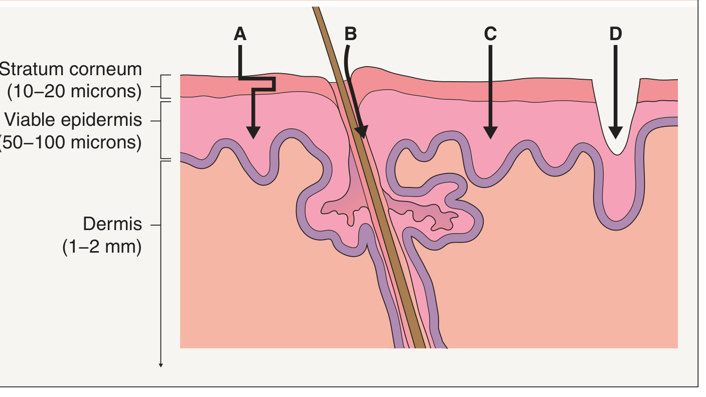
- A - Tortuous intracellular/extracellular lipid pathway (most common passive route, used with chemical enhancers)
- B - Transport through hair follicles and sweat ducts (enhanced by iontophoresis)
- C - Direct transport across the stratum corneum (enabled by electroporation)
- D - Stratum corneum disruption by microneedles, abrasion, or fractional photothermolysis
Physicochemical Requirements for Transdermal Drugs
| Property | Requirement |
|---|---|
| Molecular weight | Low (<400 Da) |
| Lipophilicity | High (octanol-water partition coefficient up to 10,000) |
| Daily dose | Low (typically <10 mg/day) |
| Skin irritation | Minimal |
| Aqueous solubility | Adequate to pass through the hydrated viable epidermis |
Theoretical Advantages of Transdermal Drug Delivery
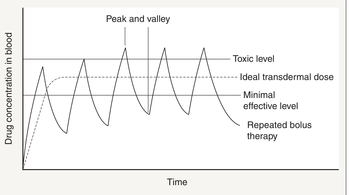
- Targeted drug delivery for dermatologic conditions (local disease site)
- Improved patient adherence - less frequent dosing
- Improved efficacy via continuous, sustained drug release
- Reduced toxicity - eliminates serum concentration "peaks" and lowers total absorbed dose
- Bypass hepatic first-pass metabolism - avoids degradation that reduces bioavailability of oral drugs
- Avoids gastrointestinal side effects and metabolism
- Decreased dosing frequency (once every 24-72 hours vs. multiple daily oral doses)
- Non-invasive - avoids painful injections
- Reduced cost due to lower total dose needed and less frequent dosing
Strategies to Enhance Transdermal Delivery
- Water (stratum corneum hydration under occlusion - distends lacunae, creates polar/non-polar pores)
- Solvents (ethanol, methanol) - extract barrier lipids and disrupt bilayer structure
- Ionic liquids, liposomes/nanoparticles, surfactants, peptides
- Iontophoresis (electrical current drives ions across skin via hair follicles)
- Electroporation (brief electrical pulses create transient pores in lipid bilayers)
- Microneedles (physically puncture the stratum corneum to create micron-scale pathways)
- Ultrasound (cavitational or thermal), mechanical abrasion, fractional photothermolysis, thermal ablation
Transdermal Fentanyl Patch - Principles and Advantages
Why Fentanyl Is Ideal for Transdermal Delivery
"The low molecular weight and high lipid solubility of fentanyl also favor transdermal absorption."
Patch Construction
- Reservoir patch (e.g., Duragesic) - fentanyl is held in a liquid drug reservoir separated from the skin by a microporous, rate-limiting membrane bonded to an adhesive polymer layer. The membrane controls the rate of drug release.
- Matrix patch - fentanyl is dispersed directly within an adhesive matrix layer; no separate reservoir or rate-limiting membrane.
Pharmacokinetics of Transdermal Fentanyl
| Parameter | Value |
|---|---|
| Available patch sizes | 12.5, 25, 50, 75, 100 mcg/h |
| Duration per patch | 72 hours (3 days) |
| Time to detectable plasma fentanyl | 1-2 hours |
| Time to full analgesic effect | 12-24 hours after application |
| Time to plateau plasma levels | 12-40 hours |
| Steady-state plasma levels | ~0.3, 0.6, 1.4, 1.7, 2.5 ng/mL (for 12.5-100 mcg/h patches) |
| Dermal reservoir effect after patch removal | Fentanyl continues absorbing for 12-24 hours |
Advantages of Transdermal Fentanyl
-
No hepatic first-pass metabolism - Oral fentanyl undergoes extensive first-pass hepatic extraction. The transdermal route delivers drug directly into systemic circulation, improving bioavailability.
-
Steady-state analgesia - Continuous delivery at a fixed rate avoids peaks and valleys. Mean plasma fentanyl concentrations are proportional to patch delivery rate and remain stable during repeated patch applications (Miller's Anesthesia 10e).
-
Non-oral route - Critical for patients who cannot take oral medications (dysphagia, severe nausea/vomiting, bowel obstruction, impaired GI absorption).
-
Improved patient compliance and comfort - Simple patch change every 72 hours replaces oral dosing multiple times daily or repeated IV/IM injections.
-
Consistent cancer pain control - Indicated primarily for persistent, unrelenting cancer pain. Katzung's Basic and Clinical Pharmacology 16e notes it "can provide more consistent blood levels of drug and potentially better pain control."
-
Fewer GI adverse events compared to oral morphine - Multiple trials have confirmed TTS fentanyl causes less constipation and fewer GI side effects than equianalgesic oral morphine (Miller's Anesthesia 10e).
-
Reversible - Patch removal is possible, though the dermal reservoir effect means levels decline gradually over 12-24 hours after removal (not immediately).
-
Available in multiple doses - Allows dose titration from 12.5 mcg/h up to 100 mcg/h; multiple patches can be combined for higher requirements.
Important Cautions and Limitations
- Slow onset - Full analgesic effect takes 12-24 hours; not suitable for acute pain or situations requiring rapid dose adjustments.
- Temperature sensitivity - Fever (40°C body temperature) or external heat sources (heating pads, warming blankets placed directly on the patch) can increase fentanyl permeation by ~33%, risking acute opioid overdose (Miller's Anesthesia 10e).
- FDA recommendation - Introduction of the 25 mcg/h patch should be reserved for opioid-tolerant patients with at least 60 mg/day oral morphine equivalent for 1 week or more (Katzung's Basic and Clinical Pharmacology 16e).
- Interpatient variability - Actual delivery rates range from 50-200 mcg/h despite the same patch size (Morgan and Mikhail 7e).
- Diversion risk - The large fentanyl depot makes patches attractive for misuse and has been associated with numerous accidental fatalities.
- Contraindicated in opioid-naive patients for chronic non-cancer pain due to heightened risk of respiratory depression.