
Give notes on tuberculosis, especially in the context of the Pakistani population and especially it's guidelines followed in Pakistan and also compare with International guidelines: - its diagnosis - treatment and management for new cases , resistant cases MDR, XDR First line drugs , TB second line , second line , last resort , recent advances drugs and their side effects with mnemonics .. - what its different types (Based on different aspect followed in guidelines and protocols ) - how to diagnose each - how to give information on each type - a section on what different protocols and guidelines are being currently implemented in Pakistani hospitals and primary and secondary healthcare centers - and also give recent updates regarding diagnsosis and management.. What are the different types of emergencies in a tuberculosis case? How to diagnose and treat each type of TB , especially based on its site and then it's treatment regimen and duration? Types of abdominal TB Different aspects of TB related to Surgery .. What is bovine TB and what are it's different clinical aspects . What are the protocols for a latent case of tuberculosis? What pet questions about history of tuberculosis are to be taken? How to guide / counsel the family household about the case and how to treat them .. What examination findings are to be expected in a case of tuberculosis? Based on each case scenario, also give algorithms, illustrations, diagrams, and images from the latest guidelines, Textbooks from Davidson, Harrison, Kumar, and Clark textbook of medicine, and the latest Pakistani local tuberculosis guidelines. Touch every clinical and exam related aspects of the TB. Also give memorable mnemonics in an easy way to remember different information wherever necessary. Don't make unnecessary mnemonics
tuberculosis Pakistan guidelines treatment
Pakistan National TB Program guidelines 2024 2025 treatment protocols NTP
tuberculosis chest X-ray findings cavitation upper lobe
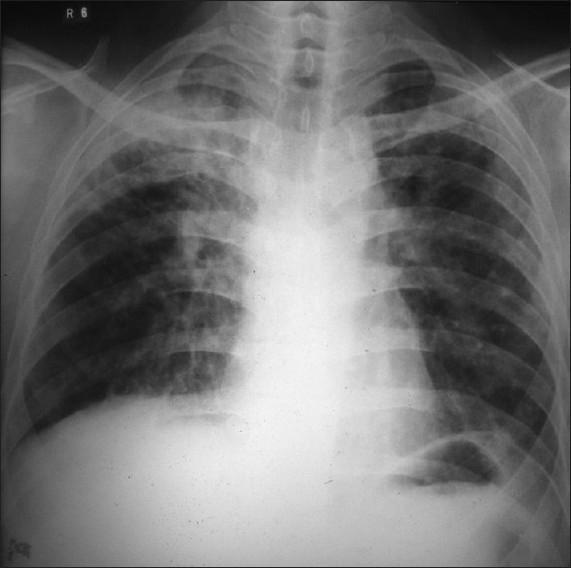
This diagnostic image is a posteroanterior (PA) chest X-ray demonstrating significant pulmonary pathology. The primary finding is located in the right upper lobe, where there are dense, heterogeneous exudative opacities consistent with lung consolidation. Within this region of increased density, a prominent, thick-walled radiolucent cavity is visible, indicating tissue necrosis or cavitation. The distribution of these findings is highly suggestive of secondary (reactivation) pulmonary tuberculosis. In contrast, the left lung field appears relatively clear, although subtle interstitial markings may be present. Key anatomical landmarks including the trachea, clavicles, and cardiac silhouette are visible, with the trachea appearing midline. This image serves as a classic educational example of cavitary lung disease and apical inflammatory processes, emphasizing the radiographic presentation of Mycobacterium tuberculosis infection in an adult patient.
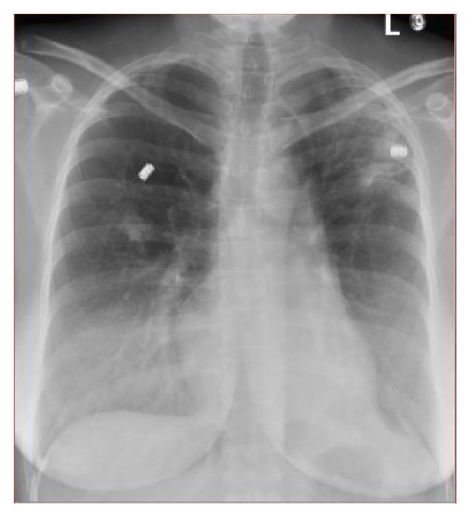
A posterior-anterior (PA) chest x-ray demonstrating bilateral pulmonary abnormalities. In the left upper lobe, there is a prominent hazy increased density or opacity, containing internal lucency suggestive of cavitary changes. The right lung shows a smaller, irregular density localized in the right middle lobe. These findings are clinically significant as they often represent infectious processes such as pulmonary tuberculosis or pyogenic pneumonia, or chronic fibrotic changes. The mediastinal contour and cardiothoracic ratio appear within normal limits. Radiopaque external markers are visible overlying the upper lung fields. This diagnostic image illustrates key radiological signs of pulmonary infiltration and cavitation, relevant for medical education regarding the diagnostic workup of mycobacterial infections and differential diagnosis of apical lung lesions.
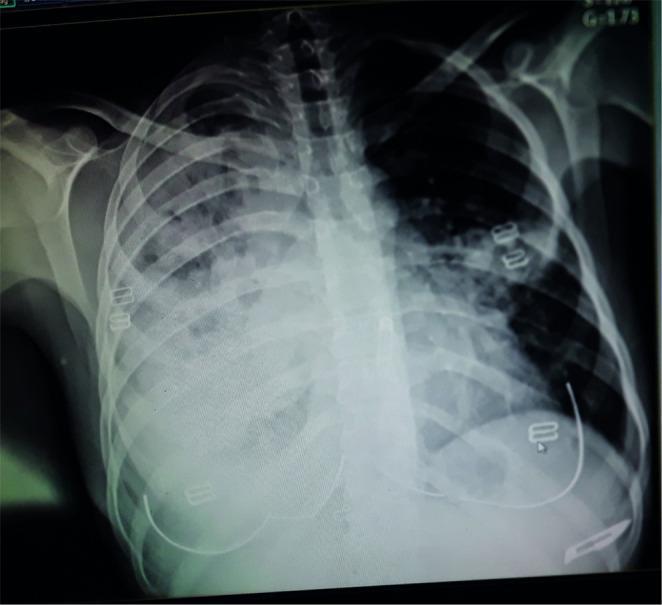
This diagnostic image is an anteroposterior (AP) chest X-ray demonstrating significant bilateral pulmonary pathology. The primary findings include diffuse, hazy opacities and patchy infiltrates throughout both lung fields, with a higher concentration and increased density in the upper lobes. Notably, the left upper lung zone exhibits lucent areas suggestive of cavitation within a region of dense consolidation. The right lung also shows extensive infiltrative changes. The cardiac silhouette is partially obscured by the adjacent pulmonary densities. Osseous structures, including the ribs and clavicles, appear intact without evidence of acute fractures or destructive lesions. Radiopaque EKG leads are visible on the chest wall. Clinically, such a pattern of cavitary lesions and upper lobe infiltrates is characteristic of chronic granulomatous infections such as pulmonary tuberculosis, or progressive malignancies like Hodgkin lymphoma with pulmonary involvement. This image serves as a key educational example for recognizing complex parenchymal lung disease and identifying cavitary features on plain film radiography.
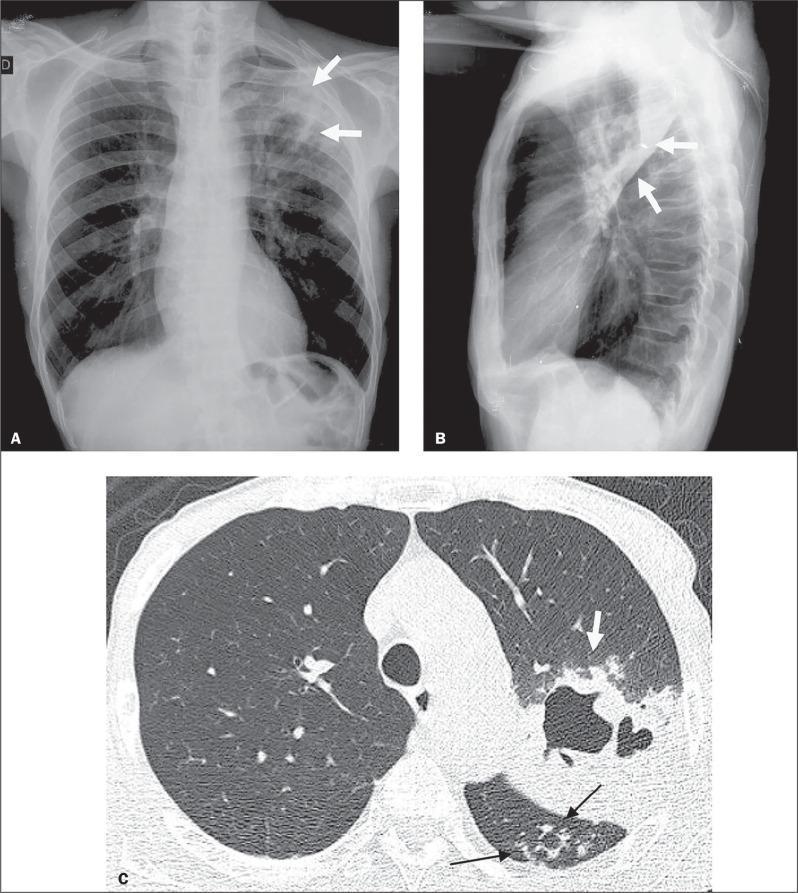
Diagnostic imaging composite of a 66-year-old male with active pulmonary tuberculosis (TB), comprising a posteroanterior chest X-ray (A), a lateral chest X-ray (B), and an axial High-Resolution Computed Tomography (HRCT) scan (C). Panels A and B demonstrate increased opacity in the left upper lung zone indicative of consolidation, with radiolucent areas representing cavitation (white arrows). Panel C, the HRCT at the level of the lower lobes, provides a detailed view of the pulmonary pathology. It reveals a focal area of dense consolidation containing a central air-filled cavity (white arrow) in the left lung. Additionally, characteristic 'tree-in-bud' patterns are visible (black arrows), consisting of small centrilobular nodules and branching linear opacities that indicate endobronchial spread of infection within the upper segment of the left lower lobe. These imaging findings are hallmark radiological features of secondary or active tuberculosis, illustrating parenchymal destruction and small airway involvement.
TB granuloma histology Langhans giant cells caseating necrosis
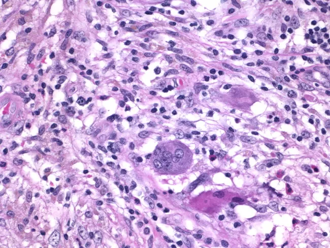
Histology of granulomatous mastitis on bright-field light microscopy. A dense inflammatory infiltrate forms granulomas centered on Langhans-type giant cells surrounded by epithelioid histiocytes, scattered lymphocytes, and occasional eosinophils. The image depicts a high-power view of the center of a granuloma within breast tissue biopsy; multinucleated giant cells with horseshoe-shaped nuclei are evident. The granulomatous architecture includes a peripheral rim of lymphocytes and histiocytes with minimal necrosis described. Staining by Hematoxylin and Eosin highlights basophilic nuclei and eosinophilic cytoplasm; pink collagenous stroma and adipose breast tissue may be visible in the background. Clinically, granulomatous mastitis can mimic infectious etiologies or neoplasia; the presence of Langhans giant cells strongly suggests a granulomatous process and warrants infectious workup for tuberculosis and fungal pathogens, as well as consideration of sarcoidosis or granulomatous reaction to foreign material. This image is diagnostic for granulomatous inflammation in breast tissue when correlated with clinical data and microbiologic studies; useful for education, differential diagnosis, and research on breast granulomatous disease. Additional educational value lies in distinguishing nonnecrotizing granulomas from caseating TB patterns, correlating histology with mammography and ultrasound, and guiding microbiologic testing and patient management in suspected granulomatous breast disease. The image supports teaching of granuloma biology and pathology.
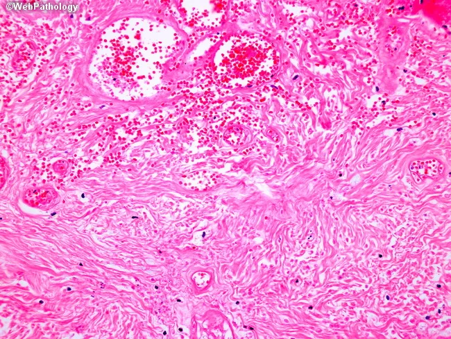
Light microscopy of an H&E-stained formalin-fixed tissue section at low to intermediate magnification reveals granulomatous inflammation characterized by well-formed nodular aggregates of epithelioid histiocytes and occasional Langhans-type multinucleated giant cells embedded in a fibrous stromal matrix. The granulomas appear in close association with accentuated lymphocytic cuffs and scattered plasma cells. Central areas show coagulative or caseating necrosis in some nodules, giving a pale eosinophilic center surrounded by a rim of epithelioid cells. The surrounding dermis/soft tissue shows delicate reticular fibers and vascular congestion with perivascular mononuclear infiltrates. There are no overt bacterial organisms evident with standard H&E, though organisms such as mycobacteria or fungi may require special stains (Ziehl-Neelsen, GMS) to confirm infection. The architecture suggests a granulomatous process rather than acute suppurative inflammation. Diagnostic significance: indicative of granulomatous disease; differential includes infectious etiologies (tuberculosis, fungal infections) and noninfectious conditions (sarcoidosis, foreign-body reaction). Clinical correlation recommended with exposure history, immunologic status, and targeted staining/cultures. Potential use cases: educational demonstration of granuloma morphology, pathology student review, differential diagnosis in infectious and granulomatous diseases, histology atlas reference. This description supports search for terms granuloma, epithelioid histiocytes, Langhans giant cells, caseation, TB, sarcoidosis, histopathology.
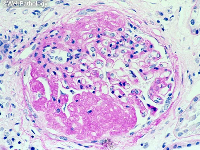
This is a histopathology image obtained with light microscopy from a formalin-fixed paraffin-embedded tissue section stained with Hematoxylin and Eosin. The field demonstrates a granulomatous inflammatory lesion, typically well circumscribed. The central area shows eosinophilic, acellular to granular necrotic material (caseous necrosis), surrounded by a dense mantle of epithelioid histiocytes with elongated, pale-staining nuclei and abundant pale cytoplasm. Multinucleated giant cells of the Langhans type may be present at the periphery. Scattered lymphocytes and occasional plasma cells form a peripheral lymphohistiocytic cuff around the granuloma. The surrounding stroma contains collagen fibers and occasional fibroblasts, with minimal additional inflammatory cells in this field. The morphology is classic for granulomatous inflammation, most commonly associated with infectious etiologies such as Mycobacterium tuberculosis or fungal infections, but can be seen in sarcoidosis or foreign body reactions depending on clinical context. Diagnostic significance: identification of a caseating granuloma raises suspicion for mycobacterial or fungal infection and warrants targeted microbiologic stains (Ziehl-Neelsen for acid-fast bacilli; GMS or PAS for fungi), cultures, and molecular testing. Clinical correlation is essential, including TB exposure history, immune status, and presentation—cough, weight loss, fever, pulmonary nodules or granulomatous lymphadenitis. This image is educational for teaching, differential diagnosis, and research into granulomatous disease pathology.
MDR-TB XDR-TB treatment bedaquiline pretomanid linezolid
miliary tuberculosis chest X-ray millet seed pattern
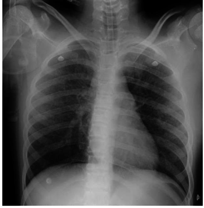
Anterior-Posterior (AP) chest X-ray of a pediatric patient demonstrating a classic 'millet seed' pattern. The image shows widespread, diffuse, and symmetric tiny nodular opacities distributed throughout both the right and left lung fields, from the apices to the bases. These micronodular densities are discrete and uniform in size, characteristic of miliary tuberculosis. The cardiac silhouette, mediastinum, and trachea appear within normal limits for the patient's age. Osseous structures, including the clavicles, ribs, and shoulders, are clearly visible. The presentation illustrates the typical radiological findings associated with hematogenous dissemination of Mycobacterium tuberculosis. This diagnostic image serves as an educational example of miliary lung disease and its manifestation in clinical radiography within a Pediatric Intensive Care Unit (PICU) context.
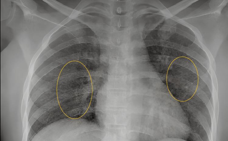
This diagnostic image is a posterior-anterior (PA) chest x-ray demonstrating classic features of miliary tuberculosis. The primary finding is a diffuse, bilateral, and symmetric distribution of small, discrete nodular opacities, approximately 1-3 mm in diameter, creating a 'miliary mottling' pattern throughout both lung fields. These millet-seed-sized nodules result in an overall granular appearance and increased density of the lung parenchyma. Yellow oval annotations highlight representative areas of the miliary pattern in the mid-lung zones. The cardiac silhouette, mediastinal contours, and diaphragmatic domes appear within normal limits without evidence of gross lymphadenopathy or pleural effusion. The visible skeletal structures, including the ribs and clavicles, show no obvious abnormalities. This radiological presentation is highly characteristic of hematogenous dissemination of Mycobacterium tuberculosis, making it a critical educational resource for understanding systemic mycobacterial disease manifestations in pulmonology and infectious disease.
tuberculin skin test Mantoux test technique reading
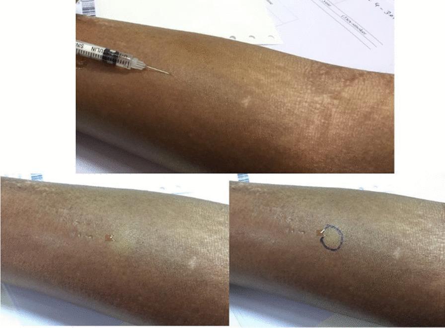
A series of three clinical photographs demonstrating the procedural steps of a Tuberculin Skin Test (TST), specifically utilizing the Mantoux technique. The image sequence illustrates the administration of purified protein derivative (PPD) on the volar aspect of the forearm in a patient with systemic sclerosis. The first frame shows a tuberculin syringe with a needle inserted at a shallow angle for intradermal injection. The second frame depicts the immediate result: the formation of a discrete, pale, raised wheal approximately 6–10 mm in diameter, indicating correct superficial placement. The third frame shows the site after a circular mark has been drawn around the wheal's perimeter to facilitate localized identification for follow-up interpretation. The skin of the patient shows characteristic features of systemic sclerosis, including localized hypopigmentation and a taut, thickened texture. This visual serves as an educational guide for infectious disease screening and diagnostic dermatology, emphasizing proper technique and the specific challenges of skin testing in patients with scleroderma.
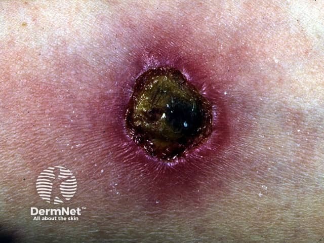
Clinical photography of a Mantoux tuberculin skin test reaction performed on the forearm. Imaging modality: clinical photography; technique: high-resolution, close-up digital photograph under standard lighting; magnification: macro. Anatomical location: forearm skin at the intradermal injection site of tuberculin purified protein derivative (PPD). Visual features: a circular, ulcerated patch with a dark necrotic center, surrounded by a violaceous to pink erythematous halo, peripheral induration, and surrounding edema. Crusting is evident at the lesion margin, with a slightly sunken center. Pathophysiology: this appearance suggests a pronounced delayed-type hypersensitivity reaction (Type IV) to tuberculin; in some cases, intense reactions may produce ulceration or necrosis, though mild induration is more common. Differential diagnosis includes an overly vigorous Mantoux reaction, superimposed infection (impetigo or cellulitis), or injection-site ulceration, and should be correlated with induration size measured after 48–72 hours and clinical history. Clinical significance: a positive reaction indicates TB exposure or BCG vaccination response, but interpretation depends on risk factors and time since exposure. Potential clinical use: TB screening, exposure assessment, vaccine response evaluation, dermatology teaching, medical education, and research training in cutaneous hypersensitivity. Note: lesions of this severity are uncommon in routine screening and require urgent clinical correlation. Documentation should include induration measurement and patient risk factors.
Pakistan NTP tuberculosis treatment guidelines 2023 2024 MDR XDR BPaL regimen
https://pmc.ncbi.nlm.nih.gov/articles/PMC12333698
TB lymphadenopathy cervical scrofula neck swelling
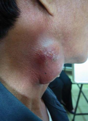
A clinical photograph of the lateral neck region of a patient, demonstrating a large, protruding mass consistent with cervical lymphadenopathy. The swelling is approximately 5-7 cm in diameter, exhibiting a lobulated and rounded morphology. The overlying skin shows significant pathological changes, including intense erythema (redness), localized edema that gives the surface a shiny and stretched appearance, and areas of focal ulceration or crusting suggestive of an impending or active fistula formation (scrofuloderma). The visual presentation illustrates paradoxical tuberculosis-associated immune reconstitution inflammatory syndrome (TB-IRIS), where existing lymph node lesions worsen after the initiation of antiretroviral therapy (ART) in HIV-infected individuals. Key educational features include the signs of acute inflammation and tissue breakdown in the context of chronic infectious lymphadenitis.
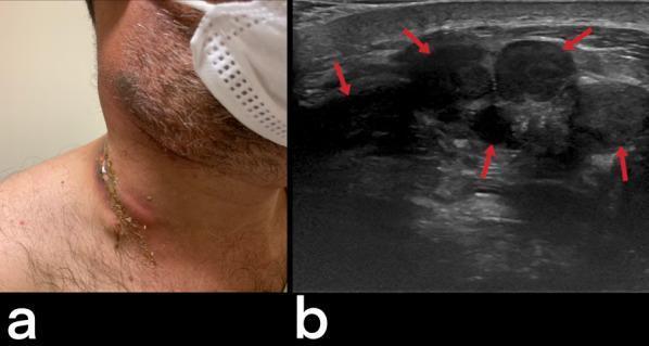
This dual-panel image demonstrates the clinical and radiological presentation of tuberculous cervical lymphadenitis, commonly known as scrofula. Panel (a) is a clinical photograph of a 32-year-old male showing progressive, discrete, and mobile lumps in the lateral neck. A notable finding is the presence of cutaneous fistulization, where an underlying nodal mass has ruptured through the skin, creating a chronic draining sinus tract. Panel (b) is a corresponding ultrasound image of the cervical region. Red arrows highlight multiple enlarged, hypoechoic lymph nodes. These nodes exhibit loss of the normal fatty hilum and internal heterogeneity, characteristic of central caseous necrosis. The correlation between the two panels illustrates the progression from internal nodal necrosis to subcutaneous abscess formation and eventual external fistulization. This presentation is a hallmark of extrapulmonary tuberculosis (EPTB) in endemic regions, emphasizing the importance of considering chronic granulomatous infections in the differential diagnosis of unilateral cervical lymphadenopathy with skin involvement.
TUBERCULOSIS - COMPREHENSIVE CLINICAL NOTES
Pakistan Context + International Guidelines | Harrison's, Murray & Nadel's, Davidson's, Surgical Textbooks + WHO/NTP 2024-2026
PART 1 - EPIDEMIOLOGY AND PAKISTAN CONTEXT
Global Burden
Pakistan - A High-Burden Country
| Indicator | Pakistan (2023 Data) |
|---|---|
| Population | 248 million |
| Total TB cases notified | 480,136 |
| Treatment success rate | 95% (SAARC best) |
| HIV co-infection in TB | 0.39% |
| MDR-TB estimated | ~35,000-40,000/year |
| Only 11% MDR-TB diagnosed and treated | Major gap |
- Overcrowding, poverty, malnutrition
- High prevalence of diabetes (TB-DM comorbidity major challenge per 2024 meta-analysis, PMID 38346381)
- Delayed diagnosis due to stigma
- High proportion of cases in rural Sindh, KPK, Balochistan
- BCGosis risk in infants post-BCG vaccination (PMID 41459544)
PART 2 - HISTORY TAKING IN TB (PET QUESTIONS)
HISTORY - TEMPLATE
S - Symptoms (PCFW): Persistent cough >2 weeks, Constitutional fever (evening rise), Fatigue/weight loss, blood-streaked sputum (haemoptysis) W - Weight loss (how much, over how long?) I - Investigation history (prior CXR, sputum, GeneXpert results) F - Family contact (household contact with known TB case?) T - Treatment history (prior TB treatment - critical for categorization) P - Past medical history (DM, HIV, steroid use, malignancy, ESRD) A - Animals (bovine TB - unpasteurized milk exposure) S - Social history (overcrowding, migration, prison, homelessness, healthcare worker) T - Travel/occupation (endemic area, healthcare worker, laboratory worker)
Cardinal Symptoms
| Symptom | Feature in TB |
|---|---|
| Cough | >2 weeks, productive |
| Fever | Low-grade, characteristically evening rise ("hectic/quotidian") |
| Night sweats | Drenching, change of clothes |
| Weight loss | >5-10% body weight |
| Haemoptysis | Variable - spotting to massive |
| Dyspnoea | Pleural effusion, pneumothorax, miliary |
| Chest pain | Pleuritic (pleuritis) |
PART 3 - CLASSIFICATION OF TUBERCULOSIS
A. By Site (Anatomical Classification)
- Upper lobe predominance (apical/posterior segments RUL, apicoposterior LUL)
- Cavitation, consolidation, nodules, bronchiectasis
| Site | Frequency | Key Features |
|---|---|---|
| Lymph nodes (Scrofula) | Most common EPTB | Cervical, supraclavicular |
| Pleural | 2nd most common | Unilateral effusion, ADA elevated |
| Bone/Joint (Pott's) | Spine most common | T8-T12, gibbus, cord compression |
| CNS (TBM) | Most serious | Basal meningitis, hydrocephalus |
| Abdominal | Terminal ileum, peritoneum | Dough belly, ascites |
| Pericardial | Constrictive pericarditis | Beck's triad, pulsus paradoxus |
| Genitourinary | Kidney most common | "Sterile pyuria" |
| Skin | Lupus vulgaris, scrofuloderma | Apple-jelly nodules |
B. By Bacteriology (WHO/NTP Classification)
| Type | Definition |
|---|---|
| Drug-Susceptible TB (DS-TB) | Sensitive to all first-line drugs |
| Isoniazid-resistant TB (Hr-TB) | Resistant to INH only |
| Rifampicin-resistant TB (RR-TB) | Resistant to Rifampicin (any) |
| MDR-TB | Resistant to both INH + Rifampicin |
| Pre-XDR-TB | MDR + resistance to any fluoroquinolone |
| XDR-TB | MDR + resistance to fluoroquinolone + at least one of bedaquiline or linezolid |
C. By Treatment History (NTP Categorization)
| Category | Definition | Regimen |
|---|---|---|
| New case | Never treated or <1 month treatment | Cat I: 2HRZE/4HR |
| Previously treated (Relapse) | Completed treatment, bacteriologically positive again | Cat II: 2HRZES/1HRZE/5HRE |
| Treatment failure | Sputum positive at month 5+ | DR-TB regimen |
| Lost to follow-up | Interrupted >2 months | Re-evaluate; restart or DR-TB |
| Treatment after failure | — | DST-guided |
D. By Latency Status
| Type | Features |
|---|---|
| Latent TB Infection (LTBI) | TST+/IGRA+ but no symptoms, no active disease |
| Active TB Disease | Symptoms + bacteriological/histological confirmation |
| Primary TB | First infection; often lower/middle lobe + hilar adenopathy |
| Post-primary (Reactivation) TB | Upper lobe, cavitation |
| Progressive Primary TB | Cavitation in primary without resolution |
| Miliary TB | Haematogenous dissemination |
PART 4 - DIAGNOSIS
Algorithm - Diagnosis of Pulmonary TB
SUSPECTED TB (cough >2wks + constitutional symptoms + CXR abnormality)
        |
        v
SPUTUM SMEAR (ZN stain) x 2
        |
    [+] ---------------------> Notify + Start HRZE (Category I)
    [-] or paucibacillary
        |
        v
GeneXpert MTB/RIF (Cartridge-Based NAAT - CBNAAT)
        |
    MTB detected, Rif Sensitive -----> DS-TB regimen
    MTB detected, Rif Resistant ------> DR-TB workup (LPA/DST)
    MTB not detected
        |
        v
    Clinical probability + CXR + Culture (LJ/MGIT)
        |
    HRCT Chest if needed
Diagnostic Tests
1. Sputum Smear Microscopy (ZN Stain)
- Ziehl-Neelsen stain - acid-fast bacilli appear bright red on blue background
- First-line, cheap, widely available in Pakistan PHC
- Sensitivity: 45-80% (lower in HIV+, paucibacillary)
- 3+ grading: Scanty (1-9 AFB/100 fields), 1+ (10-99), 2+ (1-10/field), 3+ (>10/field)
2. GeneXpert MTB/RIF (Xpert)
- WHO-recommended first-line diagnostic
- Sensitivity ~89%, Specificity ~99%
- Detects MTB AND rifampicin resistance in 2 hours
- Pakistan NTP: GeneXpert is the primary diagnostic tool at district hospitals and above
- Xpert Ultra: improved sensitivity for paucibacillary disease
3. Culture (Gold Standard)
- LJ (Lowenstein-Jensen) solid medium: 4-8 weeks
- MGIT (Mycobacterial Growth Indicator Tube): liquid, 1-3 weeks
- Required for DST in DR-TB
4. Line Probe Assay (LPA) / Hain GenoType
- FL-LPA (First Line): detects INH and Rifampicin resistance
- SL-LPA (Second Line): detects FQ + SLI resistance
- Pakistan NTP: Available at provincial reference labs (PMRC, NRL Islamabad)
5. Tuberculin Skin Test (TST / Mantoux)
- Inject 0.1 mL PPD (5 TU) intradermally, read at 48-72 hours
- Measure induration (not erythema) transverse to long axis of arm
| Threshold | Population |
|---|---|
| ≥5 mm | HIV+, recent close contact, immunosuppressed, old CXR changes |
| ≥10 mm | Healthcare workers, prisoners, migrants, diabetics, children <5 yrs, Pakistan general population |
| ≥15 mm | Low-risk population |
6. IGRA (Interferon-Gamma Release Assay)
- QuantiFERON-TB Gold Plus, T-SPOT.TB
- Not affected by BCG vaccination (unlike TST)
- Not widely available in Pakistan public sector
- Preferred for LTBI screening in BCG-vaccinated populations
- Pakistan NTP: TST preferred for household contact investigation (turns positive earlier)
7. Chest X-Ray Features
- Middle or lower lobe opacity + ipsilateral hilar adenopathy (Ghon complex + Ranke complex)
- Upper lobe, apical/posterior segments, cavitation common
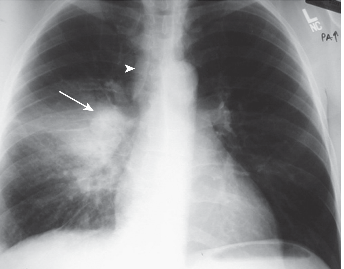
- 1-3 mm nodules diffusely throughout both lungs ("millet seeds")
8. Histopathology
- Caseating granuloma with Langhans giant cells = hallmark
- ZN stain/auramine rhodamine fluorescence for AFB
- Epithelioid histiocytes + central caseous necrosis
9. ADA (Adenosine Deaminase)
- Pleural fluid ADA >40 IU/L: highly suggestive of TB pleuritis
- CSF ADA elevated in TBM
- Ascitic fluid ADA in peritoneal TB
10. BACTEC/MGIT Culture + DST
- Required for all retreatment and DR-TB suspect cases
- Pakistan NTP: Free at National TB Reference Lab (NRL), Islamabad
PART 5 - EXAMINATION FINDINGS BY SITE
Pulmonary TB - Examination
| Finding | Significance |
|---|---|
| Reduced chest movement | Consolidation/effusion |
| Dull to percussion | Consolidation, effusion, empyema |
| Bronchial breath sounds | Consolidation |
| Crackles (crepitations) | Active consolidation |
| Amphoric breath sounds | Cavitation (hollow, bottle-like) |
| Decreased air entry | Effusion, collapse |
| Trachea deviation | Away from effusion, toward fibrosis/collapse |
| Clubbing (rare in TB) | Empyema, bronchiectasis |
Lymph Node TB (Scrofula)
- Cervical > supraclavicular > axillary
- Initially discrete, firm, mobile
- Progress to: matted, soft (caseation), collar-stud abscess, discharging sinus
- Sinuses leave characteristic "laddertracks" scars

Pott's Disease (Spinal TB)
- Gibbus deformity (angular kyphosis)
- Local vertebral tenderness
- Paraplegia/paraparesis (cold abscess compressing cord)
- Psoas abscess: fluctuant mass at femoral triangle or iliac fossa
TB Meningitis
- Signs of meningeal irritation (Kernig's, Brudzinski's)
- Cranial nerve palsies (III, IV, VI - basal exudate)
- Altered consciousness, papilloedema (raised ICP)
TB Pericarditis
- Beck's triad: hypotension + muffled heart sounds + raised JVP
- Pulsus paradoxus (tamponade)
- Friction rub (early)
- Kussmaul's sign (constrictive pericarditis)
Peritoneal TB (Abdominal)
- "Dough belly": doughy sensation on palpation (matted omentum, loops)
- Ascites (exudate, ADA elevated)
- Tenderness, usually without peritonism
- Palpable mesenteric lymph nodes (periumblical)
PART 6 - TREATMENT AND MANAGEMENT
Standard Short-Course Regimen (DS-TB New Cases)
2HRZE / 4HR
- 2 months Intensive Phase: Hisoniazid + Rifampicin + Pyrazinamide + Ethambutol
- 4 months Continuation Phase: Hisoniazid + Rifampicin
First-Line Drug Details, Doses, Side Effects
| Drug | Daily Dose (adult) | Mechanism | Key Side Effects | Mnemonic |
|---|---|---|---|---|
| Rifampicin | 10 mg/kg (max 600 mg) | RNA polymerase inhibitor | Orange secretions, Hepatotoxicity, Drug interactions (CYP450 inducer), Flu-like syndrome (intermittent) | Red/Orange urine |
| Isoniazid | 5 mg/kg (max 300 mg) | Mycolic acid synthesis inhibitor (KatG activation) | Peripheral neuropathy (pyridoxine prevents), Hepatotoxicity, Lupus-like syndrome | INH needs B6 |
| Pyrazinamide | 25 mg/kg (max 2g) | Disrupts membrane energetics in acidic pH | Hepatotoxicity, Gout (hyperuricaemia), Arthralgia | PyriZINE = joint PAIN |
| Ethambutol | 15-20 mg/kg | Arabinogalactan synthesis inhibitor | Optic neuritis (colour vision, visual acuity - red-green), Hyperuricaemia | Eyes (EMB = Eye damage) |
- Aminoglycoside, IM injection
- Side effects: Ototoxicity (8th nerve), nephrotoxicity, vestibular damage
Extended Regimen for Retreatment (Category II)
2HRZES / 1HRZE / 5HRE
- 2 months: HRZE + Streptomycin
- 1 month: HRZE
- 5 months: HRE
Special Situations - Duration Extension
| Situation | Extension |
|---|---|
| Cavitation on CXR + culture positive at 2 months | Extend total to 9 months (4HR -> 7HR) |
| CNS TB (TBM, spinal) | 9-12 months total |
| Bone/Joint TB | 9-12 months |
| Pericardial TB + steroids | 6 months (+prednisolone 60mg/d taper) |
| HIV+ with severe immunosuppression | 9 months minimum |
PART 7 - DRUG-RESISTANT TB (DR-TB)
Definitions
| Term | Definition |
|---|---|
| RR-TB | Resistant to Rifampicin (any mechanism) |
| MDR-TB | Resistant to INH + Rifampicin |
| Pre-XDR-TB | MDR-TB + resistant to any fluoroquinolone |
| XDR-TB | MDR-TB + FQ resistance + Bdq or Lzd resistance |
WHO Drug Classification for DR-TB (2022 Update)
- Levofloxacin/Moxifloxacin (Lfx/Mfx)
- Bedaquiline (Bdq)
- Linezolid (Lzd)
MDR-TB Regimens
Standard Long Oral Regimen (18-20 months)
Bdq + Lfx/Mfx + Lzd + Cfz +/- Cs (ABCL - "ABuCket of LinColn")
Shorter Oral Regimen (9-11 months) - WHO 2022
BDQ-Lfx-Lzd-Cfz-Z-E-H(high dose) [when FQ-susceptible] OR BDQ-Mfx-Lzd-Cfz-Z-E-Pto
BPaL / BPaLM Regimen (6 months) - THE NEWEST
| Aspect | BPaL | BPaLM |
|---|---|---|
| Duration | 6 months | 6 months |
| Indication | XDR-TB, pre-XDR-TB, treatment-intolerant MDR-TB | MDR-TB with FQ susceptibility |
| Approval | FDA 2019 (Nix-TB study) | WHO recommended 2022 |
| Pakistan | Piloted 2023-2024 at Rawalpindi, Multan, Lahore, Peshawar | Being rolled out under NTP + TB Alliance |
- Pakistan NTP updated guidelines to include 6-month all-oral BPaL/BPaLM regimens
- TB Alliance licensed Pretomanid to Remington Pharmaceutical Industries (Pakistan) royalty-free
- ASD (Action for Social Development) partnered with NTP for rollout in Punjab and KPK
Second-Line Drug Details and Side Effects
| Drug | Mechanism | Key Side Effects |
|---|---|---|
| Bedaquiline | ATP synthase inhibitor (diarylquinoline) | QT prolongation, hepatotoxicity, nausea. Monitor ECG |
| Pretomanid | Mycolic acid inhibitor (nitroimidazole) | Peripheral neuropathy, hepatotoxicity, QT prolongation |
| Linezolid | 50S ribosome inhibitor (oxazolidinone) | Myelosuppression, peripheral neuropathy (dose-limiting), optic neuritis, serotonin syndrome |
| Moxifloxacin | DNA gyrase/topoisomerase IV | QT prolongation, tendinopathy, CNS effects |
| Clofazimine | Free radical generation, membrane depolarization | Orange-brown skin/secretions discolouration (reversible), GI intolerance |
| Cycloserine | Alanine racemase inhibitor (cell wall) | Seizures, psychosis, depression, peripheral neuropathy |
| Delamanid | Mycolic acid inhibitor | QT prolongation, hypoalbuminaemia |
| Amikacin | Aminoglycoside (30S ribosome) | Ototoxicity, nephrotoxicity |
| Ethionamide | Mycolic acid inhibitor | GI intolerance, hypothyroidism, hepatotoxicity |
| PAS | Folate synthesis disruption | GI intolerance, hypothyroidism, hepatotoxicity |
| Imipenem/Meropenem | Beta-lactam (cell wall) | Seizures (imipenem), GI upset |
"BeDaqui-Lines Mox-ifloxacin CloFazimine" Bedaquiline + Delamanid + Levofloxacin/Moxifloxacin + Clofazimine = BDLC - "Big Danger, Long Corrected (QT)"
PART 8 - TREATMENT BY SITE (DURATION TABLE)
| Site | Regimen | Duration | Special Notes |
|---|---|---|---|
| Pulmonary (new, DS) | 2HRZE/4HR | 6 months | Extend to 9 months if cavitation + culture+ at 2m |
| Lymph node | 2HRZE/4HR | 6 months | Paradoxical reactions common; do NOT change drugs |
| Pleural | 2HRZE/4HR | 6 months | Drain large effusions; steroids reduce adhesions |
| Bone/Joint (Pott's) | 2HRZE/7-10HR | 9-12 months | Surgery for cord compression, instability, cold abscess |
| CNS (TBM) | 2HRZE/10HR | 12 months | Dexamethasone mandatory (0.4 mg/kg/d x 4 weeks taper); ethambutol poor CNS penetration - replace with Streptomycin in some protocols |
| Pericardial | 2HRZE/4HR | 6 months | Prednisolone 60 mg/d taper; pericardiectomy if constrictive |
| Abdominal (intestinal/peritoneal) | 2HRZE/4-7HR | 6-9 months | Surgery for obstruction, perforation |
| Genitourinary | 2HRZE/4HR | 6 months | Avoid catheterisation; steroids for ureteric stricture |
| Miliary | 2HRZE/7-10HR | 9-12 months | Steroids if adrenal involvement or severe hypoxia |
| Adrenal TB | 2HRZE/7HR | 9 months | Steroid replacement if adrenal insufficiency (Addisonian crisis risk) |
| Skin TB | 2HRZE/4HR | 6 months | — |
PART 9 - ABDOMINAL TUBERCULOSIS
Types (S. Das Surgical Manual + Sleisenger & Fordtran)
-
Ulcerative TB (more common)
- Results from swallowed sputum in pulmonary TB
- Multiple transverse ulcers at terminal ileum
- Thickened serosa - rarely perforates
- Healing -> stricture formation
- Symptoms: diarrhoea, weight loss, RIF pain, occult blood
- Barium: "Stierlin's sign" - non-filling of terminal ileum (fleeting filling defect)
-
Hyperplastic Ileo-caecal TB
- Most common type in South Asia including Pakistan
- Cauliflower-like mass at ileocaecal junction
- Mimics carcinoma cecum or Crohn's disease
- RIF mass + constitutional symptoms
- CT: concentric thickening, pulled up cecum
- Chronic central abdominal pain (discomfort)
- General symptoms (weight loss, fever)
- Intestinal obstruction (adherent loops)
- Mimics appendicitis (RIF pain, nausea)
- Cold abscess (pseudomesenteric cyst between mesenteric leaves)
- Calcified lymph nodes on plain X-ray (late finding)
- Wet type (exudative ascites): most common; ADA >40, protein >3 g/dL, SAAG <1.1
- Dry type (adhesive/plastic): matted loops, "dough belly"
- Fibrotic type: thick omental cake, loculated ascites
- Ascitic fluid: ADA (>40), protein, SAAG
- Colonoscopy + biopsy: ileocaecal region
- Laparoscopy + peritoneal biopsy: gold standard for peritoneal TB
- CT abdomen: mesenteric lymphadenopathy, ascites, bowel wall thickening
| Feature | Intestinal TB | Crohn's Disease |
|---|---|---|
| Geography | South Asia, Africa | Western |
| Caseating granuloma | Yes | No |
| Submucosal fibrosis | Circumferential | Transmural |
| Perianal disease | Rare | Common |
| Fistulae | Less common | Common |
| ADA | Elevated | Normal |
| AFB stain/culture | Positive | Negative |
| Response to ATT | Good (definitive) | None |
| Colonoscopy | Pseudopolyps, short segment, ileocaecal valve deformed | Cobblestone, skip lesions |
PART 10 - TUBERCULOSIS AND SURGERY
Surgical Indications in TB
- Destroyed lung/lobe (massive haemoptysis, persistent cavity, bronchiectasis)
- Diagnostic thoracotomy/VATS (persistent diagnosis uncertainty)
- Empyema necessitatis drainage
- Drug-resistant TB - adjunct to chemotherapy (segmentectomy/lobectomy/pneumonectomy for localized MDR-TB)
- Bronchopleural fistula
- Indications: Cord compression (neurological deficit), instability, abscess not responding to ATT, kyphotic deformity >40°
- Procedure: Anterior decompression + fusion (gold standard) or posterolateral approach
- Cold abscess: drain if large, fluctuant or threatening vital structures
- Intestinal obstruction (not responding to conservative management)
- Perforation: ileostomy/resection
- Bleeding: resection
- Diagnostic uncertainty (can't exclude carcinoma)
- Aspiration of fluctuant nodes for diagnosis
- Excision biopsy for diagnosis
- Avoid incision and drainage (risk of chronic sinus formation)
- Pericardiectomy for constrictive pericarditis
- Nephrectomy for non-functioning kidney
- Ureteric stenting for strictures
- Augmentation cystoplasty for contracted bladder
- Continue full course of ATT irrespective of surgery
- Surgery is adjunct, not replacement, for chemotherapy
PART 11 - BOVINE TUBERCULOSIS
Agent
- Mycobacterium bovis (M. bovis) - closely related to M. tuberculosis
- Member of M. tuberculosis complex
- Reservoir: cattle, badgers, deer
- Transmission to humans: ingestion of unpasteurized milk or milk products, direct contact with infected animals, inhalation in abattoir workers
Clinical Features in Humans
| Feature | Bovine TB (M. bovis) |
|---|---|
| Mode of infection | Oral (milk) > inhalation > direct contact |
| Primary site | GI tract (cervical/mesenteric lymph nodes), bone |
| Pulmonary disease | Less common (but can occur) |
| Extrapulmonary | Common - lymph nodes (scrofula), bone, GI, skin |
| Drug resistance | Intrinsically resistant to Pyrazinamide |
| Treatment | HRZE for 6 months (no pyrazinamide for first 2 months, use HRE+S instead; 9 months total) |
| Diagnosis | Culture on LJ: small, rough, buff-coloured colonies; no pyrazinamide susceptibility |
- Very prevalent in cattle-rearing communities (Punjab, Sindh rural)
- Unpasteurized doodh (milk) consumption widespread
- Cervical lymphadenopathy (scrofula) in children often due to M. bovis
9HRE (no PZA - intrinsic resistance) OR 2HRSE / 7HRE
PART 12 - LATENT TB INFECTION (LTBI)
Definition
- No symptoms
- Normal CXR
- No bacteriological evidence of active disease
Who to Test for LTBI (Pakistan NTP + WHO)
- Household contacts of smear-positive PTB (especially children <5)
- HIV-positive individuals
- Patients on anti-TNF therapy
- Transplant candidates
- Healthcare workers
LTBI Treatment Regimens (Harrison's 22e, 2025)
| Regimen | Schedule | Duration | Preferred Use |
|---|---|---|---|
| 3HP - Isoniazid + Rifapentine | 900 mg each weekly (>50kg) | 3 months (12 doses) | First choice (all adults, HIV+/-) |
| 4R - Rifampicin | 600 mg daily | 4 months | HIV-negative, children |
| 3HR - INH + Rifampicin | Daily | 3 months | Alternative |
| 6H/9H - Isoniazid | 300 mg daily or 900 mg twice weekly | 6-9 months | Alternative; 9 months more effective |
Household Contact Management Protocol
- All contacts - clinical assessment + CXR
- Children <5 years: If TST/IGRA not available - give isoniazid prophylaxis directly
- Children <5 with symptoms or CXR abnormality - treat as active TB
- Immunocompromised (HIV+) - treat LTBI regardless of TST
- TST+ contacts with no active disease - LTBI regimen
- BCG vaccination - for unvaccinated contacts <5 (after LTBI treatment)
PART 13 - COUNSELLING FAMILY/HOUSEHOLD CONTACTS
What to Tell the Family
C - Contagion (how TB spreads - droplet, NOT touch, sharing utensils) O - Open treatment (importance of full course, DOT) A - All contacts tested (especially children, elderly, HIV+) C - Characteristics to watch (cough >2 weeks, fever, weight loss) H - Healthy habits (ventilation, sunlight, nutrition, BCG for newborns)
Key Counselling Points
- TB is curable - complete treatment is essential
- Patient is non-infectious after 2 weeks of effective therapy (smear-negative) - can return to work/school
- Isolation at home during initial intensive phase (2 weeks) - separate room, open windows
- DOT (Directly Observed Therapy) - watch patient swallow medication daily
- No alcohol (increases hepatotoxicity risk)
- Report side effects immediately: yellow eyes (hepatitis), visual blurring (ethambutol)
- All household members must be screened (including children)
- BCG vaccination for newborns
- Nutritional support - high protein diet, vitamin supplementation
PART 14 - EMERGENCIES IN TUBERCULOSIS
M - Massive Haemoptysis A - Airway obstruction (lymph node compression) S - SIADH / Hyponatraemia (TBM) H - Hydrocephalus (TBM) P - Pneumothorax P - Pericardial Tamponade
1. Massive Haemoptysis
- Definition: >600 mL/24h or haemodynamically significant
- Causes: Rasmussen's aneurysm (pulmonary artery erosion in cavity), aspergilloma in old TB cavity, bronchiectasis
- Management: ABC, Patient on bleeding side, intubation, bronchoscopy, bronchial artery embolisation (BAE), emergency surgery
2. Airway Obstruction
- Lymph node compression in primary TB (children)
- Endobronchial TB
- Management: Bronchoscopy, corticosteroids, emergency stenting
3. TB Meningitis Emergencies
- Hydrocephalus: urgent VP shunt or external ventricular drainage
- SIADH/Hyponatraemia: fluid restrict, hypertonic saline if severe
- Seizures: anticonvulsants (note drug interactions with rifampicin)
- Cerebral herniation: mannitol, head elevation, dexamethasone
4. Pneumothorax
- Spontaneous in cavitary disease or bronchopleural fistula
- Tension pneumothorax: emergency needle decompression, chest tube
5. Cardiac Tamponade (TB Pericarditis)
- Emergency pericardiocentesis
- Then start ATT + prednisolone
6. Adrenal Crisis (TB Adrenalitis - Addison's Disease)
- Bilateral adrenal destruction by TB (most common cause of Addison's in Pakistan)
- Emergency: IV hydrocortisone 100 mg stat + fluid resuscitation
7. Liver Failure (Drug-Induced - DILI)
- Stop all four drugs if bilirubin >3 or ALT >5x normal
- Restart sequentially: R first, then H, then Z
- If severe DILI: use Streptomycin + Ethambutol + Fluoroquinolone as bridge
8. Paradoxical Reactions (IRIS)
- Worsening of TB lesions after starting ATT (immune reconstitution)
- Common in HIV+ patients starting ART
- Management: Continue ATT/ART, add NSAIDs or prednisolone
PART 15 - PAKISTAN NTP & LOCAL PROTOCOLS
National TB Control Programme (NTP Pakistan)
- Free diagnosis and treatment at all government facilities
- TB treatment integrated with PHC (Basic Health Units - BHUs, Rural Health Centres - RHCs)
- GeneXpert at district-level and above
- Directly Observed Therapy (DOT) at all levels
- Community-based DOT via community health workers (Lady Health Workers - LHWs)
- Private sector engagement through Public-Private Mix (PPM)
Pakistan Chest Society (PCS) TB Guidelines 2020
| Category | Patients | Regimen |
|---|---|---|
| Category I (New) | New PTB smear+, new severe EPTB | 2HRZE/4HR |
| Category II (Retreatment) | Relapse, treatment after failure, treatment after LTFU | 2HRZES/1HRZE/5HRE |
| Category III (New, less severe) | New PTB smear-, new less severe EPTB | 2HRZ/4HR |
| DR-TB | GeneXpert Rif-R or DST confirmed | Individualized per WHO Groups A/B/C |
Recent Pakistan NTP Updates 2023-2025
-
BPaL/BPaLM rollout: Piloted at 4 sites (Rawalpindi, Multan, Lahore, Peshawar) - now being expanded. Pakistan updated DR-TB guidelines to include 6-month all-oral regimens.
-
Pretomanid availability: TB Alliance licensed Remington Pharmaceuticals (Pakistan) to manufacture Pretomanid.
-
CBNAAT XDR test: NTP plans to introduce GeneXpert MTB/XDR cartridges at equipped labs (detects H, FQ, SLI, Eto resistance rapidly).
-
Shorter MDR-TB regimen (9-11 months): Being implemented with Linezolid replacing Ethionamide.
-
GeneXpert expansion: Hundreds of GeneXpert machines now operational; Universal DST policy being implemented.
-
PMDT (Programmatic Management of Drug-Resistant TB): Centres in all provincial capitals and major cities.
Comparison: Pakistan NTP vs WHO vs International (2024-2025)
| Aspect | Pakistan NTP | WHO 2022 | UK/NICE | USA CDC |
|---|---|---|---|---|
| DS-TB regimen | 2HRZE/4HR | 2HRZE/4HR | 2HRZE/4HR | 2HRZE/4HR |
| Shorter regimen | Not yet mainstream | 2HPZM/2HPM (4 months) available | NICE TG33 2024 | TB Trials Consortium |
| MDR-TB preferred | BPaLM (rolling out) | BPaLM (Group A+B) | BPaLM commissioning 2024 | BPaL/BPaLM 2023 |
| LTBI preferred | 6H (children), 3HP adults | 3HP or 4R | 3HP or 3HR | 3HP |
| DOT policy | Mandatory | Recommended | Selective | Selective |
| Category II (retreatment) | 2HRZES/1HRZE/5HRE | DST-guided preferred | DST-guided | DST-guided |
| IGRA vs TST | TST preferred (cost) | Either | IGRA preferred | IGRA preferred |
PART 16 - RECENT ADVANCES (2022-2026)
Diagnosis
- TB-LAM (Lipoarabinomannan) urine test: Alere Determine TB LAM Ag - for HIV+ with low CD4; WHO recommends as point-of-care test
- GeneXpert Ultra: Higher sensitivity for paucibacillary disease (>10% better than original Xpert)
- GeneXpert XDR cartridge: Detects resistance to INH, FQ, aminoglycosides, ethionamide in 90 minutes
- CLIA (Chemiluminescent immunoassay) IGRA: Automation improving throughput
Treatment
- BPaL/BPaLM regimen (6 months for MDR/XDR-TB): Game-changing; ZeNix trial showed optimal Lzd dose 600 mg
- TB Preventive Therapy (TPT) expansion: WHO push for 3HP globally including Pakistan
- Shorter DS-TB regimens: TRUNCATE-TB trial (2022) - 8-week ultra-short regimens; SHINE trial (2020) - 4 months for smear-negative non-cavitary
- 4-month DS-TB regimen (2HPZM/2HPM): Rifapentine + moxifloxacin + PZA + INH; now available as option in some guidelines (Study 31/A5349 - NEJM 2021)
- Delamanid in children <6: WHO recommends for RR-TB in younger children
- Injectable-free MDR-TB regimens: WHO 2022 moved all injectables to Group C (last resort)
Vaccines
- M72/AS01E: Phase 2b trial showed 50% efficacy in latent TB; Phase 3 now underway
- BCG revaccination: REVAC-B+C trial in adolescents showed benefit in some populations
PART 17 - DIAGNOSTIC APPROACH BY SITE
TB Meningitis Diagnosis
- LP: Turbid/clear/xanthochromic CSF; lymphocytic pleocytosis (100-500 cells); high protein (1-5 g/L); low glucose (<45 mg/dL or CSF:serum glucose <0.5)
- ADA elevated
- India ink negative (differentiates from cryptococcal meningitis)
- GeneXpert on CSF (sensitivity ~60-80%)
- Culture (gold standard - 6-8 weeks)
- MRI: Basal meningeal enhancement, hydrocephalus, infarcts (common)
| Stage | Features |
|---|---|
| I | Fully conscious, no focal deficit |
| II | Altered consciousness or focal deficit |
| III | Coma or dense focal deficits |
Pleural TB Diagnosis
- Thoracocentesis: Exudate (Light's criteria), lymphocytic (>70%), ADA >40 IU/L
- Pleural biopsy (Abrams): Caseating granuloma (most sensitive histological test)
- GeneXpert on pleural fluid: low sensitivity (~16-50%)
- Culture of pleural fluid: low yield
Pericardial TB Diagnosis
- Echo: Effusion, pericardial thickening, constrictive pattern
- Pericardiocentesis: Exudate, ADA elevated
- Pericardial biopsy: Caseating granuloma
Genitourinary TB Diagnosis
- "Sterile pyuria" (urine WBC positive, routine culture negative)
- EMU (Early Morning Urine) x 3 for AFB culture
- IVP/CT urogram: Irregular calices, "beaded" ureter, strictures, autonephrectomy
- Cystoscopy: "Golf hole" ureteric orifice (fibrosis)
QUICK REVISION MNEMONICS SUMMARY
| Topic | Mnemonic |
|---|---|
| First-line drugs | RIPE (Rifampicin, Isoniazid, Pyrazinamide, Ethambutol) |
| Standard regimen | 2 RIPE / 4 RI |
| Side effects - INH | INH = Neuropathy (needs B6) + Hepatitis |
| Side effects - Ethambutol | Ethambutol = Eyes (optic neuritis) |
| Side effects - Rifampicin | Red/Orange urine, Rifampicin Revs up liver (induces CYP) |
| Side effects - PZA | Pain in Ze joints (arthralgia + gout) |
| QT-prolonging TB drugs | BeDa MoL Clo (Bedaquiline, Delamanid, Moxifloxacin, Linezolid, Clofazimine) |
| TB emergencies | MASH-PP (Massive haemoptysis, Airway obstruction, SIADH, Hydrocephalus, Pneumothorax, Pericardial tamponade) |
| History taking | SWIFT-PAST |
| Family counselling | COACH |
| BPaLM drugs | Bedaquiline + Pretomanid + Linezolid + Moxifloxacin |
SOURCES
- Murray & Nadel's Textbook of Respiratory Medicine (9780323655873) - TB Chapters 53, 54
- Harrison's Principles of Internal Medicine 22E (2025, McGraw Hill) - Chapter 186, p. 1457-1522
- S. Das Manual on Clinical Surgery, 13th Edition - Abdominal TB
- Sleisenger & Fordtran's GI and Liver Disease - Intestinal/Peritoneal TB
- Lippincott Illustrated Reviews: Pharmacology - TB drug mechanisms
- Red Book 2021 (AAP) - Paediatric TB treatment regimens
- Pakistan Chest Society (PCS) TB Guidelines 2020 - pakistanchestsociety.pk
- WHO Global TB Report 2025 - who.int
- SAARC TB Fact Sheet 2024 - saarctb.org
- Pakistan's path forward in DR-TB management: BPaL/BPaLM - Ann Med Surg 2025
- TB-DM comorbidity Pakistan (PMID 38346381), BCGosis (PMID 41459544)