
explain anurysm in brifly for short note by help of flow chart
A clean medical flowchart titled "ANEURYSM - Overview" with the following structure: Box 1 (top, blue): DEFINITION - Localized dilatation of arterial wall (≥50% increase in diameter) Arrow down to: Box 2 (green, 3 branches side by side): TYPES Branch A: TRUE ANEURYSM (all 3 layers: intima, media, adventitia) Branch B: FALSE ANEURYSM (single fibrous tissue layer, e.g. traumatic) Branch C: AV ANEURYSM (artery-vein communication) Arrow from TRUE ANEURYSM down to Box 3 (yellow): SHAPES - Fusiform (entire circumference expands) - Saccular (part of wall expands) - Dissecting (blood between intimal layers) Arrow down to Box 4 (orange): CAUSES Left column - CONGENITAL: Berry aneurysm (Circle of Willis), Marfan syndrome, Ehlers-Danlos Right column - ACQUIRED: Atherosclerosis (most common), Syphilis, Trauma, Mycotic/Infective Arrow down to Box 5 (red): SYMPTOMS - Pulsatile swelling (expansile) - Dull aching pain / Acute pain on stretching - Severe pain + hematoma (rupture) - Ischemia (thrombosis/emboli) - Venous obstruction Arrow down to Box 6 (purple): COMPLICATIONS - Rupture → hemorrhage - Thrombosis → distal ischemia - Embolism - Pressure on adjacent structures Arrow down to Box 7 (dark blue): TREATMENT - Surgical resection + graft - Endovascular stenting - Ligation (proximal + distal) Clean white background, color-coded boxes, clear arrows, professional medical diagram style
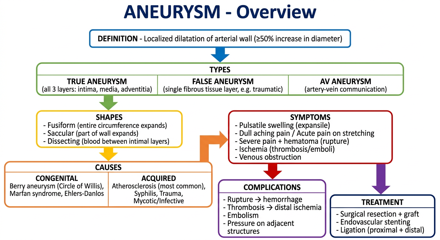
ANEURYSM - Short Note
Definition
- S Das Manual on Clinical Surgery, 13th Ed.; Bailey & Love's Short Practice of Surgery, 28th Ed.
Types
| Type | Wall Structure | Example |
|---|---|---|
| True Aneurysm | All 3 layers (intima, media, adventitia) | Atherosclerotic aortic aneurysm |
| False Aneurysm | Single fibrous tissue layer only | Post-traumatic/iatrogenic |
| AV Aneurysm | Artery-vein communication | Cirsoid aneurysm, AV fistula |
Shapes (of True Aneurysm)
- Fusiform - uniform expansion of the entire circumference
- Saccular - expansion of only part of the wall (usually traumatic)
- Dissecting - intima ruptures (usually under an atheromatous plaque) and blood is forced between the layers of tunica media
Causes
- Berry aneurysm (Circle of Willis) - due to deficiency of elastic lamina at branch points
- Marfan syndrome / Ehlers-Danlos syndrome
- Cirsoid aneurysm, Congenital AV fistula
- Degenerative - Atherosclerosis (by far the most common cause overall)
- Traumatic - Penetrating wounds, irradiation, cervical rib compression
- Infective (Mycotic) - Syphilis, bacterial infection, mycotic emboli, polyarteritis
Common Sites
Symptoms
- Often asymptomatic (detected incidentally)
- Dull aching pain (commonest symptom)
- Acute pain when vessel suddenly stretches
- Pulsatile expansile mass (hallmark finding on exam)
- Severe bursting pain + hematoma on rupture
- Distal ischaemia (from thrombosis or embolism)
- Venous obstruction - painful, swollen, blue limb
Signs on Examination
- Expansile pulsatile swelling in the course of an artery
- Pulsation diminishes on proximal pressure
- Compressible swelling
- Palpable thrill + systolic bruit on auscultation
- Swelling moves sideways but NOT along the artery axis
Complications
- Rupture - most feared, life-threatening hemorrhage
- Thrombosis - distal ischemia (especially popliteal aneurysm)
- Embolism - from thrombus in aneurysm sac
- Pressure effects - on nerves (neuralgic pain), veins (edema), adjacent organs
Treatment
| Option | Indication |
|---|---|
| Surgical resection + graft | Standard for large/symptomatic aneurysms |
| Endovascular stent graft (EVAR) | Aortic aneurysms, high surgical risk |
| Proximal + distal ligation | Mycotic or inaccessible aneurysms |
| Coil embolization | Intracranial (Berry) aneurysms |