
Hi I want to be a gastroenterologist I want you to teach me each and everything from scratch
What is your current level of medical knowledge? · What would you like to start with first?
gastroenterology full curriculum MBBS medical student learning roadmap 2024
🏥 Your Complete Gastroenterology Learning Roadmap
📚 Your Primary Textbooks (Available in Our Library)
| Textbook | Purpose |
|---|---|
| Yamada's Textbook of Gastroenterology, 7th ed (3 volumes) | The gold-standard comprehensive GI reference |
| Sleisenger & Fordtran's GI and Liver Disease | Clinical disease management bible |
| Clinical Gastrointestinal Endoscopy, 3rd ed | Procedures and endoscopy techniques |
| Harrison's Principles of Internal Medicine 22E (2025) | GI sections + systemic disease connections |
| Robbins & Cotran Pathologic Basis of Disease | GI pathology |
| Gray's Anatomy for Students | GI anatomy foundations |
🗺️ The Full Curriculum - 10 Modules
MODULE 1 - Foundations (Start Here)
- Embryological development of the GI tract (foregut, midgut, hindgut)
- Gross anatomy: esophagus, stomach, small intestine, large intestine, liver, biliary system, pancreas
- Histological layers of the GI wall (mucosa, submucosa, muscularis, serosa)
- Blood supply, lymphatics, and innervation (enteric nervous system)
- Peritoneum and mesentery
- Motility: peristalsis, migrating motor complex, sphincter function
- Secretion: gastric acid, pepsin, bile, pancreatic enzymes
- Digestion and absorption: carbohydrates, proteins, fats, vitamins, minerals
- Gut-brain axis and enteric nervous system (the "second brain")
- Gut microbiome basics
MODULE 2 - Esophageal Diseases
- GERD (Gastroesophageal Reflux Disease) - most common GI complaint worldwide
- Barrett's Esophagus and surveillance
- Achalasia and motility disorders
- Esophageal cancer (squamous cell vs. adenocarcinoma)
- Eosinophilic esophagitis
- Esophageal varices (portal hypertension)
- Boerhaave syndrome, Mallory-Weiss tear
MODULE 3 - Gastric & Duodenal Diseases
- Peptic ulcer disease (H. pylori, NSAIDs)
- H. pylori: diagnosis and eradication regimens
- Gastric cancer
- Gastritis (acute, chronic, autoimmune)
- Gastroparesis
- Functional dyspepsia
- Zollinger-Ellison syndrome (gastrinoma)
MODULE 4 - Small Intestinal Diseases
- Celiac disease (pathophysiology, serological markers, biopsy findings)
- Crohn's disease (vs. UC - key differences)
- Small intestinal bacterial overgrowth (SIBO)
- Carcinoid tumors / neuroendocrine tumors
- Malabsorption syndromes (tropical sprue, short bowel, Whipple disease)
- Intestinal obstruction, volvulus, intussusception
MODULE 5 - Colorectal Diseases
- Ulcerative colitis (vs. Crohn's - know this comparison cold)
- Colorectal cancer - screening, staging, molecular pathways (APC, KRAS, TP53)
- Colorectal polyps: adenomatous, hyperplastic, serrated
- Diverticular disease (diverticulosis vs. diverticulitis)
- Irritable bowel syndrome (Rome IV criteria)
- Microscopic colitis
- Anorectal disorders: hemorrhoids, fissures, fistulas, abscesses
MODULE 6 - Liver Diseases (Hepatology)
- Liver anatomy, function tests interpretation (ALT, AST, ALP, GGT, bilirubin)
- Viral hepatitis: A, B, C, D, E - complete understanding
- Cirrhosis: causes, Child-Pugh and MELD scoring
- Complications of cirrhosis: ascites, SBP, hepatic encephalopathy, HRS, esophageal varices
- Alcoholic liver disease and MASLD (formerly NAFLD)
- Autoimmune hepatitis
- Primary biliary cholangitis (PBC) and PSC
- Hepatocellular carcinoma (HCC)
- Acute liver failure
- Wilson disease, hemochromatosis, alpha-1 antitrypsin deficiency
MODULE 7 - Biliary System Diseases
- Cholelithiasis (gallstones) - types, risk factors, complications
- Cholecystitis (acute and chronic)
- Choledocholithiasis and cholangitis (Charcot's triad, Reynolds pentad)
- Primary sclerosing cholangitis (PSC)
- Cholangiocarcinoma
- ERCP - indications, technique, complications
MODULE 8 - Pancreatic Diseases
- Acute pancreatitis - causes, Ranson/Atlanta criteria, management
- Chronic pancreatitis - etiology, complications
- Pancreatic cancer (ductal adenocarcinoma) - very poor prognosis
- Pancreatic cystic lesions (IPMN, mucinous cystadenoma, serous)
- Autoimmune pancreatitis
MODULE 9 - GI Procedures & Endoscopy
- Upper GI endoscopy (EGD) - indications, technique, findings
- Colonoscopy - prep, technique, polyp removal
- ERCP - biliary and pancreatic interventions
- Endoscopic ultrasound (EUS)
- Capsule endoscopy
- Liver biopsy
- Paracentesis and hepatic procedures
- GI bleeding management (upper vs. lower)
MODULE 10 - GI Pharmacology & Clinical Skills
- PPIs, H2 blockers, antacids
- Antibiotics for H. pylori (triple/quadruple therapy)
- Antivirals for Hepatitis B and C (DAAs)
- IBD medications (aminosalicylates, corticosteroids, thiopurines, biologics)
- Laxatives and prokinetics
- Anti-diarrheals
- Reading GI imaging: CT abdomen, MRCP, abdominal X-ray
- Interpreting liver function tests, stool studies, breath tests
- Clinical approach to: GI bleeding, jaundice, ascites, dysphagia, diarrhea, constipation
🎯 How We Will Work Together
- Tell me which module you want to start with (I recommend Module 1 - Foundations)
- Each session, I will teach you the topic with:
- Core concepts explained clearly
- Diagrams and visual aids where available
- Key clinical pearls and mnemonics
- High-yield exam points
- Case-based examples
- After each topic, I can quiz you to check your understanding
- We will reference Yamada's, Sleisenger's, and other authoritative textbooks directly from our library
✅ Recommended Starting Order
I am pursuing BAMS And i am in 3rd prof So please teach me all type of diseases of gastroenterolist I want to be that
🩺 Gastroenterology Course for BAMS 3rd Prof
Welcome - Your Personal GI Masterclass
📖 LESSON 1 - Understanding the GI Tract (The Master System)
What is the GI Tract?
Ayurvedic parallel: In BAMS, you know this as Annavaha Srotas + Purishavaha Srotas. The same system, described from a different lens.
🔵 The GI Tract - Organ by Organ
1. MOUTH (Oral Cavity)
- Job: Mechanical breakdown (chewing) + chemical digestion begins
- Key secretion: Salivary amylase - breaks starch into maltose
- Clinical relevance: Oral ulcers may signal Crohn's disease, vitamin deficiencies, or autoimmune conditions
2. ESOPHAGUS
- Length: ~25 cm
- Job: Transport food from mouth to stomach - no digestion happens here
- Key structures:
- Upper Esophageal Sphincter (UES) - prevents food reflux into throat
- Lower Esophageal Sphincter (LES) - MOST IMPORTANT - prevents stomach acid from coming up
- Movement: Peristalsis (wave-like muscular contractions)
- Clinical relevance: When LES is weak → GERD (acid reflux) - one of the most common GI diseases worldwide
3. STOMACH
- Job: Storage + churning + chemical digestion
- Key secretions:
- Hydrochloric acid (HCl) - kills bacteria, activates pepsin
- Pepsin - digests proteins
- Intrinsic Factor - essential for Vitamin B12 absorption (loss = pernicious anemia)
- Mucus - protects stomach lining from its own acid
- Capacity: 1-1.5 litres after a meal
- Clinical relevance: When acid damages the mucus layer → Peptic Ulcer Disease
4. SMALL INTESTINE (The Absorption Powerhouse)
- Length: ~6-7 metres
- 3 Parts:
- Duodenum (25 cm) - receives bile and pancreatic juice; most active digestion
- Jejunum (2.5 m) - maximum nutrient absorption
- Ileum (3.5 m) - absorbs Vitamin B12 and bile salts
- Special structures: Villi + Microvilli (brush border) - increase surface area to the size of a tennis court (200 m²)!
- Key fact: 90% of all digestion and absorption happens here
- Clinical relevance: Damage to villi → Malabsorption (e.g., Celiac disease)
5. LARGE INTESTINE (Colon)
- Length: ~1.5 metres
- Parts: Cecum → Ascending → Transverse → Descending → Sigmoid → Rectum → Anus
- Job:
- Water and electrolyte absorption (turns liquid stool solid)
- Houses 100 trillion bacteria (gut microbiome)
- No significant nutrient absorption
- Daily volume: 1000-1500 mL enters, only 100-200 mL exits as stool
- Clinical relevance: Inflammation → Ulcerative Colitis; Cancer → Colorectal Cancer (3rd most common cancer worldwide)
6. LIVER
- Weight: 1.5 kg - largest internal organ
- Jobs (500+ functions!):
- Produces bile (for fat digestion)
- Metabolizes drugs, toxins, alcohol
- Produces clotting factors, albumin
- Stores glucose (as glycogen)
- Processes all nutrients absorbed from intestine (via portal vein)
- Clinical relevance: Viral hepatitis, cirrhosis, fatty liver disease, liver cancer
7. GALLBLADDER
- Job: Stores and concentrates bile made by the liver
- Releases bile when fatty food enters duodenum (CCK hormone triggers this)
- Clinical relevance: Bile + cholesterol → Gallstones (cholelithiasis) - extremely common, especially in women
8. PANCREAS
- Has TWO completely different functions:
- Exocrine (digestive): Secretes amylase, lipase, protease into duodenum
- Endocrine (hormonal): Insulin and glucagon (blood sugar control) → diabetes
- Clinical relevance: Acute pancreatitis (5/10 very painful emergency), Pancreatic cancer (silent killer)
🔵 The 4 Layers of the GI Wall
| Layer | What It Does | Clinical Importance |
|---|---|---|
| Mucosa (innermost) | Absorption, secretion, protection | Ulcers, cancers start here |
| Submucosa | Blood vessels, nerves (Meissner's plexus), lymphatics | Tumors invade this layer |
| Muscularis | Circular + longitudinal muscle; Auerbach's plexus | Motility disorders |
| Serosa/Adventitia (outermost) | Protective covering | Perforation goes through all layers |
🔵 Symptoms - The GI Tract's "Language"
| Symptom | What It Means |
|---|---|
| Heartburn / Pyrosis | Burning behind sternum - acid reflux |
| Dysphagia | Difficulty swallowing - esophageal disease |
| Odynophagia | Painful swallowing - esophageal ulcer/infection |
| Dyspepsia | Upper abdominal discomfort/bloating after eating |
| Nausea & Vomiting | Many causes - gastric, hepatic, pancreatic |
| Hematemesis | Vomiting blood - URGENT - upper GI bleed |
| Melena | Black tarry stools - digested blood from upper GI |
| Hematochezia | Fresh red blood in stool - lower GI bleed |
| Diarrhea | >3 loose stools/day |
| Constipation | <3 stools/week or straining |
| Jaundice | Yellow skin/eyes - liver or bile duct disease |
| Ascites | Fluid in abdomen - liver failure |
Mnemonics for GI bleeding: "MELENA = blood digested (UPPER GI)" | "HEMATOCHEZIA = FRESH blood (LOWER GI)"
🔵 The 4 Mechanisms of GI Disease
- Structural damage - erosion, ulceration, perforation, obstruction
- Bleeding - from ulcers, varices, tumors, inflammation
- Inflammation - IBD, hepatitis, pancreatitis
- Motility/Functional disorder - IBS, gastroparesis, achalasia
🔵 How Gastroenterologists Approach Every Patient
Step 1: HISTORY
  - Location of pain/symptom
  - Onset, duration, severity
  - Relation to food and bowel movements
  - Alarm symptoms? (weight loss, bleeding, dysphagia, fever)
  - Medications, alcohol, travel history

Step 2: PHYSICAL EXAM
  - Abdominal inspection → auscultation → percussion → palpation
  - Check for jaundice, pallor, clubbing (liver disease)
  - Check for tenderness, guarding, masses
  - Rectal exam (never skip!)

Step 3: INVESTIGATIONS
  - Blood: CBC, LFT, LFT, amylase, lipase, H. pylori
  - Stool: culture, occult blood, parasites
  - Imaging: X-ray, USG abdomen, CT scan, MRCP
  - Endoscopy: OGD (upper), Colonoscopy (lower)
  - Biopsy when needed

Step 4: DIAGNOSIS & TREATMENT
📋 Master List of ALL GI Diseases (Your Full Syllabus)
🔴 ESOPHAGUS
- GERD (Gastroesophageal Reflux Disease)
- Barrett's Esophagus
- Achalasia
- Esophageal Cancer
- Eosinophilic Esophagitis
- Esophageal Varices
- Mallory-Weiss Tear
- Boerhaave Syndrome
- Diffuse Esophageal Spasm
- Esophagitis (Candidal, Herpetic, CMV)
🟠 STOMACH & DUODENUM
- Peptic Ulcer Disease (PUD)
- H. pylori infection
- Acute Gastritis
- Chronic Gastritis (Type A & B)
- Gastroparesis
- Gastric Cancer
- Zollinger-Ellison Syndrome (Gastrinoma)
- Autoimmune Gastritis (Pernicious Anemia)
- Functional Dyspepsia
- Gastric Outlet Obstruction
- Menetrier's Disease
🟡 SMALL INTESTINE
- Celiac Disease (Gluten Enteropathy)
- Crohn's Disease
- Small Intestinal Bacterial Overgrowth (SIBO)
- Whipple's Disease
- Tropical Sprue
- Short Bowel Syndrome
- Carcinoid Tumor / NET
- Small Bowel Obstruction
- Meckel's Diverticulum
- Intestinal Tuberculosis
- Giardiasis & Parasitic infections
🟢 LARGE INTESTINE & ANORECTAL
- Ulcerative Colitis (UC)
- Colorectal Cancer
- Colorectal Polyps (Adenomas, Hyperplastic, Serrated)
- Diverticulosis & Diverticulitis
- Irritable Bowel Syndrome (IBS)
- Microscopic Colitis
- Pseudomembranous Colitis (C. difficile)
- Ischemic Colitis
- Volvulus (Sigmoid & Cecal)
- Intussusception
- Hemorrhoids (Internal & External)
- Anal Fissure
- Perianal Fistula & Abscess
- Anal Cancer
🔵 LIVER
- Viral Hepatitis A, B, C, D, E
- Alcoholic Liver Disease
- MASLD / MASH (Fatty Liver Disease)
- Autoimmune Hepatitis
- Primary Biliary Cholangitis (PBC)
- Primary Sclerosing Cholangitis (PSC)
- Cirrhosis
- Portal Hypertension
- Ascites
- Spontaneous Bacterial Peritonitis (SBP)
- Hepatic Encephalopathy
- Hepatorenal Syndrome (HRS)
- Esophageal Varices & Variceal Bleeding
- Hepatocellular Carcinoma (HCC)
- Acute Liver Failure (ALF)
- Wilson's Disease
- Hemochromatosis
- Alpha-1 Antitrypsin Deficiency
- Drug-Induced Liver Injury (DILI)
- Budd-Chiari Syndrome
🟣 BILIARY SYSTEM
- Cholelithiasis (Gallstones)
- Acute Cholecystitis
- Chronic Cholecystitis
- Choledocholithiasis (CBD stones)
- Acute Cholangitis (Charcot's triad)
- Cholangiocarcinoma (Bile duct cancer)
- Primary Sclerosing Cholangitis
- Biliary Strictures
🟤 PANCREAS
- Acute Pancreatitis
- Chronic Pancreatitis
- Pancreatic Adenocarcinoma
- Pancreatic Cystic Lesions (IPMN, MCN)
- Autoimmune Pancreatitis
- Pancreatic Neuroendocrine Tumors (NETs)
- Pancreatic Exocrine Insufficiency
✅ Session Summary - What You Learned Today
- The GI tract: 8 organs, each with a specific job
- The 4 wall layers and their clinical significance
- The master symptom list of GI disease
- The 4 mechanisms of all GI disease
- The systematic clinical approach
- All 81 GI diseases organized by organ (your full syllabus!)
🎯 What's Coming in Lesson 2
GERD - Gastroesophageal Reflux Disease
- Exact mechanism (why the acid goes up)
- Causes and risk factors
- Symptoms, complications
- How to diagnose it
- How to treat it (lifestyle + medicines)
- Barrett's esophagus - when GERD becomes dangerous
- Case studies
Next
📖 LESSON 2 - GERD (Gastroesophageal Reflux Disease)
The Most Common GI Disease in the World
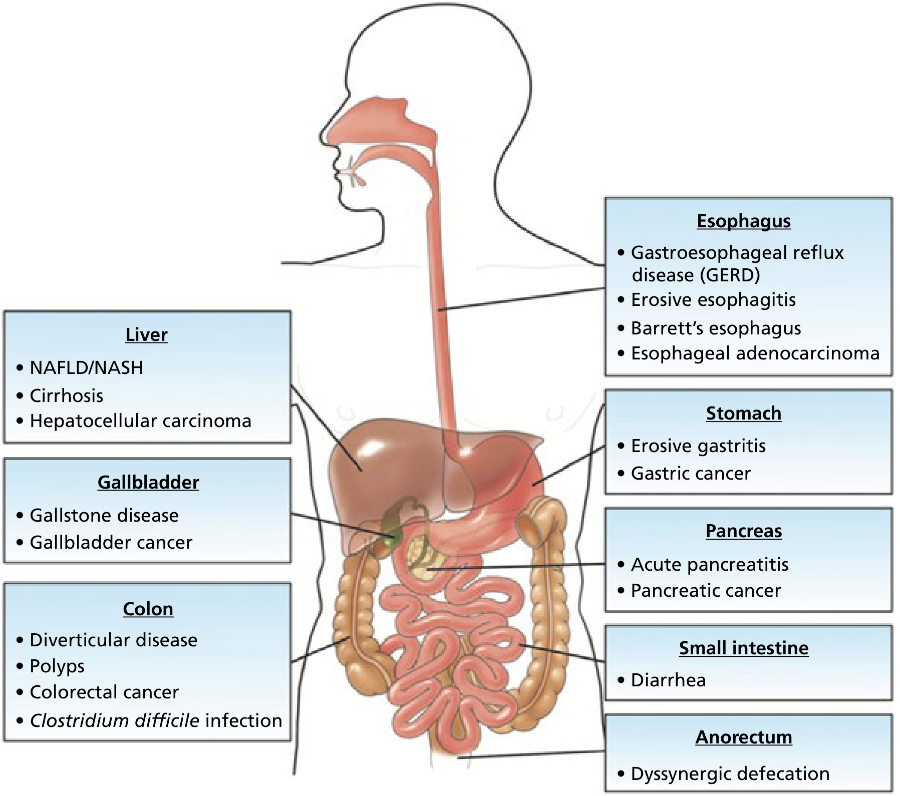
🔵 WHAT IS GERD?
Ayurvedic Parallel (for your BAMS background): GERD correlates closely with Amlapitta in Ayurveda - the condition of excess/vitiated Pitta in Amashaya (stomach) with urdhvaga (upward movement) pattern. The Ushna, Tikshna, Amla qualities of Pitta producing daha (burning) in Uras (chest) is your existing reference point.
- Affects ~20% of adults in Western countries
- Affects ~8-15% in Asia (but rapidly rising with urbanization and obesity)
- One of the top 3 reasons people visit a gastroenterologist
🔵 NORMAL ANATOMY - What PREVENTS Reflux?
MOUTH
  ↓  (swallowing)
ESOPHAGUS (25 cm pipe - NO acid here normally)
  ↓
[LOWER ESOPHAGEAL SPHINCTER = LES] ← THE GATEKEEPER
  ↓  (one-way valve - opens only to let food down)
STOMACH (full of HCl acid, pH 1.5-3.5)
- A thick ring of circular smooth muscle at the bottom of the esophagus
- Normally closed (resting pressure 15-30 mmHg) - keeps acid IN the stomach
- Opens briefly when you swallow to let food into stomach
- Supported by diaphragmatic crura (external LES) and the phreno-esophageal ligament
- High LES resting pressure (15-30 mmHg)
- Adequate LES length (at least 2 cm must be inside the abdomen)
- Normal angle of His (sharp angle between esophagus and stomach)
🔵 PATHOPHYSIOLOGY - Why Does Reflux Happen?
Mechanism 1 - Transient LES Relaxations (tLESRs) ← MOST COMMON
- The LES relaxes at the wrong time - NOT during swallowing
- These are reflex relaxations triggered by gastric distension
- Account for ~80% of all reflux episodes in GERD patients
- This is the main mechanism in mild to moderate GERD
Mechanism 2 - Low Resting LES Pressure
- The sphincter is simply too weak
- Pressure drops below 6 mmHg (normal = 15-30 mmHg)
- Causes continuous reflux, especially when lying flat
- Seen in severe GERD and scleroderma patients
Mechanism 3 - Hiatal Hernia
- The upper part of the stomach slides above the diaphragm into the chest
- This disrupts both the LES and the diaphragmatic crura (external sphincter)
- The diaphragm no longer helps squeeze the LES shut
- Seen in most patients with severe GERD and Barrett's esophagus
Additional Contributing Factors:
| Factor | How It Causes GERD |
|---|---|
| Obesity | High intraabdominal pressure pushes acid upward |
| Pregnancy | Progesterone relaxes LES + enlarging uterus increases abdominal pressure |
| Smoking | Nicotine directly reduces LES tone |
| Alcohol | Relaxes LES, increases acid secretion |
| High-fat meals | Delays gastric emptying, distends stomach |
| Caffeine, chocolate, mint | Reduce LES pressure |
| NSAIDs | Directly damage esophageal mucosa |
| Lying down after meals | Gravity no longer helps keep acid down |
🔵 WHY DOES ACID DAMAGE THE ESOPHAGUS?
- pH of esophagus drops from normal (6-7) to below 4
- Acid + pepsin together cause mucosal injury
- Repeated exposure → inflammation → erosions → ulcers → scarring
- Long-term repeated injury → Barrett's Esophagus (pre-cancerous change)
🔵 SYMPTOMS OF GERD
Typical (Esophageal) Symptoms:
| Symptom | Description | Key Point |
|---|---|---|
| Heartburn (Pyrosis) | Burning sensation behind the sternum (breastbone) | Worsens after meals, lying down, bending forward |
| Regurgitation | Sour/bitter liquid coming up into mouth or throat | Patient says "acid taste in mouth" |
| Water Brash | Sudden flood of saliva in mouth | Reflex hypersalivation to neutralize acid |
| Dysphagia | Difficulty swallowing | Alarm symptom - suggests stricture or cancer |
| Odynophagia | Pain on swallowing | Suggests severe esophagitis or ulcer |
Atypical (Extra-Esophageal) Symptoms:
| Symptom | Explanation |
|---|---|
| Chronic cough | Acid microaspiration irritates airways |
| Asthma / wheezing | Acid triggers bronchospasm via vagal reflex |
| Hoarseness / voice change | Acid damages vocal cords (laryngopharyngeal reflux) |
| Chronic sore throat | Acid irritates pharynx |
| Dental erosions | Acid destroys tooth enamel |
| Non-cardiac chest pain | GERD mimics heart attack! |
| Sinusitis / ear problems | In some patients |
Clinical Pearl: When a patient comes with "heart attack-like" chest pain and the cardiac workup is negative - always think GERD! GERD is one of the top causes of non-cardiac chest pain.
🔵 ALARM SYMPTOMS - Red Flags (URGENT Investigation Needed!)
- 🚨 Dysphagia (difficulty swallowing) - may mean stricture or cancer
- 🚨 Odynophagia (painful swallowing)
- 🚨 Unexplained weight loss
- 🚨 Hematemesis (vomiting blood)
- 🚨 Melena (black tarry stools)
- 🚨 Anemia (iron deficiency)
- 🚨 Age >50 with new onset symptoms
- 🚨 Symptoms not responding to treatment
🔵 COMPLICATIONS OF GERD
NORMAL ESOPHAGUS
     ↓ (repeated acid injury over years)
REFLUX ESOPHAGITIS (Grade A-D)
     ↓ (continued injury)
PEPTIC STRICTURE (scarring narrows the esophagus → dysphagia)
     ↓ (in some patients with severe long-standing GERD)
BARRETT'S ESOPHAGUS ← PRE-CANCEROUS CHANGE
     ↓ (in ~0.5% per year)
ESOPHAGEAL ADENOCARCINOMA ← CANCER
Complication 1 - Reflux Esophagitis
- Graded by the Los Angeles Classification (LA Grade A-D):
| Grade | Finding |
|---|---|
| Grade A | Mucosal breaks < 5 mm, not continuous between folds |
| Grade B | Mucosal breaks > 5 mm, not continuous between folds |
| Grade C | Mucosal breaks continuous between folds, < 75% circumference |
| Grade D | Mucosal breaks involving > 75% of esophageal circumference |
Complication 2 - Peptic Stricture
- Repeated inflammation → fibrosis and narrowing
- Patient presents with progressive dysphagia (first to solids, then liquids)
- Treatment: endoscopic dilation + PPIs
Complication 3 - Barrett's Esophagus ← MOST IMPORTANT COMPLICATION
- Definition: Normal squamous epithelium of the distal esophagus is replaced by columnar epithelium (intestinal metaplasia with goblet cells)
- This is called metaplasia - one type of cell replacing another
- Affects 5-15% of patients who undergo endoscopy for GERD symptoms
- Risk factors: Long-standing GERD, obesity, male sex, white race, smoking, age >50
- Importance: Barrett's is a pre-malignant condition - it can progress to esophageal adenocarcinoma
- Annual cancer risk from Barrett's: ~0.5% per year (low in any single year, but significant over a lifetime)
- Surveillance: Regular endoscopy + biopsy every 3-5 years (or more frequently if dysplasia found)
Key Histology: You look for goblet cells on biopsy - their presence confirms intestinal metaplasia = Barrett's esophagus
🔵 DIAGNOSIS OF GERD
Step 1 - Clinical Diagnosis (Most common approach)
- Classic heartburn + regurgitation = GERD diagnosis can be made clinically
- PPI trial: Give a proton pump inhibitor for 4-8 weeks
- If symptoms improve significantly → confirms GERD (therapeutic trial)
- This is the standard first approach in typical cases without alarm symptoms
Step 2 - Investigations (When needed)
| Investigation | What It Shows | When Used |
|---|---|---|
| Upper GI Endoscopy (OGD) | Esophagitis grading, Barrett's, ulcers, strictures | Alarm symptoms, treatment failure, surveillance |
| 24-hour pH monitoring | Most sensitive test - measures how often acid drops below pH 4 | When diagnosis uncertain, before surgery |
| pH-Impedance monitoring | Detects both acid AND non-acid reflux | Best for complete reflux assessment |
| Esophageal Manometry | Measures LES pressure and esophageal motility | Before anti-reflux surgery |
| Barium Swallow | Shows strictures, hiatal hernia | Less used now |
Key Exam Fact: The most sensitive test for GERD diagnosis = 24-hour ambulatory pH + impedance monitoring - Harrison's 22E
🔵 TREATMENT OF GERD
Step 1 - Lifestyle Modifications (Always first!)
| Modification | Reason |
|---|---|
| Lose weight | Reduces intraabdominal pressure |
| Elevate head of bed by 6-8 inches | Uses gravity to prevent nighttime reflux |
| Don't lie down for 3 hours after meals | Keeps acid in stomach while digesting |
| Avoid trigger foods: fatty food, chocolate, mint, coffee, alcohol, citrus, tomato | All reduce LES pressure or irritate directly |
| Small, frequent meals | Prevents excessive gastric distension |
| Stop smoking | Nicotine relaxes LES |
| Avoid tight clothing | Reduces abdominal pressure |
Step 2 - Antacids (For mild, occasional symptoms)
- Examples: Aluminum hydroxide, Magnesium hydroxide, Calcium carbonate (Tums)
- Mechanism: Neutralize acid already in stomach (don't reduce acid production)
- Onset: Fast (minutes) but short-lasting (30-60 min)
- Use: On-demand for mild heartburn
Step 3 - H2 Blockers (Moderate symptoms)
- Examples: Ranitidine (withdrawn due to NDMA), Famotidine, Cimetidine
- Mechanism: Block H2 receptors on parietal cells → reduce acid secretion
- Reduce acid by ~70%
- Problem: Tolerance develops (tachyphylaxis) with regular use
Step 4 - Proton Pump Inhibitors (PPIs) - THE GOLD STANDARD
- Examples: Omeprazole, Pantoprazole, Rabeprazole, Lansoprazole, Esomeprazole
- Mechanism: Irreversibly block H+/K+ ATPase pump (the final step in acid production) on parietal cells
- Reduce acid by >90% - most powerful acid suppressants available
- Taken 30-60 minutes before meals (so drug reaches parietal cells while they are being activated by food)
- Duration: 4-8 weeks for esophagitis; long-term for Barrett's and severe GERD
- Side effects (long-term): Hypomagnesemia, B12 deficiency, increased infection risk, osteoporosis
Mnemonic for PPIs: "Omeprazole Pantoprazole Rabeprazole Lansoprazole Esomeprazole" = OPRLE or just remember they all end in "-prazole"
Step 5 - Prokinetics (Adjunct therapy)
- Examples: Metoclopramide, Domperidone
- Mechanism: Increase LES pressure + speed up gastric emptying
- Reduce gastric distension - less reflux trigger
- Limited long-term use due to side effects (extrapyramidal effects with metoclopramide)
Step 6 - Anti-Reflux Surgery (When all else fails)
- Nissen Fundoplication - the stomach fundus is wrapped 360° around the lower esophagus to mechanically strengthen the LES
- Indicated when: Young patient who doesn't want lifelong PPIs, large hiatal hernia, symptoms persist despite max medical therapy
- LINX procedure: A magnetic ring placed around the LES to reinforce it (newer, reversible option)
🔵 GERD IN SPECIAL SITUATIONS
| Situation | Key Points |
|---|---|
| Pregnancy | Use antacids/alginate first; PPIs generally safe but use cautiously |
| Children/Infants | Very common in infants (immature LES); usually resolves by age 1; lifestyle first |
| Elderly | Often present atypically (no heartburn!); present with silent esophagitis, anemia |
| Asthma patients | GERD worsens asthma; treat GERD to improve asthma control |
🔵 CLINICAL CASE (Practice Your Thinking!)
- Burning sensation in chest after meals for 2 years
- Worse when lying down at night
- Sour taste in mouth in the mornings
- Occasional dry cough
- No weight loss, no difficulty swallowing, no blood in stools
- What is your diagnosis?
- What alarm symptoms are ABSENT here?
- What will you do first - investigate or treat?
- What medication and what advice will you give?
- What complication is this patient at risk of developing?
- GERD - classic presentation (heartburn, regurgitation, nocturnal symptoms, cough)
- No dysphagia, no weight loss, no bleeding, no anemia - absence of alarm symptoms means no urgent endoscopy needed
- Treat empirically first - PPI trial for 4-8 weeks (no need for immediate endoscopy since no alarm symptoms)
- Omeprazole 20 mg once daily, 30 min before breakfast + Lifestyle: lose weight, elevate head of bed, stop smoking, avoid late meals, reduce fatty food
- Barrett's Esophagus - he is a middle-aged obese male smoker with long-standing GERD = high-risk profile
✅ Lesson 2 Summary - Key Points to Never Forget
| # | Key Point |
|---|---|
| 1 | GERD = failure of the LES (Lower Esophageal Sphincter) |
| 2 | Most common mechanism = transient LES relaxations (tLESRs) |
| 3 | Hiatal hernia is the main structural cause |
| 4 | Typical symptoms = heartburn + regurgitation |
| 5 | Atypical symptoms = cough, hoarseness, asthma, chest pain |
| 6 | Alarm symptoms = refer for urgent endoscopy |
| 7 | Most sensitive test = 24-hour pH-impedance monitoring |
| 8 | Gold standard treatment = PPIs ("-prazole" drugs) |
| 9 | GERD → Barrett's Esophagus → Esophageal Adenocarcinoma |
| 10 | Barrett's = squamous → columnar metaplasia (goblet cells on biopsy) |
🎯 Coming Up in Lesson 3
- Why stomach acid eats through its own wall
- H. pylori - the Nobel Prize-winning discovery
- How to diagnose and eradicate H. pylori
- Complications: perforation, bleeding, obstruction
- All the drugs used to treat it
You did great Lekin mujhe ye sb Hinglish me chahiye aur clinical cases chahiye achche achche se 3 kam se kam aur bs jo main aur important points h gastroenterologist ke loye vohi details jo jyada mt do aur Is baar make it best aur treatment lifestyle diet ye sb bhi achche se smjhao
🔥 LESSON 2 - GERD (Acid Reflux Disease)
Hinglish mein - Maximum Clarity, Minimum Bakwaas
🎯 EK LINE MEIN GERD KYA HAI?
"Stomach ka acid wapas esophagus mein aana - aur esophagus ko jalana - yahi GERD hai."
🔑 MOST IMPORTANT CONCEPT - LES (Lower Esophageal Sphincter)
ESOPHAGUS  (food pipe - koi acid nahi hota yahan)
     ↓
  [LES] ← YE DARWAZA HAI
     ↓
STOMACH  (acid factory - pH 1.5 to 3.5)
LES kyun fail hota hai? (3 main reasons)
| Reason | Kya hota hai | Kahan zyada |
|---|---|---|
| Transient LES relaxations | LES galat time pe khul jaata hai | 80% cases - most common |
| Weak LES pressure | Pressure <6 mmHg ho jaata hai (normal 15-30) | Severe GERD |
| Hiatal Hernia | Stomach ka part diaphragm ke upar aa jaata hai - LES support khatam | Barrett's risk high |
⚡ RISK FACTORS - "Ye log GERD ke liye bane hain"
| Risk Factor | Reason |
|---|---|
| Obesity | Pet mein pressure badh jaata hai, acid upar dhakelta hai |
| Pregnancy | Progesterone LES relax karta hai + uterus pressure deta hai |
| Smoking | Nicotine directly LES tone kam karta hai |
| Alcohol | LES relax + acid secretion increase |
| Fatty food, chocolate, mint, coffee | Sab LES pressure reduce karte hain |
| Late night khana | Lete waqt gravity help nahi karta |
| Tight clothes | Abdominal pressure increase |
| NSAIDs (aspirin, ibuprofen) | Directly esophageal mucosa damage karte hain |
🩺 SYMPTOMS - Kaise Pehchanein GERD?
Typical Symptoms (Classic GERD):
| Symptom | Hindi mein | Key point |
|---|---|---|
| Heartburn / Pyrosis | Seene mein jalan | Khane ke baad worse, raat ko worse |
| Regurgitation | Khatta/kadwa taste aana | "Muh mein acid aa jaata hai" |
| Water Brash | Muh mein suddenly paani bhar jaata hai | Saliva reflex - acid neutralize karne ke liye |
Atypical Symptoms (Confusing - GERD dusri bimari jaise lagta hai!):
| Symptom | Explanation |
|---|---|
| Chronic cough | Acid microaspiration se airway irritate hoti hai |
| Hoarseness / voice change | Acid vocal cords tak pahunch jaata hai |
| Asthma jaise symptoms | Vagal reflex se bronchospasm hota hai |
| Non-cardiac chest pain | GERD heart attack jaisa feel kara sakta hai! |
| Dental erosions | Acid daant gala deta hai |
🔴 Clinical Pearl: Jab patient aaye chest pain se aur cardiac workup normal ho - always think GERD! Ye ek top GI trap hai boards mein bhi.
🚨 ALARM SYMPTOMS - Ye Dekho Toh Seedha Endoscopy!
- 🚩 Dysphagia (nigalne mein takleef) - stricture ya cancer?
- 🚩 Unexplained weight loss
- 🚩 Hematemesis (khoon ki ulti)
- 🚩 Melena (kaala poth jaisa stool)
- 🚩 Anemia (iron deficiency)
- 🚩 Age >50 mein naye symptoms
- 🚩 4-8 weeks PPI ke baad bhi symptoms - response nahi
Mnemonic: "ABCDE" - Anemia, Bleeding, Chest pain persists, Dysphagia, Emesis (vomiting) + weight loss = Endoscopy karo!
⚠️ COMPLICATIONS - GERD Ka Dangerous Safar
NORMAL ESOPHAGUS
     ↓ (months-years of acid exposure)
REFLUX ESOPHAGITIS (Grade A → B → C → D)
     ↓
PEPTIC STRICTURE (scarring → narrowing → dysphagia)
     ↓
BARRETT'S ESOPHAGUS ← DANGER ZONE
     ↓ (0.5% per year)
ESOPHAGEAL ADENOCARCINOMA
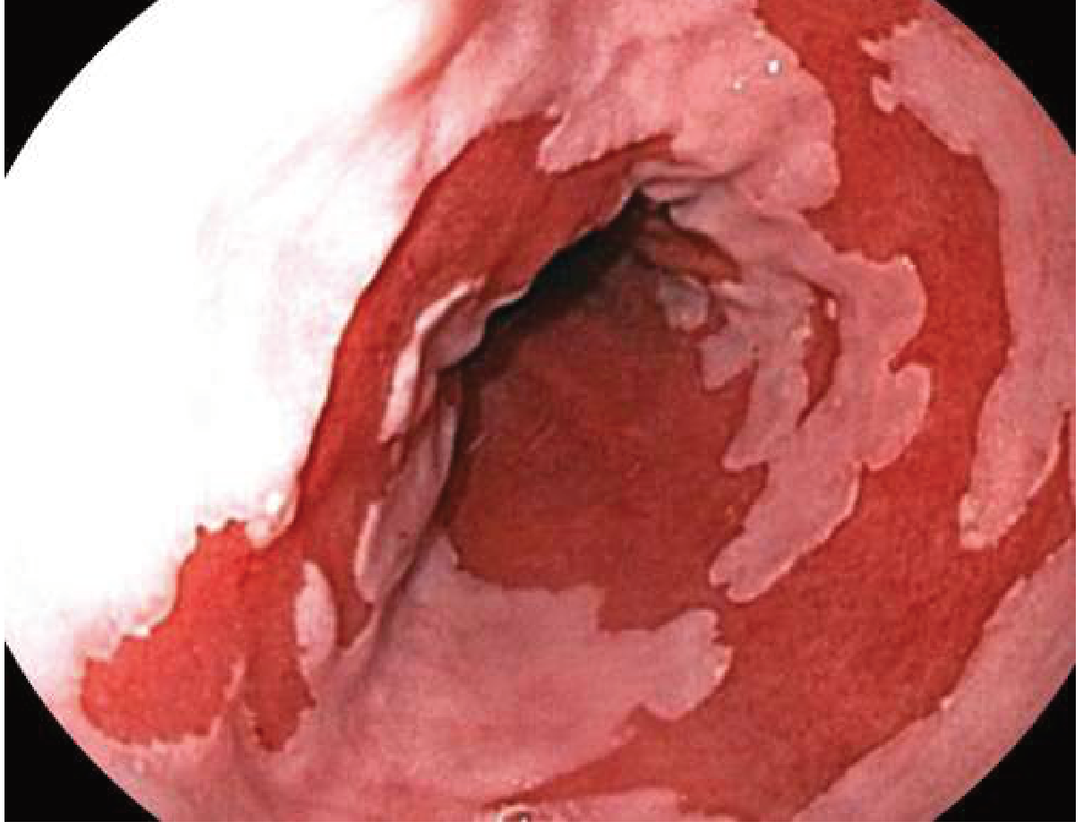
Barrett's Esophagus - Samjho Achi Tarah
- Normal esophagus mein squamous cells hote hain (flat cells)
- Repeated acid injury ke baad ye cells columnar cells (goblet cells) mein badal jaate hain
- Yahi metaplasia hai - ek cell type dusre mein replace hona
- Ye change pre-cancerous hai
- Endoscopy pe kaisa dikhta hai? - Salmon-pink coloured mucosa normal white mucosa ki jagah
Yahan dekho - actual endoscopy image of Barrett's Esophagus:
🔬 DIAGNOSIS - Kaise Confirm Karte Hain?
| Test | Kya batata hai | Kab karte hain |
|---|---|---|
| Clinical + PPI Trial | Symptoms + 4-8 wk PPI response | First step - typical symptoms, no alarm |
| Upper GI Endoscopy (OGD) | Esophagitis grade, Barrett's, stricture | Alarm symptoms, treatment failure |
| 24-hr pH + Impedance monitoring | Most sensitive test - actual acid exposure measure karta hai | Diagnosis doubtful, pre-surgery |
| Esophageal Manometry | LES pressure, motility | Anti-reflux surgery se pehle |
| Barium Swallow | Stricture, hiatal hernia | Limited use ab |
(Harrison's 22E: "Most sensitive test for GERD = 24-hour ambulatory pH and impedance monitoring")
💊 TREATMENT - Step by Step
🥗 STEP 1 - LIFESTYLE + DIET (Sabse Pehle Yahi!)
| ✅ Khao | ❌ Bilkul Avoid Karo |
|---|---|
| Daliya, oats, banana, apple | Fried/oily food - LES relax karta hai |
| Lean protein (chicken, fish, dal) | Chocolate - LES tone kam karta hai |
| Green vegetables | Mint/peppermint - LES relax karta hai |
| Low-fat dairy | Coffee/tea zyada - acid production increase |
| Whole grains | Alcohol - dual effect (LES + acid) |
| Ginger (anti-inflammatory) | Citrus fruits, tomatoes - directly irritate |
| Coconut water | Carbonated drinks - gas se pressure |
| Curd/yogurt (thodi matra) | Spicy masaledaar food |
| Rule | Reason |
|---|---|
| Khane ke 3 ghante baad leto | Gravity se acid neeche rehta hai |
| Bed ka head end 15-20 cm utha do | Raat ko gravity help karta hai |
| Chhoti chhoti meals lena, 5-6 baar | Stomach zyada distend na ho |
| Weight loss | Abdominal pressure kam hota hai - GERD dramatically better |
| Smoking band karo | Nicotine ka LES pe direct effect |
| Tight belt/clothing avoid karo | Abdominal pressure kam karo |
| Stress management karo | Stress gastric acid secretion badhata hai |
💊 STEP 2 - MEDICATIONS (Ladder Approach)
Mild symptoms  →  ANTACIDS
       ↓ (if not enough)
Moderate       →  H2 BLOCKERS
       ↓ (gold standard)
Standard/Severe →  PPIs ← CORNERSTONE OF TREATMENT
       ↓ (adjunct)
Prokinetics     →  METOCLOPRAMIDE (limited use)
       ↓ (surgery last resort)
NISSEN FUNDOPLICATION
🔵 ANTACIDS
- Examples: Gelusil, Digene, Eno, Milk of Magnesia
- Mechanism: Acid ko neutralize karte hain (production nahi rokate)
- Onset: 5-10 minutes - bahut fast
- Duration: 30-60 minutes only
- Use: Kabhi kabhi heartburn ke liye - on-demand
- Dose: 15 mL after meals and at bedtime
🔵 H2 BLOCKERS (H2 Receptor Antagonists)
- Examples: Famotidine (most common), Cimetidine, Ranitidine (withdrawn)
- Mechanism: Parietal cells pe H2 receptor block karo → acid secretion ~70% kam
- Problem: Tachyphylaxis - regular use se effectiveness kam hoti hai
- Use: Mild-moderate GERD, nighttime acid breakthrough
🔴 PPIs - PROTON PUMP INHIBITORS ← KING OF GERD TREATMENT
- Examples: Omeprazole 20mg, Pantoprazole 40mg, Rabeprazole 20mg, Lansoprazole 30mg, Esomeprazole 40mg
- Mechanism: H+/K+ ATPase pump (proton pump) ko irreversibly block karta hai - yahi acid banane ki final step hai
- Acid reduction: >90% - sabse powerful
- Timing: Khane se 30-60 min pehle - issi waqt parietal cells activate hote hain
- Duration: 4-8 weeks esophagitis ke liye; long-term Barrett's ke liye
- Vitamin B12 deficiency
- Hypomagnesemia
- C. difficile infection risk
- Osteoporosis / hip fracture risk
- SIBO (small intestinal bacterial overgrowth)
🔵 SURGERY - Nissen Fundoplication
- Kab: Young patient, surgery prefer karta ho; large hiatal hernia; PPIs se symptoms nahi jaate
- Kya hota hai: Stomach ka fundus esophagus ke around 360° wrap kar dete hain - mechanical LES banana
- New option: LINX device - magnetic ring LES ke around lagaate hain
🏥 CLINICAL CASES - 3 Real-World Scenarios
🟢 CASE 1 - The Classic Presentation
🔴 CASE 2 - The Alarm Symptom Case
- Peptic stricture (scarring se narrowing - benign)
- Barrett's esophagus with dysplasia
- Esophageal adenocarcinoma (cancer - worst case)
🟡 CASE 3 - The Tricky Atypical Presentation
- No heartburn (patient specifically bolta hai - "seene mein jalan nahi hoti")
- Subah uthke thodi hoarseness hoti hai
- Raat ko khana khane ke baad kuch ghante baad theek se so nahi pata
📊 GERD - Quick Revision Table (Exam Ready!)
| Topic | Key Point |
|---|---|
| Most common mechanism | Transient LES relaxations (tLESRs) |
| Most common structural cause | Hiatal Hernia |
| Most sensitive test | 24-hr pH + impedance monitoring |
| Gold standard diagnosis in routine | Clinical + PPI trial response |
| Endoscopy kab | Alarm symptoms ya treatment failure |
| Gold standard treatment | PPIs (omeprazole, pantoprazole) |
| Timing of PPI | Khane se 30-60 min pehle |
| Barrett's kya hai | Squamous → Columnar metaplasia (goblet cells) |
| Barrett's ka cancer risk | ~0.5% per year |
| Barrett's treatment | Radiofrequency Ablation (RFA) |
| Surgery | Nissen fundoplication |
| Atypical GERD | Cough, hoarseness, asthma, chest pain |
✅ Lesson 2 Complete!
- LES fail → GERD → Barrett's → Cancer - ye chain yaad rakho
- Alarm symptoms → Endoscopy seedha - PPI trial mat dena
- Heartburn nahi matlab GERD nahi - galat! Silent GERD hota hai
🎯 Lesson 3 Mein Aayega:
Peptic Ulcer Disease (PUD)
- H. pylori kya hai - Nobel Prize wali discovery
- Stomach apna aap kyun nahi khata (normally)?
- Ulcer kab khata hai - mechanism samjhenge
- Classic presentation + 3 clinical cases (Hinglish mein!)
- Complications: bleeding, perforation, obstruction
- H. pylori eradication regimens - exactly kaunsi drugs, kitni dose
Next
🔥 LESSON 3 - Peptic Ulcer Disease (PUD)
Hinglish mein | 3 Clinical Cases | Full Treatment Guide
🎯 EK LINE MEIN PUD KYA HAI?
"Stomach ya duodenum ki wall mein ek gaddha (ulcer) ban jaata hai jab acid + pepsin mucosal defenses ko tod dete hain."
🔑 SABSE PEHLE - Balance Theory Samjho
⚔️ ATTACK FORCES          vs        🛡️ DEFENSE FORCES
- HCl (gastric acid)              - Mucus layer
- Pepsin (enzyme)                 - Bicarbonate secretion
- H. pylori                       - Mucosal blood flow
- NSAIDs                          - Prostaglandins (PGE2)
- Bile reflux                     - Tight junctions
- Stress                          - Epithelial renewal
- Attack forces zyada ho jaayein YA Defense forces kam ho jaayein
Ayurvedic parallel: Ye exactly Parinama Shoola ka mechanism hai - Ushna + Tikshna Pitta jab Kapha (protective layer = mucus) ko consume kar leta hai → Shotha + Kshata (inflammation + ulceration)
🔵 ULCER KAHAN BANTA HAI?
| Site | % Cases | Reason |
|---|---|---|
| Duodenal ulcer (DU) | ~70% | Acid directly duodenum ko attack karta hai |
| Gastric ulcer (GU) | ~25% | Defense failure - acid normal bhi ho sakta hai |
| Esophageal | Rare | GERD complication |
| Meckel's diverticulum | Very rare | Ectopic gastric mucosa |
Key difference: DU = acid zyada hoti hai | GU = defense kam hoti hai
⚡ CAUSES - "PUD Kyun Hota Hai?"
CAUSE 1 - H. pylori Infection ← KING OF PUD CAUSES
- >70% PUD cases mein H. pylori responsible hai (Robbins Pathology)
- Ye ek gram-negative spiral bacterium hai
- Stomach ki mucus layer mein rehta hai - jahaan koi aur bacteria nahi jee sakta
- Urease enzyme produce karta hai → urea → ammonia → mucosal damage
- Puri duniya ki ~50% population infected hai (India mein ~70-80%!)
- Lekin infected logon mein se sirf 5-10% ko ulcer hota hai - matlab host factors bhi important hain
- Oral-oral route (kissing, shared utensils)
- Fecal-oral route (contaminated water/food)
- India mein common kyun? - Sanitation issues + joint family living
CAUSE 2 - NSAIDs / Aspirin
- Ibuprofen, Diclofenac, Naproxen, Aspirin - sab common NSAIDs
- Mechanism (2 ways):
- Topical: Directly mucosa ko damage karte hain (acid ya pill form mein)
- Systemic: COX-1 block karte hain → Prostaglandin (PGE2) kam hoti hai → mucus + bicarbonate production kam → mucosa unprotected
- NSAID ulcers mein pain nahi hota aksar - "silent ulcers" - seedha bleeding se present!
- Risk zyada: Age >60, previous ulcer, steroid + NSAID combination
CAUSE 3 - Rarer Causes
| Cause | Mechanism |
|---|---|
| Zollinger-Ellison Syndrome | Gastrinoma tumor → unlimited gastrin → massive acid → multiple ulcers |
| Stress (ICU patients) | Reduced mucosal blood flow |
| Smoking | Mucosal blood flow kam, healing slow |
| Alcohol | Direct mucosal damage |
| Steroids | Prostaglandin synthesis suppress |
🩺 SYMPTOMS - Kaise Pehchanein?
Typical (Uncomplicated) Ulcer:
| Symptom | Detail |
|---|---|
| Epigastric pain | "Pet ke upar beech mein dard" - burning ya gnawing |
| Food relation | DU: Khane se relief (food acid buffer karta hai) → 2-3 hr baad wapas dard |
| Nocturnal pain | Raat 2-3 baje uthna - DU ka classic feature |
| "Painful hunger" | Khaali pet dard - khane se thodi der ke liye theek |
| Nausea, belching | Common |
| Heartburn | GERD associated ya hyperacidity se |
🔴 Critical Point: Up to 40% ulcers SILENT hote hain - koi symptom nahi! Seedha complication se present hote hain (Goldman-Cecil Medicine)
Duodenal vs Gastric Ulcer - Quick Compare:
| Feature | Duodenal Ulcer | Gastric Ulcer |
|---|---|---|
| Pain with food | Relieved by food | Worsened by food |
| Nocturnal pain | Classic | Less common |
| Acid secretion | High | Normal/Low |
| H. pylori | 90% associated | 70% associated |
| Cancer risk | Very low | ⚠️ Higher - biopsy zaroori! |
| Weight | May gain (eating relieves pain) | May lose (eating causes pain) |
🚨 COMPLICATIONS - "Jab Ulcer Khatarnak Ho Jaata Hai"
Complication 1 - BLEEDING (Sabse Common)
- Ulcer kisi blood vessel ko erode kar deta hai
- Presentation: Hematemesis (khoon ki ulti) + Melena (kaala poth stool)
- Duodenal ulcer → Gastroduodenal artery erode → massive bleed
- Management: Emergency endoscopy → cauterize/clip the vessel + IV PPI drip
Complication 2 - PERFORATION (Sabse Dangerous)
- Ulcer puri wall ke through chala jaata hai → hole ban jaata hai
- Stomach ka content peritoneal cavity mein jaata hai → Chemical peritonitis
- Presentation:
- Sudden, severe, "knife-like" epigastric pain
- Pain shoulder tak radiate (diaphragm irritation)
- Board-like rigid abdomen (defense guarding)
- X-ray: Free air under diaphragm ← PATHOGNOMONIC!
- Management: EMERGENCY SURGERY - patch repair (Graham's patch)
Complication 3 - GASTRIC OUTLET OBSTRUCTION (Rare ab)
- Pyloric area mein ulcer → repeated inflammation → fibrosis → narrowing → obstruction
- Presentation:
- Projectile non-bilious vomiting of old food
- Vomitus mein previously eaten food (12-24 hrs pehle ka)
- "Succussion splash" on examination
- Weight loss, dehydration
- Management: Endoscopic dilation ya surgery
Complication 4 - PENETRATION
- Ulcer adjacent organ mein ghus jaata hai (pancreas most common)
- Presentation: Pain back ki taraf radiate karta hai + constant (khane se relief nahi)
- Raised serum amylase/lipase (pancreas involved)
🔬 DIAGNOSIS
| Test | Kya batata hai | Kab |
|---|---|---|
| Upper GI Endoscopy (OGD) | Gold standard - ulcer directly dekhte hain + biopsy | First choice |
| Biopsy | Gastric ulcer mein ZAROORI - malignancy rule out karo | Har gastric ulcer mein |
| Barium Meal (Double contrast) | Ulcer crater dikhta hai | Endoscopy available nahi ho toh |
| Urea Breath Test | H. pylori detect karna - best non-invasive test | Diagnosis + post-treatment confirmation |
| Stool Antigen Test | H. pylori antigen in stool | Cheap, easy |
| Rapid Urease Test (RUT) | Endoscopy ke time biopsy pe karte hain | At time of endoscopy |
| H. pylori Serology (IgG) | Past exposure - can't differentiate active infection | Limited use |
Exam Pearl: Urea Breath Test = Best non-invasive test for H. pylori detection. Treatment ke baad confirm karne ke liye bhi yahi use karo (at least 4 weeks baad - PPIs band karke)
💊 TREATMENT - Complete Breakdown
🥗 DIET + LIFESTYLE (Pehle Yahi!)
| ✅ Theek hai | ❌ Avoid Karo |
|---|---|
| Dahi/yogurt (probiotic - H. pylori suppress karta hai) | Spicy masaledaar khana |
| Banana (mucus layer support karta hai) | Alcohol - direct mucosal damage |
| Boiled/steamed khana | Smoking - healing slow karta hai |
| Oats, daliya | Strong chai, coffee - acid stimulate |
| Coconut water | NSAIDs - ibuprofen, aspirin |
| Cabbage juice (traditional) | Carbonated drinks |
| Small, frequent meals | Khaali pet rehna - acid attack karta hai |
| Ample paani peena | Very hot khana/paani |
| Rule | Reason |
|---|---|
| Smoking band karo | Mucosal blood flow kam hoti hai, healing slow |
| NSAIDs band karo ya PPI saath lo | Prostaglandin protection |
| Stress manage karo | Stress → cortisol → acid increase |
| Regular meals lo | Khaali pet mat rehna |
| Alcohol avoid | Direct mucosal irritant |
💊 STEP-BY-STEP MEDICAL TREATMENT
STEP 1 - Acid Suppress Karo (PPIs - Always)
- Tab. Omeprazole 20mg (ya Pantoprazole 40mg) - twice daily during treatment, then once daily maintenance
- Acid suppress karo → ulcer ko heal karne ka mauka do
- 4 weeks duodenal ulcer ke liye
- 8 weeks gastric ulcer ke liye (zyada time chahiye)
STEP 2 - H. pylori Eradicate Karo (If positive)
🔴 H. PYLORI ERADICATION REGIMENS
REGIMEN 1 - Standard Triple Therapy (14 days)
| Drug | Dose |
|---|---|
| PPI (Omeprazole/Pantoprazole) | Standard dose - twice daily |
| Amoxicillin | 1g - twice daily |
| Clarithromycin | 500mg - twice daily |
Penicillin allergy mein? → Amoxicillin ki jagah Metronidazole use karo → PMC regimen
REGIMEN 2 - Bismuth Quadruple Therapy (14 days)
| Drug | Dose |
|---|---|
| PPI | Twice daily |
| Bismuth subsalicylate | 4 times daily |
| Tetracycline | 500mg - 4 times daily |
| Metronidazole | 400mg - 3-4 times daily |
REGIMEN 3 - Concomitant Quadruple Therapy (Non-bismuth)
| Drug | Dose |
|---|---|
| PPI | Twice daily (high dose) |
| Amoxicillin | 1g twice daily |
| Clarithromycin | 500mg twice daily |
| Metronidazole | 400mg twice daily |
ERADICATION CONFIRM KAISE KARO?
- PPIs 2 weeks pehle band karo (warna false negative)
- Antibiotics 4 weeks pehle band karo
- Positive → eradication fail → second-line therapy
STEP 3 - NSAID-Induced Ulcer Mein
- NSAID band karo (agar possible)
- Agar NSAID continue karna zaroori ho → Selective COX-2 inhibitor (Celecoxib) use karo - kam harmful
- PPI add karo as gastroprotection jab bhi long-term NSAID dena ho
- Misoprostol (Prostaglandin analogue) - mucosal protection karta hai, lekin side effects (diarrhea) ki wajah se kam use hota hai
🏥 CLINICAL CASES - 3 Real Scenarios
🟢 CASE 1 - "Raat Wala Dard"
- Tab. Omeprazole 40mg BD + Tab. Amoxicillin 1g BD + Tab. Clarithromycin 500mg BD → Bidi band karo → Diet: Regular small meals, spicy/oily avoid → 4 weeks baad Urea Breath Test - eradication confirm karo
🔴 CASE 2 - "Seene mein dard - Heart attack ya kuch aur?"
- 2 large-bore IV lines
- Normal saline/Ringer's lactate fast daal do
- Blood group + cross-match
- Haemoglobin, coagulation check
- NPO (nil by mouth)
- Injection (adrenaline inject karo vessel ke around)
- Thermal coagulation (heat se band karo)
- Hemoclip lagao → IV Pantoprazole infusion - 80mg bolus + 8mg/hr continuous drip (72 hours)
🟡 CASE 3 - "Najaat nahi mil rahi"
- Weight loss
- Pain constant + back mein radiate karta hai (pancreas penetration?)
- Khane se worse hona (gastric ulcer ya cancer?)
- Antacids se response nahi
📊 QUICK REVISION TABLE - Exam Ready!
| Topic | High-Yield Point |
|---|---|
| Most common cause | H. pylori (>70%) |
| Most common ulcer | Duodenal ulcer (70%) |
| DU ka classic symptom | Nocturnal pain, relieved by food |
| GU ka classic symptom | Pain worsens with food |
| Silent ulcers | Up to 40% PUD cases! |
| NSAID mechanism | COX-1 block → PG↓ → mucosa unprotected |
| Most dangerous complication | Perforation |
| Most common complication | Bleeding |
| Perforation X-ray finding | Free air under diaphragm |
| Best non-invasive H. pylori test | Urea Breath Test |
| Gold standard diagnosis | Upper GI Endoscopy |
| Gastric ulcer rule | Always biopsy - rule out cancer! |
| Standard H. pylori treatment | PAC Triple therapy 14 days |
| Confirm eradication | Urea Breath Test 4 weeks post-treatment |
| Quadruple therapy kab | High resistance area ya triple failure |
| Zollinger-Ellison | Gastrinoma → massive acid → multiple ulcers |
✅ Lesson 3 Complete!
- H. pylori = Most common cause - test karo, eradicate karo, confirm karo
- Gastric ulcer = ALWAYS biopsy - cancer never miss karo
- Perforation = Free air under diaphragm = Emergency surgery
🎯 Lesson 4 Mein Aayega:
Inflammatory Bowel Disease (IBD)
Crohn's Disease vs Ulcerative Colitis
- Ye dono diseases confuse karte hain sabko - hum clearly distinguish karenge
- "Skip lesions vs continuous" - ye kya hota hai?
- Extraintestinal manifestations - aankhon, joints, skin tak kaise pahunchti hai?
- Biologics kya hote hain (Infliximab, Adalimumab) - modern treatment
- 3 clinical cases Hinglish mein!
Next
🔥 LESSON 4 - Inflammatory Bowel Disease (IBD)
Crohn's Disease vs Ulcerative Colitis
Hinglish mein | 3 Clinical Cases | Full Treatment Guide
🎯 EK LINE MEIN IBD KYA HAI?
"Intestine mein chronic, relapsing inflammation - jiska exact cause immune system aur gut microbiome ka galat reaction hai - ye IBD hai."
| Crohn's Disease (CD) | Ulcerative Colitis (UC) | |
|---|---|---|
| Short name | CD | UC |
| Yaad rakhne ka tarika | Crohn = Complete GI tract | Ulcerative = Under (colon only) |
🔑 THE GOLDEN COMPARISON TABLE
| Feature | Crohn's Disease | Ulcerative Colitis |
|---|---|---|
| GI tract involvement | Mouth se anus tak - anywhere | Sirf colon (large intestine) |
| Most common site | Terminal ileum + right colon | Rectum se shuru → upar badhta hai |
| Pattern | "Skip lesions" - beech mein normal gut bhi | Continuous - koi gap nahi |
| Wall involvement | Transmural - poori wall tak | Mucosa + submucosa tak only |
| Fistula | ✅ Yes - hallmark | ❌ No |
| Stricture | ✅ Yes - common | ❌ Rare |
| Perianal disease | ✅ Yes - fissures, fistulas, tags | ❌ No |
| Granulomas | ✅ Yes (non-caseating) | ❌ No |
| Bleeding | Less common | ✅ Hallmark - bloody diarrhea |
| Tenesmus | Less | ✅ Yes - urgency to pass stool |
| Cancer risk | ⬆️ Increased | ⬆️ Higher than Crohn's |
| Surgery = Cure? | ❌ No - recurs | ✅ Yes - colectomy cures UC |
| Smoking effect | ⬆️ Smoking worsens CD | 🔽 Smoking protects UC (paradox!) |
Mnemonic - Crohn's disease yaad karne ke liye: "FISTULA GRANULOMA SKIP" - Fistulas, Granulomas, Skip lesions = Crohn's
🔬 PATHOLOGY - Andar Kya Hota Hai?
Crohn's Disease:
- Inflammation poori wall mein ghus jaati hai (transmural)
- Non-caseating granulomas - TB se differentiate karo (TB = caseating)
- Cobblestone appearance on endoscopy - ulcers ke beech normal mucosa
- Skip areas - diseased gut ke beech normal gut hota hai
- Baar baar inflammation → fibrosis → stricture → obstruction
- Fistulas ban jaate hain - ek organ se doosre organ mein channel
Ulcerative Colitis:
- Inflammation sirf mucosa aur submucosa mein
- Crypt abscesses - histology mein characteristic finding
- Pseudopolyps - healing ke time ban jaate hain
- Continuous inflammation - rectum se shuru, upar jaata hai
- No granulomas, no fistulas
⚡ CAUSES - IBD Kyun Hota Hai?
GENETIC PREDISPOSITION
         +
ENVIRONMENTAL TRIGGERS (diet, smoking, infections, antibiotics)
         +
ABNORMAL GUT MICROBIOME
         =
DYSREGULATED IMMUNE RESPONSE → Chronic intestinal inflammation
- Family history (10-15x risk increase agar first-degree relative affected)
- Western diet (high fat, low fiber)
- Antibiotics overuse (gut microbiome disturb hoti hai)
- Urban living - hygiene hypothesis
- Age: Crohn's - 15-30 saal (young adults) | UC - 20-40 saal
- Smoking: Crohn's mein risk badhaata hai | UC mein paradoxically protective
🩺 SYMPTOMS - Kaise Pehchanein?
Crohn's Disease Symptoms:
| Symptom | Detail |
|---|---|
| Abdominal pain - RLQ | Terminal ileum involved hoti hai most often - right lower quadrant dard |
| Diarrhea | Non-bloody usually (unless colon involved) |
| Weight loss | Malabsorption se - especially fat + B12 + iron |
| Fever | Active inflammation ya abscess |
| Perianal disease | Fissures, fistulas, skin tags around anus |
| Mouth ulcers | Aphthous ulcers |
| Abdominal mass | Inflamed terminal ileum + thickened mesentery |
| Obstruction symptoms | Cramping pain + bloating after meals - stricture formation |
Ulcerative Colitis Symptoms:
| Symptom | Detail |
|---|---|
| Bloody diarrhea | ← HALLMARK of UC - ye dekho toh seedha UC socho |
| Mucus in stool | Inflammation se mucus production |
| Tenesmus | Urgency + incomplete evacuation feeling |
| Abdominal cramping | Left side zyada (sigmoid/rectum involved) |
| Nocturnal diarrhea | Raat ko uthna - important symptom |
| Weight loss, fatigue | Severe disease mein |
| Anemia | Chronic blood loss se |
🌍 EXTRAINTESTINAL MANIFESTATIONS (EIM)
"Jab IBD Sirf Pet Tak Nahi Rehti"
| System | Manifestation | Note |
|---|---|---|
| Joints | Peripheral arthritis, Ankylosing spondylitis, Sacroiliitis | Most common EIM (~20%) |
| Skin | Erythema nodosum (painful red nodules on shin), Pyoderma gangrenosum (deep ulcers) | |
| Eyes | Uveitis (eye pain, red eye, photophobia), Episcleritis | 5-15% patients |
| Liver/Bile | Primary Sclerosing Cholangitis (PSC) - UC ke 2-7.5% mein | PSC wale 70-80% patients mein UC hoti hai! |
| Kidneys | Calcium oxalate stones (Crohn's mein fat malabsorption se) | |
| Bones | Osteoporosis (steroids + inflammation) |
High Yield: Jab koi young patient aaye with arthritis + skin rash + eye redness + diarrhea - think IBD!
🔬 DIAGNOSIS - Kaise Confirm Karte Hain?
Lab Tests:
| Test | Crohn's | UC |
|---|---|---|
| ASCA (Anti-Saccharomyces cerevisiae Ab) | ✅ Positive 40-70% | ❌ <15% |
| pANCA (perinuclear ANCA) | ❌ 20% only | ✅ Positive 55% |
| CRP, ESR | Elevated in active disease | Elevated in active disease |
| Fecal Calprotectin | Elevated | Elevated - excellent for monitoring |
| CBC | Anemia, leukocytosis | Anemia, leukocytosis |
| B12, Iron, Folate | Deficient (Crohn's - malabsorption) | Iron deficient (blood loss) |
Mnemonic: ASCA = Crohn's | pANCA = UC
Endoscopy + Biopsy = GOLD STANDARD:
| Finding | Crohn's | UC |
|---|---|---|
| Endoscopy | Skip lesions, cobblestone, fissuring ulcers | Continuous from rectum, friable mucosa, pseudopolyps |
| Histology | Non-caseating granulomas, transmural inflammation | Crypt abscesses, mucosal/submucosal inflammation only, NO granulomas |
Imaging:
- CT/MRI Enterography - Crohn's ke liye best - bowel wall thickening, fistulas, abscesses dikhaata hai
- "Comb sign" on MRI - engorged blood vessels around inflamed bowel - Crohn's ka classic
- X-ray - Toxic megacolon rule out karne ke liye (UC emergency)
⚠️ COMPLICATIONS
Crohn's Complications:
| Complication | Kya hota hai |
|---|---|
| Strictures + Obstruction | Fibrosis se lumen narrow → bowel obstruction |
| Fistulas | Enteroenteric (bowel-bowel), rectovaginal, enterocutaneous, enterovesicular |
| Abscesses | 15-20% patients mein - fever + pain + leukocytosis |
| Malabsorption | B12, iron, fat-soluble vitamins |
| Short bowel syndrome | Multiple surgeries ke baad |
| Cancer | Small bowel + colon cancer risk increase |
UC Complications:
| Complication | Kya hota hai |
|---|---|
| Toxic Megacolon | Most dangerous emergency - colon dilate ho jaata hai >6cm, transmural inflammation, perforation risk |
| Perforation | Urgent surgery |
| Massive GI bleed | Colectomy needed |
| Colorectal Cancer | Risk badh jaati hai - especially pancolitis + >10 years disease |
| PSC | Bile duct fibrosis → cirrhosis |
Toxic Megacolon yaad karo: "HALT" = High fever, Abdominal distension, Leukocytosis, Tachycardia + X-ray mein colon >6cm
💊 TREATMENT - Step by Step
🥗 DIET + LIFESTYLE
| ✅ Helpful | ❌ Avoid |
|---|---|
| Low-fiber diet during flare (roti, khichdi, boiled chicken) | Raw vegetables, whole grains during flare |
| Elemental/polymeric liquid nutrition (EN) - especially children mein | Alcohol |
| Small, frequent meals | Smoking - disease badhaata hai |
| Adequate hydration | NSAIDs - flare trigger kar sakte hain |
| Probiotics (yogurt, buttermilk) | High-fat food during malabsorption |
| Omega-3 supplements |
| ✅ Helpful | ❌ Avoid |
|---|---|
| Low-residue diet during flare | Dairy (kuch patients lactose intolerant ho jaate hain) |
| Cooked soft vegetables | Spicy food |
| Banana, apple sauce | Raw salads during flare |
| Adequate protein | NSAIDs |
| Iron-rich food (khoon kam hota hai) | Alcohol |
- Stress management - yoga, meditation (stress = flare trigger!)
- Adequate sleep
- Regular follow-up colonoscopy for cancer surveillance
- Smoking band karo (CD ke liye especially)
- Vaccinations zaroori hain (biologics pe hain toh live vaccines mat do)
💊 MEDICATIONS - Complete Ladder
STEP 1 - 5-ASA (Aminosalicylates) ← First Line, Mild-Moderate UC
- Examples: Mesalazine (5-ASA) 2-4g/day, Sulfasalazine 3-6g/day
- Mechanism: Local anti-inflammatory action in gut mucosa - prostaglandin synthesis inhibit karta hai
- Best for: Mild-moderate UC (colonic disease)
- Forms: Oral tablets + rectal suppositories + enemas (proctitis mein rectal form zyada effective)
- Crohn's mein: Limited benefit - sirf colonic Crohn's mein kuch kaam karta hai
- Sulfasalazine side effects: Headache, nausea, oligospermia (male fertility affect)
STEP 2 - CORTICOSTEROIDS ← Acute Flare ke liye
- Examples: Prednisolone 40-60mg/day oral, IV Methylprednisolone/Hydrocortisone (severe cases)
- Budesonide 9mg/day - ileal Crohn's mein - less systemic side effects
- Use: Acute flares induce karne ke liye - NOT for maintenance!
- Side effects (long-term): Osteoporosis, diabetes, hypertension, Cushing's, adrenal suppression, infection risk
- Rule: "Steroids se remission induce karo, biologics/immunomodulators se maintain karo"
STEP 3 - IMMUNOMODULATORS ← Maintenance
-
Azathioprine (AZA) 2-2.5mg/kg/day OR 6-Mercaptopurine (6-MP)
- Mechanism: Purine synthesis inhibit → T-cell proliferation kam
- Use: Steroid-dependent cases, maintenance therapy
- Side effects: Myelosuppression (CBC monitor karo), hepatotoxicity, pancreatitis
- Onset: 3-6 months lagta hai - slow drug hai
-
Methotrexate (Crohn's mein, especially young males <35 yrs)
- Injection form zyada effective
- Side effects: Hepatotoxicity, teratogenic (pregnancy mein contraindicated!)
STEP 4 - BIOLOGICS ← Moderate-Severe Disease, Game Changer!
Anti-TNF-α Agents (Sabse Common):
| Drug | Route | Use |
|---|---|---|
| Infliximab (Remicade) | IV infusion - 5mg/kg at 0, 2, 6 weeks → every 8 weeks | CD + UC |
| Adalimumab (Humira) | SC injection - self-inject kar sakte hain | CD + UC |
| Certolizumab pegol | SC - pregnancy mein safer | CD |
- Mechanism: TNF-α ko block karo → inflammation cascade rokao
- Side effects: Infection risk (TB reactivation - MUST do TB test before starting!), lymphoma risk, demyelination
Anti-integrin (Gut-selective):
| Drug | Use | Advantage |
|---|---|---|
| Vedolizumab | CD + UC | Gut-specific - systemic infection risk kam |
Anti-IL-12/23:
| Drug | Use |
|---|---|
| Ustekinumab | CD + UC - especially TNF failure ke baad |
| Risankizumab | CD |
Exam Pearl: Infliximab + Azathioprine combination > either alone - ye "combo therapy" hai aur best results deta hai moderate-severe CD mein
STEP 5 - SURGERY
- Stricture → bowel obstruction
- Abscess/Fistula not responding to treatment
- Failure of medical therapy
- Cancer
- Emergency surgery: Toxic megacolon, massive bleed, perforation
🏥 CLINICAL CASES - 3 Real Scenarios
🟢 CASE 1 - "Young Student ka Dard"
🔴 CASE 2 - "Bathroom Se Nahi Nikal Raha"
-
6 bloody stools/day + systemic features (fever, tachycardia, anemia, high ESR) = SEVERE UC
- Ye patient severe category mein hai (10-15 stools + weight loss + anemia)
🟡 CASE 3 - "Ajeeb Symptoms Sab Jagah"
📊 MASTER COMPARISON - EXAM READY TABLE
| Point | Crohn's | UC |
|---|---|---|
| Site | Mouth to anus | Colon only |
| Pattern | Skip lesions | Continuous |
| Inflammation depth | Transmural | Mucosal/submucosal |
| Granulomas | ✅ Yes (non-caseating) | ❌ No |
| Fistula | ✅ Hallmark | ❌ No |
| Bloody diarrhea | Less | ✅ Hallmark |
| Tenesmus | Less | ✅ Yes |
| Perianal disease | ✅ Yes | ❌ No |
| ASCA positive | ✅ 40-70% | ❌ <15% |
| pANCA positive | ❌ 20% | ✅ 55% |
| Cancer risk | Increased | Higher than CD |
| Surgery | NOT curative | ✅ CURATIVE |
| Smoking | ⬆️ Worsens | 🔽 Protects (paradox!) |
| First line mild | Budesonide/5-ASA | 5-ASA (Mesalazine) |
| Severe/biologic | Infliximab + AZA | Infliximab/Vedolizumab |
| Emergency | Obstruction, fistula | Toxic megacolon |
✅ Lesson 4 Complete!
- CD = Skip + Transmural + Fistula | UC = Continuous + Mucosal + Bloody diarrhea
- EIMs yaad karo: Joints, Skin (EN + PG), Eyes (Uveitis), Liver (PSC) - ye sab ek patient mein aa sakte hain
- Infliximab se pehle HAMESHA TB screen karo - TB reactivation life-threatening hai
🎯 Lesson 5 Mein Aayega:
LIVER DISEASES (Hepatology)
Viral Hepatitis A, B, C + Cirrhosis + Portal Hypertension
- Hepatitis A, B, C ka pura comparison - kaise spread, kaise diagnose, kaise treat
- Cirrhosis ka mechanism - liver kaise "rock" ban jaata hai
- Portal hypertension aur uski complications - ascites, varices, encephalopathy
- Jaundice ka complete approach
- 3 clinical cases Hinglish mein!
Next
🔥 LESSON 5 - LIVER DISEASES (Hepatology)
Viral Hepatitis + Cirrhosis + Portal Hypertension
Hinglish mein | 3 Clinical Cases | Full Treatment Guide
🎯 LIVER KYA KARTA HAI? (30 Second Recap)
"Liver body ka CHEMICAL FACTORY hai - 500+ kaam karta hai. Agar ye band ho jaaye toh poora body fail ho jaata hai."
| Function | Matlab |
|---|---|
| Bile production | Fat digest karna |
| Detoxification | Alcohol, drugs, toxins clean karna |
| Protein synthesis | Albumin (oncotic pressure), clotting factors (PT/INR) |
| Glycogen storage | Sugar store karna |
| Drug metabolism | First-pass effect |
| Urea cycle | Ammonia → Urea (nahi kiya toh encephalopathy!) |
| Bilirubin processing | Jaundice yahan se aata hai |
| Immune function | Kupffer cells |
| Hormone metabolism | Estrogen breakdown (fail → gynecomastia in males!) |
| Vitamin storage | A, D, E, K, B12 |
PART 1 - VIRAL HEPATITIS
🔵 HEPATITIS A, B, C, D, E - Master Comparison Table
| Feature | Hep A | Hep B | Hep C | Hep D | Hep E |
|---|---|---|---|---|---|
| Virus | RNA | DNA | RNA | RNA (defective) | RNA |
| Transmission | Fecal-oral | Blood, sex, mother→baby | Blood (mainly) | Blood (only with HBV) | Fecal-oral |
| Incubation | 2-6 wks | 6 wks - 6 months | 2-26 wks | 3-7 wks | 2-9 wks |
| Chronic infection? | ❌ Never | ✅ 5-10% adults, 90% neonates | ✅ 50-80% | ✅ Yes | ❌ Mostly no |
| Fulminant liver failure | Rare | Rare | Very rare | ⬆️ High risk | ⬆️ Pregnant women! |
| Vaccine available? | ✅ Yes | ✅ Yes | ❌ No | Prevented by HBV vaccine | ✅ (limited) |
| Cancer risk | ❌ | ✅ HCC | ✅ HCC | ✅ HCC | ❌ |
| Treatment | Supportive only | Tenofovir/Entecavir | DAAs - 95%+ cure! | Peg-Interferon | Supportive |
| India relevance | Very common | Endemic | Common | Co-infects HBV | High in pregnant! |
Mnemonic - Transmission yaad karna:
- "A aur E" = "Aur khao" = Fecal-Oral (contaminated food/water)
- "B, C, D" = "Blood, sex, needle" = Parenteral route
🔴 HEPATITIS B - SABSE IMPORTANT (India mein endemic!)
HBV Ka Pura Lifecycle - Serological Markers
| Marker | Kya hai | Kya matlab |
|---|---|---|
| HBsAg | Surface antigen | ✅ Active infection (acute ya chronic) |
| Anti-HBs | Surface antibody | ✅ Immunity (vaccination ya past infection) |
| HBcAg | Core antigen | Blood mein nahi milta |
| Anti-HBc IgM | Core antibody IgM | ✅ ACUTE HBV infection |
| Anti-HBc IgG | Core antibody IgG | Past infection (lifelong marker) |
| HBeAg | Envelope antigen | Active viral replication - highly contagious! |
| Anti-HBe | Envelope antibody | Seroconversion - viral replication kam ho raha |
| HBV DNA | Viral load | Replication directly measure karta hai |
Important Scenarios - Samjho:
| Scenario | HBsAg | Anti-HBs | Anti-HBc | Matlab |
|---|---|---|---|---|
| Acute infection | ✅ + | ❌ - | IgM + | Naya infection |
| Chronic infection | ✅ + | ❌ - | IgG + | >6 months se positive |
| Vaccinated | ❌ - | ✅ + | ❌ - | Sirf vaccine se immunity |
| Past infection + recovered | ❌ - | ✅ + | IgG + | Natural immunity |
| "Window period" | ❌ - | ❌ - | IgM + | HBsAg gayab ho gaya, Anti-HBs abhi aaya nahi |
Critical Exam Pearl: Window period mein sirf Anti-HBc IgM positive hota hai - agar sirf yahi positive mile toh bhi acute HBV socho!
Chronic HBV ke Phases:
Immune Tolerant → Immune Active → Inactive Carrier → Reactivation
(HBeAg+, high DNA, (ALT raised, active (HBeAg-, low DNA, (HBV DNA
normal ALT)       hepatitis)         normal ALT)        rises again)
HBV Treatment:
| Drug | Dose | Note |
|---|---|---|
| Tenofovir disoproxil (TDF) | 300mg once daily | Preferred first-line |
| Tenofovir alafenamide (TAF) | 25mg once daily | Kidney + bone friendly, better than TDF |
| Entecavir | 0.5mg once daily | Excellent resistance barrier |
| Pegylated Interferon-α | Weekly injection 48 wks | Finite treatment, HBsAg loss possible |
- Goal: HBV DNA suppress karo → liver damage rokao → cirrhosis + HCC prevent karo
- Kab treat karein: HBeAg positive + ALT raised + HBV DNA >20,000 IU/mL → treat!
- Duration: Usually lifelong (oral antivirals) - HBsAg loss rare hai
🟡 HEPATITIS C - "The Silent Killer" (But Now Curable!)
- Duniya mein 170 million log chronically infected
- Blood-to-blood transmission - shared needles, tattoo needles, blood transfusion (pre-1992)
- Sexual transmission kam but possible
- No vaccine - isliye prevention = harm reduction only
- 50-80% acute HCV → chronic ho jaata hai (Goldman-Cecil Medicine)
HCV Diagnosis:
Step 1: Anti-HCV antibody test
   ↓ Positive?
Step 2: HCV RNA (PCR) - confirm active infection
   ↓ Positive?
Step 3: HCV Genotype - treatment choice ke liye
   ↓
Treat with DAAs!
Important: Anti-HCV positive = past exposure, but HCV RNA positive = active infection. Dono check karo!
HCV Treatment - DAAs (Direct-Acting Antivirals) - REVOLUTIONARY!
| Regimen | Drugs | Duration | Genotype |
|---|---|---|---|
| Sofosbuvir + Velpatasvir (Epclusa) | 1 tablet daily | 12 weeks | Pan-genotypic (sab mein kaam karta hai) |
| Glecaprevir + Pibrentasvir (Maviret) | 3 tablets daily | 8 weeks | Pan-genotypic |
| Ledipasvir + Sofosbuvir (Harvoni) | 1 tablet daily | 12 weeks | Genotype 1, 4 |
- Cure rate: >95% - ye ek medical miracle hai!
- SVR (Sustained Virological Response) = Cure - treatment ke 12 weeks baad HCV RNA undetectable = cured!
- Koi HCV vaccine nahi - ye ek badi problem hai
- Needlestick se HCV exposure mein: Koi post-exposure prophylaxis nahi! - monitor karo, treat karo agar infection ho
🟤 HEPATITIS E - India mein Special Importance!
- Fecal-oral, contaminated water se
- Usually self-limiting
- BUT pregnant women mein FULMINANT LIVER FAILURE - mortality 20-25%!
- 3rd trimester mein sabse dangerous
- Koi specific treatment nahi - supportive care + ICU
- Ye India boards mein bahut aata hai!
PART 2 - CIRRHOSIS
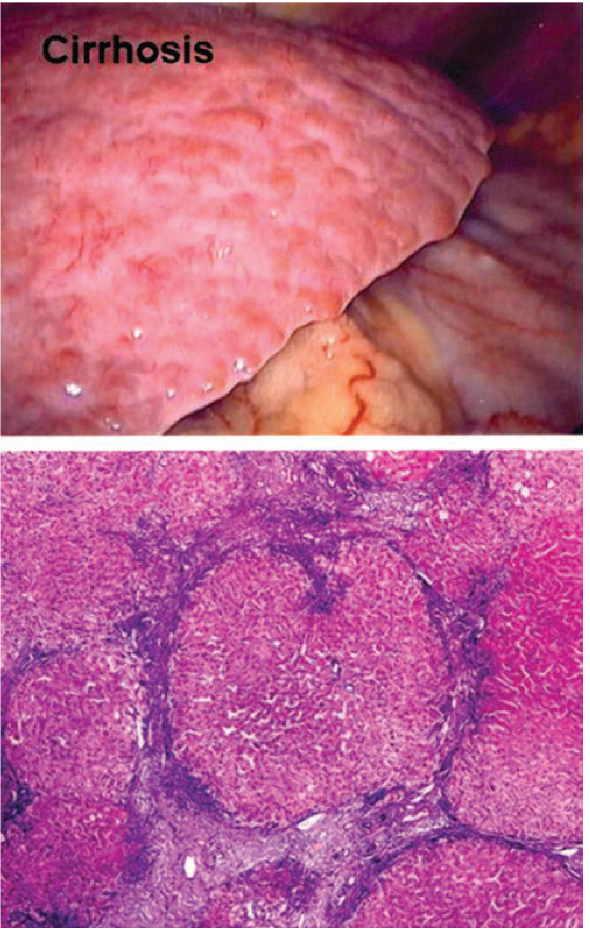
🔵 CIRRHOSIS KYA HAI?
"Liver ka normal architecture destroy ho jaata hai - healthy cells fibrotic scar tissue se replace ho jaati hain - ye cirrhosis hai."
Causes of Cirrhosis:
| Cause | India mein frequency |
|---|---|
| Viral Hepatitis B + C | Most common in India |
| Alcohol | Very common |
| MASLD/NAFLD (Fatty liver) | Fastest growing cause worldwide |
| Autoimmune hepatitis | Less common |
| Wilson's disease | Young patients |
| Hemochromatosis | Iron overload |
| Primary Biliary Cholangitis | Middle-aged women |
| Primary Sclerosing Cholangitis | Young men, UC association |
Cirrhosis Stages:
COMPENSATED CIRRHOSIS          DECOMPENSATED CIRRHOSIS
(Liver still managing)    →    (Complications appear)
- Asymptomatic mostly          - Ascites
- 40% patients                 - Variceal bleeding
- 10-13 yr median survival     - Hepatic encephalopathy
                               - Jaundice
                               - 5-year mortality 85% without transplant!
Physical Exam Findings - Cirrhosis ke Signs:
| Sign | Reason |
|---|---|
| Jaundice | Bilirubin process nahi ho raha |
| Spider naevi | Estrogen not metabolized → superficial vasodilation |
| Palmar erythema | Same - estrogen |
| Gynecomastia (males) | Estrogen nahi break ho raha |
| Caput medusae | Dilated periumbilical veins - portal hypertension |
| Splenomegaly | Portal hypertension se blood backup |
| Ascites | Fluid in peritoneal cavity |
| Asterixis (liver flap) | Hepatic encephalopathy - hand tremor |
| Fetor hepaticus | Sweet-musty breath - ammonia |
| Leukonychia (white nails) | Hypoalbuminemia |
| Dupuytren's contracture | Alcohol-related |
| Parotid enlargement | Alcoholic cirrhosis |
| Testicular atrophy | Hormonal imbalance |
⚡ PORTAL HYPERTENSION - "Jab Portal Vein Ka Pressure Badh Jaata Hai"
Normal Portal Pressure: 5-10 mmHg
Portal Hypertension: > 12 mmHg
Clinically Significant: > 10-12 mmHg (varices form, ascites develop)
Cirrhosis → Fibrosis → Resistance to portal blood flow ↑
         ↓
Portal vein pressure ↑ (Portal Hypertension)
         ↓
Blood alternate routes dhundta hai (collateral vessels = VARICES)
         ↓
Splanchnic vasodilation → Kidney vasoconstriction → Sodium + water retention
         ↓
ASCITES + Edema
Complications of Portal Hypertension:
1. VARICES (Esophageal + Gastric)
- Portal blood esophageal veins se jaata hai → dilated tortuous veins = varices
- Esophageal varices - most common, most dangerous
- Risk: Variceal bleeding = 10-15% mortality per episode! (Rosen's Emergency Medicine)
- Prevention: Non-selective beta-blockers (Propranolol/Carvedilol) - portal pressure kam karte hain
- Acute bleed treatment:
- IV Terlipressin/Octreotide (splanchnic vasoconstriction)
- Emergency endoscopy → Band ligation (rubber bands se varices band karo)
- Antibiotics prophylaxis (Norfloxacin/Ceftriaxone) - SBP prevent karo
- Sengstaken-Blakemore tube - last resort
2. ASCITES
- Peritoneal cavity mein fluid accumulate ho jaata hai
- Mechanism: Portal hypertension + hypoalbuminemia + sodium retention
- Diagnosis: SAAG (Serum-Ascites Albumin Gradient)
- SAAG ≥ 1.1 g/dL = Portal hypertension (cirrhosis)
- SAAG <1.1 g/dL = Malignancy, TB, pancreatic
- Treatment:
- Salt restriction - <2g sodium/day (namak bilkul band!)
- Spironolactone 100mg/day (first-line diuretic - aldosterone antagonist) ± Furosemide 40mg
- Large-volume paracentesis (>5L) + albumin 8g per liter drained
- Refractory ascites → TIPS procedure (Transjugular Intrahepatic Portosystemic Shunt)
3. SPONTANEOUS BACTERIAL PERITONITIS (SBP)
- Ascitic fluid mein bacteria aa jaate hain bina kisi obvious source ke
- Presentation: Fever + abdominal pain + worsening encephalopathy
- Diagnosis: Ascitic fluid PMN >250 cells/mm³
- Treatment: Ceftriaxone 2g IV daily × 5 days + Albumin (prevents hepatorenal syndrome)
- Prophylaxis: Norfloxacin 400mg daily (agar previous SBP ya low protein ascites)
4. HEPATIC ENCEPHALOPATHY (HE)
- Liver ammonia detoxify nahi kar pa rahi → blood mein ammonia → brain effect
- Grades:
| Grade | Symptoms |
|---|---|
| Grade 1 | Slight confusion, sleep disturbance |
| Grade 2 | Drowsy, disorientation, asterixis (liver flap) |
| Grade 3 | Marked confusion, somnolent but arousable |
| Grade 4 | Coma |
- Precipitating factors (HHGIFS mnemonic): Hemorrhage, Hypokalaemia, GI bleed, Infection, Failure renal, Sedatives/constipation
- Treatment:
- Precipitating cause remove karo
- Lactulose - first line! 3-4 times daily → 2-3 soft stools/day target
- Rifaximin 550mg BD - lactulose ke saath add karo (gut bacteria se ammonia production kam)
- Protein restrict mat karo (old concept - galat hai!) - adequate protein dena zaroori hai
5. HEPATORENAL SYNDROME (HRS)
- Cirrhosis → Kidney blood supply fail → Acute kidney injury
- No structural kidney damage - functional problem
- HRS-AKI (Type 1 - rapid) vs HRS-CKD (Type 2 - gradual)
- Treatment: Terlipressin + Albumin → Liver transplant ultimate solution
📊 SCORING SYSTEMS - Child-Pugh + MELD
Child-Pugh Score (5 parameters):
| Parameter | 1 point | 2 points | 3 points |
|---|---|---|---|
| Bilirubin (mg/dL) | <2 | 2-3 | >3 |
| Albumin (g/dL) | >3.5 | 2.8-3.5 | <2.8 |
| PT (seconds prolonged) | <4 | 4-6 | >6 |
| Ascites | None | Mild | Moderate-Severe |
| Encephalopathy | None | Grade 1-2 | Grade 3-4 |
| Score | Class | Prognosis |
|---|---|---|
| 5-6 | Class A | Compensated, Good prognosis |
| 7-9 | Class B | Moderate - consider transplant listing |
| 10-15 | Class C | Decompensated - Poor, urgent transplant |
MELD Score:
- Model for End-stage Liver Disease
- Formula: 3.78×ln(Bilirubin) + 11.2×ln(INR) + 9.57×ln(Creatinine) + 6.43
- Higher score = Worse prognosis = Higher transplant priority
- MELD >15 = Liver transplant consider karo
- MELD >40 = Very high mortality
Exam Pearl: Child-Pugh = Clinical use | MELD = Transplant organ allocation
💊 TREATMENT + DIET in CIRRHOSIS
🥗 DIET:
| ✅ Khaao | ❌ Avoid |
|---|---|
| High protein (1.2-1.5g/kg/day) - muscle maintain karo | Alcohol - bilkul nahi! |
| Small frequent meals (6 meals/day) | Salt (namak <2g/day if ascites) |
| Late evening snack (LES) - muscle wasting rokta hai | Raw/undercooked food (infection risk - immune compromised) |
| Branch chain amino acids (BCAAs) | NSAIDs - kidney function worsen karte hain |
| Zinc supplements | Sedatives/benzodiazepines - encephalopathy trigger |
| Adequate calories (35-40 kcal/kg/day) |
🏥 CLINICAL CASES - 3 Real Scenarios
🟢 CASE 1 - "Peele Aankhein"
🔴 CASE 2 - "Blood vomiting wala patient"
🟡 CASE 3 - "Ajeeb ajeeb baatein karna"
- UTI ke liye appropriate antibiotic (culture pe based)
- Electrolytes correct karo
- Lactulose 30mL 3-4 times daily - target 2-3 soft stools/day
- Ammonia ka gut mein conversion + excretion badhata hai
- Agar severe → Lactulose enema bhi de sakte hain
- Rifaximin 550mg twice daily - gut bacteria se ammonia production kam karo
- Standard of care hai ab Lactulose ke saath
- Protein restrict mat karo! (old myth) - 1.2g/kg/day continue karo
- Small frequent meals
- Branched chain amino acids (BCAAs) supplement karo
📊 MASTER REVISION TABLE - Hepatology
| Topic | Key Point |
|---|---|
| Hep A transmission | Fecal-oral |
| Hep A chronic? | Never - always acute |
| Hep B window period marker | Anti-HBc IgM only positive |
| Hep B chronic diagnosis | HBsAg positive >6 months |
| Hep B treatment | Tenofovir/Entecavir lifelong |
| Hep C treatment | DAAs (Sofosbuvir+Velpatasvir) - >95% cure |
| Hep E danger | Pregnant women - fulminant liver failure |
| Cirrhosis histology | Fibrous septa + regenerative nodules |
| Child-Pugh Class A | Score 5-6 - compensated |
| MELD use | Transplant organ allocation priority |
| Portal hypertension cutoff | >12 mmHg |
| Ascites diagnosis | SAAG ≥1.1 = portal hypertension |
| Ascites first-line treatment | Salt restriction + Spironolactone |
| Variceal bleed management | Terlipressin + Band ligation + Antibiotics |
| SBP diagnosis | Ascitic fluid PMN >250/mm³ |
| SBP treatment | Ceftriaxone + Albumin |
| Encephalopathy treatment | Lactulose + Rifaximin |
| Encephalopathy Grade 4 | Coma |
| Asterixis | Liver flap = Grade 2 encephalopathy |
✅ Lesson 5 Complete!
- Hep C = 95%+ curable with DAAs - ye ek revolution hai medicine mein
- Cirrhosis ki complications yaad karo: AVHE = Ascites, Varices, Hepatic encephalopathy, Hepatorenal syndrome
- Hepatic encephalopathy mein precipitant dhundo aur treat karo - sirf lactulose dena kaafi nahi
🎯 Lesson 6 Mein Aayega:
PANCREATIC DISEASES + GALLBLADDER DISEASES
- Acute Pancreatitis - "worst pain in life" - mechanism, Ranson criteria, management
- Chronic Pancreatitis + complications
- Gallstones - types, why bante hain, cholecystitis, Charcot's triad
- ERCP kya hota hai - biliary intervention
- Pancreatic cancer - "silent killer"
- 3 clinical cases Hinglish mein!
Not bad but i want you to start from scratch basic to advanced Let’s start the journey from starting
- GI anatomy: mouth se anus tak
- Digestion and absorption physiology
- Liver, gallbladder, pancreas basics
- Symptoms ka approach: pain, vomiting, diarrhea, constipation, bleeding, jaundice
- Common diseases
- Emergencies
- Investigations: LFT, USG, endoscopy, colonoscopy, CT, MRCP
- Treatment principles, diet, lifestyle, drugs, and clinical cases
Bas reply karo: Start Lesson 1
Let’s start
gastrointestinal anatomy physiology
Merck Manual gastrointestinal tract anatomy physiology digestion absorption
Lesson 1: GI System ka Basic Map
Food mouth se stool tak kaise travel karta hai?
1. GI System kya hota hai?
- Food lena
- Food ko small particles mein todna
- Nutrients absorb karna
- Waste ko stool bana ke bahar nikalna
- Liver: bile banata hai, toxins/drugs process karta hai
- Gallbladder: bile ko store karta hai
- Pancreas: digestive enzymes aur bicarbonate deta hai
Food journey
Mouth
  ↓
Pharynx
  ↓
Esophagus
  ↓
Stomach
  ↓
Duodenum
  ↓
Jejunum
  ↓
Ileum
  ↓
Cecum → Colon
  ↓
Rectum
  ↓
Anus
2. Mouth: Digestion yahin start hoti hai
Mouth ke main kaam
- Teeth: food ko mechanically break karte hain
- Tongue: food ko mix karke bolus banati hai
- Saliva: food ko wet banata hai aur starch digestion start karta hai
Important enzyme
- Salivary amylase: starch/carbohydrates ko break karna start karta hai.
Clinical connection
3. Esophagus: Food pipe
Isme digestion nahi hoti
Peristalsis kya hai?
Food bolus
   ↓
Circular muscles contract behind it
   ↓
Food forward move karta hai
Lower Esophageal Sphincter, LES
- Normal: food neeche jaane deta hai, acid ko upar aane se rokta hai.
- LES weak: acid esophagus mein wapas aata hai.
- Result: GERD, heartburn, acid reflux.
Yaad rakho: LES failure = GERD
4. Stomach: Storage + Acid Factory
- Food ko temporarily store karna
- Food ko churn aur mix karna
- Protein digestion start karna
Stomach kya banata hai?
| Substance | Kaam |
|---|---|
| HCl acid | Bacteria kill karta hai, pepsin activate karta hai |
| Pepsin | Protein digestion start karta hai |
| Mucus + bicarbonate | Stomach wall ko acid se bachata hai |
| Intrinsic factor | Vitamin B12 absorption ke liye important |
Most important concept: Acid versus protection
Acid + Pepsin = Attack
Mucus + Bicarbonate + Blood supply = Defense
Clinical connection
- H. pylori bacteria aur NSAIDs such as diclofenac, ibuprofen, aspirin mucosal defense ko damage karte hain.
- Isliye ulcer disease samajhne ke liye pehle ye acid-defense balance samajhna zaroori hai.
5. Small Intestine: Absorption ka Main Organ
Duodenum → Jejunum → Ileum
A. Duodenum
- Bile from liver/gallbladder
- Pancreatic juice from pancreas
Duodenum ka role
- Acidic stomach food ko neutralize karna
- Fat, protein, carbohydrate digestion ko strongly start karna
- Iron aur calcium absorption ka important site
Duodenum mein ulcer common hota hai because yahan stomach acid first time enter karta hai.
B. Jejunum
- Carbohydrates
- Proteins
- Fats
- Folate
- Most vitamins and minerals
- Water
Villi kya hote hain?
Normal intestine: flat surface
Villi: finger projections
Microvilli: microscopic brush border
Clinical connection
- Chronic diarrhea
- Weight loss
- Iron deficiency anemia
- Weakness
- Bloating
C. Ileum
Ileum ki special absorption
- Vitamin B12
- Bile salts
Super high-yield: Terminal ileum disease or resection → B12 deficiency + bile salt malabsorption.
Clinical connection
- B12 deficiency
- Fat malabsorption
- Gallstones
- Kidney stones
mil sakte hain.
6. Liver, Gallbladder, Pancreas: GI ke Helper Organs
A. Liver
GI-related liver functions
- Bile production
- Absorbed nutrients ko process karna
- Drugs/alcohol metabolize karna
- Albumin banana
- Clotting factors banana
- Bilirubin process karna
- Ammonia ko urea mein convert karna
Portal vein concept
Intestine → Portal vein → Liver → Heart
B. Gallbladder
Fatty food duodenum mein aaya
       ↓
CCK release hua
       ↓
Gallbladder contract hua
       ↓
Bile duodenum mein aayi
       ↓
Fat digestion help hui
Clinical connection
C. Pancreas
| Function | Secretion | Kaam |
|---|---|---|
| Exocrine | Amylase, lipase, proteases, bicarbonate | Digestion |
| Endocrine | Insulin, glucagon | Blood glucose control |
Important enzymes
- Amylase: carbohydrates digest karta hai
- Lipase: fats digest karta hai
- Proteases: proteins digest karte hain
- Bicarbonate: stomach acid neutralize karta hai
Clinical connection
Severe epigastric pain radiating to back + vomiting.
7. Large Intestine: Water Save Karna aur Stool Banana
Cecum → Ascending colon → Transverse colon
→ Descending colon → Sigmoid colon
→ Rectum → Anus
Colon ka main kaam
- Water absorb karna
- Electrolytes absorb karna
- Stool form karna
- Gut bacteria ko house karna
- Waste ko rectum tak move karna
Small intestine vs colon
| Feature | Small intestine | Colon |
|---|---|---|
| Main work | Nutrient absorption | Water absorption + stool formation |
| Length | About 6 m | About 1.5 m |
| Important disease | Celiac, Crohn's | UC, colon cancer, diverticular disease |
| Bacteria | Relatively fewer | Huge microbiome present |
Clinical connection
- Water absorption kam: diarrhea
- Slow movement or excessive water absorption: constipation
- Colonic mucosal inflammation: bloody diarrhea, as in ulcerative colitis.
8. Rectum and Anus: Defecation ka Control
- Internal anal sphincter: involuntary
- External anal sphincter: voluntary control
9. Entire GI Tract ka One-Line Function Map
| Organ | One main job |
|---|---|
| Mouth | Chewing + starch digestion start |
| Esophagus | Food transport |
| Stomach | Acid, protein digestion, food mixing |
| Duodenum | Bile + pancreatic juice mixing |
| Jejunum | Maximum nutrient absorption |
| Ileum | B12 + bile salt absorption |
| Colon | Water absorption + stool formation |
| Rectum/Anus | Stool storage and expulsion |
| Liver | Bile + metabolism + synthesis |
| Gallbladder | Bile storage |
| Pancreas | Digestive enzymes + bicarbonate |
10. Three Basic Clinical Connections
Case 1: Acid reflux
LES weak hai, isliye stomach acid esophagus mein reflux ho raha hai.
Case 2: Chronic diarrhea and anemia
Small intestine, especially duodenum/jejunum, nutrient absorption ka major site hai. Agar mucosa/villi damaged hain, iron absorption kam ho sakta hai.
Case 3: Yellow eyes + pale stool + dark urine
Gallstone common bile duct block kar raha ho sakta hai. Bile intestine mein nahi aa rahi, isliye stool pale. Conjugated bilirubin urine mein jaa kar dark urine bana raha hai.
Today’s Must-Remember Points
- LES acid reflux se protect karta hai.
- Stomach acid banata hai but mucus-bicarbonate barrier se protected hota hai.
- Duodenum receives bile and pancreatic juice.
- Jejunum is the major nutrient absorption site.
- Terminal ileum absorbs vitamin B12 and bile salts.
- Colon water absorb karke stool banata hai.
- Liver makes bile, gallbladder stores it, pancreas enzymes deta hai.
- Portal vein intestine se blood liver tak le jaati hai.
Next lesson
Hum carbohydrates, proteins, fats, bile, pancreatic enzymes, villi, microbiome, aur malabsorption ko simple Hinglish mein samjhenge.
Sath sath me lifestyle advice healthy eating etc aur treatment bhi btate rho Aur mujhe ek best gastroenterologist bnna h + diabetes specialist
I want a complete and detailed learning guide in gastroenterology from scratch to advanced level. Please teach me about all major and minor gastrointestinal diseases, including their causes, symptoms, diagnosis methods, and treatment options. Cover diseases like GERD, peptic ulcers, inflammatory bowel diseases (Crohn’s, ulcerative colitis), liver diseases (hepatitis, cirrhosis), pancreatitis, GI infections, GI cancers, functional disorders (IBS, motility issues), and more. Explain clinical presentations with real-life case examples, diagnostic techniques (endoscopy, imaging, lab tests), and step-by-step treatment plans including medications, endoscopic procedures, surgery, and lifestyle modifications (diet, exercise, stress management). Also, provide guidance on preventive care, patient counseling, follow-up management, and handling complications. Include the latest advances and evidence-based practices. Make the content practical, clear, and clinically relevant so I can confidently understand, diagnose, and manage GI diseases like a skilled gastroenterologist. Please structure the information logically, starting from basic anatomy and physiology, moving to diseases and treatments, and finishing with advanced clinical knowledge and case-based learning.
Sath sath me lifestyle advice healthy eating etc aur treatment bhi btate rho Aur mujhe ek best gastroenterologist bnna h + diabetes specialist