
Explain the cause of anginal chest pain and cardiac failure in hyperthyroidism
Anginal Chest Pain and Cardiac Failure in Hyperthyroidism
Underlying Haemodynamic Changes
| Parameter | Change |
|---|---|
| Heart rate | Increased (sinus tachycardia) |
| Stroke volume | Increased |
| Cardiac output | Increased 2-3x normal (high-output state) |
| Systemic vascular resistance (SVR) | Decreased (peripheral vasodilation) |
| Blood volume / preload | Increased (via renin-aldosterone stimulation) |
| Myocardial contractility | Enhanced |
| Myocardial oxygen demand | Markedly increased |
1. Cause of Anginal Chest Pain
A. In Patients with Pre-existing CAD (Type 2 MI / Demand Ischemia)
- Tachycardia shortens diastolic filling time, further reducing coronary perfusion
- The increased cardiac work (rate x pressure product) escalates oxygen consumption
- In older hyperthyroid patients with known or suspected CAD, this increased cardiac workload reliably triggers anginal episodes
B. In Patients with Normal Coronary Arteries (Coronary Vasospasm)
2. Cause of Cardiac Failure
A. High-Output Failure (Predominantly Right Heart)
- Pulmonary hypertension develops - Systemic vascular resistance falls in hyperthyroidism but pulmonary vascular resistance does not. Because of the greatly increased output delivered to the pulmonary circulation, pulmonary arterial pressure rises.
- This leads to elevated mean venous pressure, hepatic congestion, and peripheral edema - the picture of right-sided heart failure.
- The kidneys respond to the high-output state by increasing sodium reabsorption (via renin and aldosterone stimulation), further expanding plasma volume and worsening congestion.
Note: The exercise intolerance seen in many hyperthyroid patients is not purely cardiac - it also results from skeletal and respiratory muscle weakness, and the reduced cardiac reserve (cardiac output cannot rise further during exercise because it is already near maximum at rest).
B. Tachycardia-Mediated (Low-Output) Cardiomyopathy
- The persistently elevated heart rate creates a tachycardia-mediated cardiomyopathy - a well-recognized cause of dilated cardiomyopathy
- The left ventricle dilates and ejection fraction falls
- As the LV dilates, mitral regurgitation may develop, further reducing forward output
- Pulmonary congestion develops - producing dyspnea, orthopnea, and paroxysmal nocturnal dyspnea
- The clinical picture becomes indistinguishable from conventional heart failure with reduced ejection fraction
Atrial Fibrillation as an Amplifying Factor
- Increases ventricular rate irregularly, worsening tachycardia-mediated injury
- Reduces effective cardiac output (loss of atrial kick)
- Creates risk of systemic embolism and stroke
- May be the first presenting symptom of hyperthyroidism in older adults, in whom the classic sympathomimetic features (anxiety, tremor) are less prominent
Summary Table
| Mechanism | Consequence |
|---|---|
| Increased O2 demand from tachycardia + high contractility | Angina (demand ischemia) in CAD patients |
| Coronary vasospasm | Angina at rest with normal coronaries |
| Pulmonary vascular resistance unchanged while output rises | Pulmonary hypertension → right heart failure, edema |
| Renin-aldosterone activation | Volume expansion, worsening congestion |
| Sustained tachycardia / AF | Tachycardia-mediated cardiomyopathy → low-output failure |
| LV dilation from cardiomyopathy | Mitral regurgitation → further failure |
- Braunwald's Heart Disease, Chapter 96 (Heart Failure in Overt and Subclinical Hyperthyroidism; CHD in Hyperthyroidism)
- Tietz Textbook of Laboratory Medicine, 7th Ed. (Signs and Symptoms of Thyrotoxicosis - Cardiovascular System)
- Fuster and Hurst's The Heart, 15th Ed. (Thyroid Cardiomyopathy)
- Harrison's Principles of Internal Medicine, 22nd Ed. (High-Output States)
- Katzung's Basic and Clinical Pharmacology, 16th Ed. (Pathophysiology of Heart Failure)
Summarise to write for md exam
Angina & Cardiac Failure in Hyperthyroidism
MD Exam Summary
Core Mechanism (Learn This First)
- ↑ Heart rate + ↑ contractility + ↑ stroke volume → ↑ Cardiac output (2-3x normal)
- ↓ Systemic vascular resistance (peripheral vasodilation)
- ↑ Blood volume (T3 stimulates renin-aldosterone → Na+ and water retention)
- Net effect: High-output state with massively increased myocardial oxygen demand
T3 targets: β-adrenergic receptors, contractile apparatus, Na+/K+-ATPase, sarcoplasmic reticulum Catecholamines are actually normal - but β-blockers still work by blunting T3's adrenergic-mimicking effects
I. Anginal Chest Pain - Two Mechanisms
Type 1 - Demand Ischemia (Pre-existing CAD)
- Most common mechanism
- ↑ Heart rate + ↑ contractility → ↑ myocardial O2 demand
- Tachycardia shortens diastolic filling time → ↓ coronary perfusion
- Fixed coronary stenosis cannot meet the demand → ischemia
- Seen in: older patients with known/suspected CAD
- Rx: Beta-blockers (symptomatic relief) + restoration of euthyroidism (definitive)
Type 2 - Coronary Vasospasm (Normal Coronaries)
- Seen in: younger women, chest pain at rest, ischemic ECG changes
- Angiography shows normal coronary arteries but vasospasm (like Prinzmetal's angina)
- MI is rare
- Rx: Calcium channel blockers / nitroglycerin
II. Cardiac Failure - Two Mechanisms
Mechanism A - High-Output Right Heart Failure
| Step | Explanation |
|---|---|
| SVR falls | T3-driven peripheral vasodilation |
| Pulmonary vascular resistance (PVR) does NOT fall | Unlike SVR |
| Increased output hits the pulmonary bed | Pulmonary arterial pressure rises |
| Right heart overloaded | Elevated JVP, hepatic congestion, peripheral oedema |
| Volume overload worsens it | Renin-aldosterone expands plasma volume |
Key exam point: SVR ↓ but PVR unchanged - this asymmetry explains pulmonary hypertension and right heart failure
Mechanism B - Tachycardia-Mediated Cardiomyopathy (Low-Output Failure)
- Sustained tachycardia or AF → tachycardia-mediated cardiomyopathy
- LV dilates → EF falls → low cardiac output
- LV dilation causes functional mitral regurgitation → further ↓ forward output
- Pulmonary congestion → dyspnea, orthopnea, PND
- Picture of dilated cardiomyopathy / HFrEF
Key exam point: This is reversible - rate control alone (beta-blocker) can improve LV function even before antithyroid therapy
III. Role of Atrial Fibrillation
- T3 shortens action potential duration + refractory period of atrial myocardium → substrate for AF
- AF may be the first presentation in elderly (classic sympathomimetic features absent in old age)
- AF amplifies failure: irregular rate, loss of atrial kick, risk of embolism/stroke
One-Line Mechanisms for MCQs
| Finding | Mechanism |
|---|---|
| Angina with CAD | ↑ O2 demand > supply (demand ischemia) |
| Angina without CAD | Coronary vasospasm |
| Right heart failure / oedema | PVR unchanged while CO rises → pulmonary hypertension |
| Dilated cardiomyopathy | Tachycardia-mediated (rate-related) cardiomyopathy |
| Reversible cardiomyopathy | Rate control / antithyroid therapy restores function |
| AF common | T3 shortens atrial refractory period |
High-Yield Exam Points
- Cardiovascular complications = highest morbidity/mortality in hyperthyroidism
- Catecholamines are normal - symptoms are T3-mediated, not catecholamine excess
- High-output failure = NOT the same as typical HF - CO is elevated, SVR is low
- Tachycardia-mediated cardiomyopathy is the mechanism for true LV failure
- Rate control precedes antithyroid therapy in managing LV failure
- Thyroid cardiomyopathy is potentially reversible with treatment
- Elderly patients present with apathetic hyperthyroidism - AF and cardiac failure without classic features
Metabolic changes in thyrotoxicosis
Metabolic Changes in Thyrotoxicosis
MD Exam Summary
Core Principle
↑ Basal metabolic rate (BMR) by 30-60% in severe thyrotoxicosis
Molecular Basis of Increased BMR
-
Na+/K+-ATPase upregulation - T3 transcriptionally induces both α and β subunits of the Na/K pump in muscle, liver, and kidney. Increased pump activity consumes more ATP → ↑ O2 consumption and heat generation. A compensatory Na+/K+ leak creates a futile cycle (energy consumed, no useful work done).
-
Mitochondrial uncoupling - T3 upregulates uncoupling proteins (thermogenin/UCP) in brown adipose tissue. Mitochondria consume O2 and produce heat without generating ATP.
-
↑ β-adrenergic receptor expression - Catecholamine levels are normal, but T3 increases tissue sensitivity by upregulating β-receptors in heart, skeletal muscle, and adipose tissue → amplified adrenergic thermogenesis.
1. Carbohydrate Metabolism
| Change | Mechanism |
|---|---|
| ↑ Gluconeogenesis | T3 induces key gluconeogenic enzymes: PEPCK, pyruvate carboxylase, glucose-6-phosphatase |
| ↑ Glycogenolysis | T3 stimulates hepatic glycogen breakdown |
| Blood glucose usually normal | Pancreas compensates with ↑ insulin secretion |
| ↑ Glucose absorption from gut | T3 enhances intestinal glucose uptake |
Exam point: Despite driving gluconeogenesis AND glycogenolysis, plasma glucose is usually normal because insulin rises in parallel. However, thyrotoxicosis can unmask/worsen diabetes mellitus.
2. Protein Metabolism
| Change | Mechanism |
|---|---|
| ↑ Protein synthesis | T3 stimulates anabolism |
| ↑ Protein degradation (proteolysis) | Exceeds synthesis - net catabolic effect |
| Muscle wasting + weakness | Net loss of muscle protein; amino acids shunted to liver for gluconeogenesis |
| Negative nitrogen balance | ↑ Urea production and urinary nitrogen excretion |
| Weight loss | Loss of muscle bulk contributes alongside fat loss |
Exam point: Proteolysis > synthesis → net catabolism → negative nitrogen balance. This is why thyrotoxic patients have proximal muscle weakness.
3. Lipid Metabolism
| Change | Mechanism |
|---|---|
| ↑ Lipolysis | T3 stimulates lipolysis in adipose → releases free fatty acids + glycerol |
| ↑ Lipogenesis | T3 also stimulates FA synthesis in liver (but at very high T3, lipolysis dominates) |
| Net: fat stores depleted | Lipolysis > lipogenesis at high T3 → generalized fat mobilization |
| ↓ Total cholesterol | T3 upregulates hepatic LDL receptors → ↑ LDL clearance |
| ↓ LDL, ↓ HDL | Both reduced (LDL more than HDL) |
| ↓ Triglycerides | Increased hepatic clearance |
| ↑ Free fatty acids | Released from adipose lipolysis; fuel for hepatic gluconeogenesis |
Exam point: Thyrotoxicosis → ↓ cholesterol (opposite of hypothyroidism). Cholesterol rises when patient becomes hypothyroid after treatment.
4. Energy Metabolism / Thermogenesis
| Feature | Detail |
|---|---|
| ↑ BMR 30-60% | Signature metabolic finding |
| Heat intolerance | Excess heat production from futile cycles |
| Sweating | Compensatory heat dissipation |
| Weight loss | Despite ↑ appetite (polyphagia); caloric expenditure exceeds intake |
| ↑ O2 consumption | All tissues affected - heart, liver, muscle, kidney |
5. Bone / Calcium Metabolism
| Change | Mechanism |
|---|---|
| ↑ Bone turnover | T3 directly stimulates osteoclast activity |
| ↑ Bone resorption > formation | Net bone loss |
| Hypercalcaemia (mild, in ~10%) | Increased osteoclastic bone resorption → Ca2+ released into blood |
| Osteoporosis | Chronic thyrotoxicosis → reduced bone mineral density → fracture risk |
| ↑ Urinary calcium | Hypercalciuria from increased bone resorption |
| ↓ PTH (secondary) | PTH suppressed by mild hypercalcaemia |
Exam point: Thyrotoxicosis is a recognised secondary cause of osteoporosis (important in post-menopausal women on T4 replacement).
6. Vitamin and Drug Metabolism
| Change | Implication |
|---|---|
| ↑ Degradation of fat-soluble + water-soluble vitamins | Increased vitamin requirements |
| ↑ Drug metabolism | Higher drug clearance rates |
| ↓ Warfarin requirement | T3 increases degradation of clotting factors → anticoagulant effect amplified |
High-yield exam point: Thyrotoxicosis potentiates warfarin - dose reduction needed. Mechanism: increased catabolism of vitamin K-dependent clotting factors.
Summary Table for Revision
| System | Key Change | Net Result |
|---|---|---|
| BMR | ↑ 30-60% | Heat intolerance, sweating |
| Carbohydrate | ↑ Gluconeogenesis + ↑ glycogenolysis | Normal blood glucose (insulin compensates) |
| Protein | Proteolysis > synthesis | Muscle wasting, -ve nitrogen balance |
| Fat | Lipolysis > lipogenesis | Fat loss, ↓ cholesterol, ↓ TG |
| Bone | ↑ Osteoclast activity | Osteoporosis, mild hypercalcaemia |
| Vitamins/Drugs | ↑ Catabolism | ↓ Warfarin dose needed |
| Thermogenesis | Na/K-ATPase + mitochondrial uncoupling | Weight loss despite ↑ appetite |
High-Yield MCQ Points
- BMR rises 30-60% in severe thyrotoxicosis
- Catecholamines are normal - but β-receptor expression is upregulated
- Blood glucose is usually normal despite ↑ gluconeogenesis (pancreas compensates)
- Negative nitrogen balance due to net protein catabolism
- Cholesterol is low (not high) - hypothyroidism causes hypercholesterolaemia
- Osteoporosis occurs from direct osteoclast stimulation by T3
- Warfarin dose must be reduced in thyrotoxicosis (increased clotting factor catabolism)
- Weight loss occurs despite increased appetite (polyphagia) - net energy expenditure exceeds intake
- Futile cycles (simultaneous synthesis + degradation) are the core molecular mechanism for ↑ O2 consumption
In relation to Anaesthesia
Thyrotoxicosis in Relation to Anaesthesia
MD Exam Summary
Why This Matters Perioperatively
- Haemodynamic instability under anaesthesia
- Thyroid storm precipitated by surgery/stress
- Airway difficulty from goitre
- Drug interactions (altered drug clearance, catecholamine sensitivity)
- Postoperative complications (RLN injury, hypocalcaemia after thyroidectomy)
I. Preoperative Assessment and Preparation
Goals
- Achieve euthyroid state before elective surgery - essential to minimise risk
- Assess and control end-organ effects (arrhythmias, heart failure)
- Evaluate airway - degree of tracheal compression/deviation (CT/MRI)
Preoperative Drug Regimen
| Drug | Mechanism | Role |
|---|---|---|
| Propylthiouracil (PTU) | Blocks thyroid hormone synthesis + inhibits T4→T3 conversion | First-line antithyroid |
| Methimazole | Blocks thyroid hormone synthesis | Alternative to PTU |
| Propranolol | Beta-blockade + also inhibits T4→T3 conversion | Controls tachycardia, tremor, anxiety; given 7-14 days pre-op |
| Potassium iodide (SSKI) | Wolff-Chaikoff effect: blocks hormone synthesis and reduces gland vascularity | Added 7-10 days pre-op for faster control; must start antithyroid drugs first to avoid worsening thyrotoxicosis |
| Dexamethasone (8-12 mg/day) | Reduces thyroid hormone secretion + inhibits T4→T3 conversion | Severe thyrotoxicosis requiring emergency surgery |
Key exam point: Iodide must never be given before antithyroid drugs - it can initially worsen thyrotoxicosis before the Wolff-Chaikoff effect kicks in.
Key exam point: Propranolol is the beta-blocker of choice - it is the only one that also blocks T4→T3 peripheral conversion.
Fitness for Surgery
- Marker of adequate control: normalised free T4 + absence of overt symptoms (TSH lags and may not normalise for weeks)
- All antithyroid medications are continued on the morning of surgery
- Emergency surgery in uncontrolled thyrotoxicosis: achieve heart rate <90 bpm with beta-blockers; add dexamethasone + SSKI
II. Airway Considerations
| Issue | Detail |
|---|---|
| Difficult intubation | Incidence 5-8% in goitre surgery |
| Size ≠ risk | Thyroid cancer (not goitre size) is the stronger predictor of difficult airway |
| Tracheal deviation | Lateral displacement from goitre vs. anterior mediastinal mass |
| Large substernal goitre | Can behave like anterior mediastinal mass - risk of intrathoracic airway collapse after induction |
| Management | CT/MRI review pre-op; awake fibreoptic intubation if signs of difficult airway |
| Tracheomalacia | Long-standing goitre may weaken tracheal rings; extubate under optimal conditions for reintubation |
| Transoral thyroidectomy | Requires nasal intubation |
III. Intraoperative Anaesthetic Management
Choice of Agents
| Agent | Use | Notes |
|---|---|---|
| Propofol | Safe - preferred induction agent | ↑ clearance and ↑ distribution volume in hyperthyroidism; increase TIVA infusion rates |
| Volatile agents | All acceptable | MAC is NOT altered by hyperthyroidism |
| Ketamine | AVOID | Stimulates sympathetic NS → aggravates tachycardia, hypertension |
| Pancuronium | AVOID | Sympathomimetic → aggravates tachycardia |
| Anticholinergic drugs | Avoid if possible | Can worsen tachycardia |
| Regional anaesthesia | Excellent alternative | Avoid epinephrine-containing solutions |
Key Intraoperative Principles
- Induction may be slower than in euthyroid patients and may require higher volatile agent concentrations - because of the elevated cardiac output (increased drug distribution and clearance)
- Maintain adequate anaesthetic depth to blunt sympathetic responses to surgical stimulation
- Vasopressors: use direct-acting (phenylephrine) rather than indirect-acting (ephedrine) - avoids catecholamine release in a catecholamine-sensitised patient
- Muscle relaxants: the incidence of myasthenia gravis is increased in hyperthyroidism → reduce initial NMB dose; use a nerve stimulator (twitch monitor) to guide dosing
- NIM tube for RLN monitoring: avoid muscle relaxants and topical laryngeal anaesthesia if using NIM endotracheal tube
Haemodynamic Profile to Anticipate
- Systolic hypertension + widened pulse pressure
- ↓ Systemic vascular resistance
- Sinus tachycardia / AF / high-output heart failure
- Increased sensitivity to catecholamines (upregulated β-receptors)
Beta-Blockers Intraoperatively (Mainstay)
- Control tachycardia, arrhythmias
- Esmolol infusion (50-350 mcg/kg/h, titrated) for acute rate control
- Test dose: 0.1-0.2 mg/kg esmolol; if heart rate slows without worsening heart failure - continue infusion
- Remember: beta-blockers do NOT prevent thyroid storm
IV. Thyroid Storm - The Critical Emergency
What Triggers It
Clinical Features
| Feature | Detail |
|---|---|
| Hyperthermia | Often ≥40°C |
| Tachycardia / AF | Rapid ventricular rate |
| Hypotension | Late feature; cardiovascular collapse |
| Agitation / delirium | CNS hyperactivity |
| Congestive heart failure | High-output failure |
| Hypokalemia | Characteristic (distinguishes from MH) |
Thyroid Storm vs. Malignant Hyperthermia (Critical Exam Distinction)
| Feature | Thyroid Storm | Malignant Hyperthermia |
|---|---|---|
| Timing | Usually postoperative | Intraoperative (during volatile/sux exposure) |
| ↑ End-tidal CO2 | No | Yes (early marker) |
| ↑ CK | No | Yes (massive) |
| Metabolic acidosis | Mild | Severe |
| Rigidity | Absent | Present |
| K+ | Hypokalemia | Hyperkalemia |
| Trigger | Surgery/stress in hyperthyroid pt | Volatile agents / succinylcholine |
| Treatment | Antithyroid drugs, iodide, β-blockers | Dantrolene |
Management of Thyroid Storm
| Step | Drug/Action |
|---|---|
| 1. Inhibit synthesis | PTU 200-400 mg PO/NGT every 6h |
| 2. Block hormone release | Sodium iodide 250 mg IV/PO every 6h (give AFTER PTU) |
| 3. Control heart rate | Esmolol infusion (first-line intraoperatively) or propranolol |
| 4. Reduce T4→T3 conversion | Propranolol + dexamethasone/hydrocortisone |
| 5. Cooling | Ice packs over major arteries, cooling blankets, iced saline lavage |
| 6. Antipyretic | Paracetamol (avoid aspirin - displaces T4 from binding proteins) |
| 7. IV fluids | Replace losses; correct electrolytes |
| 8. Refractory cases | Therapeutic plasma exchange (plasmapheresis) |
V. Postoperative Complications (After Thyroidectomy)
| Complication | Signs | Management |
|---|---|---|
| Bilateral RLN injury | Stridor, aphonia, glottic closure | Immediate reintubation → tracheostomy |
| Unilateral RLN injury | Hoarseness (transient, often compensates) | Observe; laryngoscopy |
| Hypocalcaemia (hypoparathyroidism) | Symptoms 24-96h post-op; perioral tingling, stridor, laryngospasm | IV calcium chloride or gluconate; check Mg |
| Tracheomalacia | Airway collapse on extubation | Re-intubate; monitor post-extubation |
| Thyroid storm | See above | See above |
| Haematoma | Neck swelling, stridor | Urgent surgical decompression |
| Pneumothorax | During substernal goitre resection | Chest drain |
Exam point: Laryngospasm after thyroidectomy should make you think hypocalcaemia first.
VI. Special Situations
Emergency Surgery in Uncontrolled Hyperthyroidism
- Optimise with antithyroid drugs + SSKI + propranolol (oral) pre-induction
- Target HR <90 bpm
- Use direct-acting vasopressors only
- Avoid ketamine, ephedrine, anticholinergics
- Have esmolol infusion ready
- Glucocorticoids (dexamethasone) for severe disease
Subclinical Hyperthyroidism
- Still carries increased risk of AF and MACE perioperatively
- Monitor and control heart rate
High-Yield Exam Points
- Achieve euthyroidism before elective surgery - free T4 is the operative marker, not TSH
- MAC is unchanged in hyperthyroidism; induction may be slower (high cardiac output)
- Propofol infusion rate must be increased (increased clearance and Vd)
- Avoid ketamine, pancuronium, anticholinergics, ephedrine, adrenaline-containing LAs
- Use phenylephrine (direct-acting) for hypotension
- Reduce NMB dose - association with myasthenia gravis
- Propranolol = beta-blocker of choice (also blocks T4→T3 conversion)
- Beta-blockers do NOT prevent thyroid storm
- Thyroid storm = postoperative, hypokalemia, no ↑ ETCO2 → distinguishes from MH
- After thyroidectomy: laryngospasm = hypocalcaemia until proven otherwise
Complete anaesthetic management from millers and stoelting
Complete Anaesthetic Management of Hyperthyroidism
From Miller's Anesthesia (10e) & Barash/Stoelting's Clinical Anesthesia (9e)
PREOPERATIVE MANAGEMENT
1. Primary Goal
"The most important goal in managing the hyperthyroid patient is to make the patient euthyroid before surgery, if possible." — Stoelting's (Barash), Ch. 47
2. Preoperative Assessment
- Cardiovascular: arrhythmias (especially AF), heart failure, systolic hypertension, widened pulse pressure
- Airway: degree of tracheal compression/deviation from goitre; review CT or MRI
- End-organ effects: muscle weakness (myopathy), bone loss, electrolytes
- Subclinical hyperthyroidism: even with normal T3/T4 and low TSH (<0.5 mU/L) - still increased risk of AF, MACE, heart failure
- Operative fitness criterion: normalisation of free T4 + absence of overt symptoms (TSH lags - do not wait for TSH normalisation)
3. Drug Therapy - Preoperative Preparation
Antithyroid Drugs (6-8 weeks required)
| Drug | Mechanism | Notes |
|---|---|---|
| Propylthiouracil (PTU) | Inhibits iodide organification (thyroid hormone synthesis) + blocks peripheral T4→T3 conversion | First-line; additional benefit of blocking T4→T3 |
| Methimazole | Inhibits iodide organification only | Alternative to PTU |
- Both are thiourea derivatives
- Normal glands store enough hormone to maintain euthyroidism for months even after synthesis is abolished - hence 6-8 weeks needed
- Toxic reactions (uncommon): skin rash, nausea, fever, agranulocytosis, hepatitis, arthralgias
- Continue all antithyroid medications on the morning of surgery
Beta-Blockers
| Drug | Role | Special Feature |
|---|---|---|
| Propranolol | Drug of choice | Blocks tachycardia, tremor, anxiety, heat intolerance within 12-24 hours; ALSO impairs T4→T3 conversion (over 1-2 weeks) |
| Any beta-blocker | Symptommatic control | Long-acting agents more convenient |
- Dose: Propranolol titrated to effect + potassium iodide (2-5 drops every 8 hours)
- Preparation time: 7-14 days
- Beta-blockers do NOT inhibit hormone synthesis and do NOT prevent thyroid storm
- In heart failure: use with caution; small test dose of esmolol (0.1-0.2 mg/kg) - if HR slows without worsening failure, continue infusion at 50-350 mcg/kg/h
Potassium Iodide (SSKI) - Wolff-Chaikoff Effect
- Mechanism: inhibits iodide organification AND reduces synthesis and release of thyroid hormones; also reduces size and vascularity of the hyperplastic gland
- Onset: faster symptom control (7-10 days) vs. 2-6 weeks with antithyroid drugs alone
- Use: added when rapid preparation needed (e.g. before urgent thyroidectomy)
- CRITICAL: Antithyroid drugs must be started before iodide - iodide alone may initially worsen thyrotoxicosis
- Iopanoic acid (iodide-based contrast agent) has similar effects
Glucocorticoids
- Dexamethasone 8-12 mg/day (or hydrocortisone 50-100 mg IV every 6h)
- Indications: severe thyrotoxicosis; emergency surgery in uncontrolled hyperthyroid patient
- Mechanism: reduces thyroid hormone secretion + inhibits peripheral T4→T3 conversion
Emergency Surgery Protocol (Uncontrolled Hyperthyroid)
- Antithyroid drug (PTU/methimazole)
- SSKI after antithyroid drug
- Beta-blocker to HR <90 bpm
- Dexamethasone
- Target: free T4 normalised + symptomatic control
INTRAOPERATIVE MANAGEMENT
4. Airway
| Scenario | Management |
|---|---|
| Routine goitre | Standard laryngoscopy; difficult airway incidence 5-8% |
| Thyroid cancer | Higher risk of difficult airway (not goitre size) |
| Large goitre with airway compression/tracheal deviation | Awake fibreoptic intubation |
| Large substernal goitre | Behaves like anterior mediastinal mass - risk of intrathoracic airway collapse after induction; awake technique; CT/MRI must be reviewed |
| Transoral thyroidectomy | Nasal intubation required |
| Long-standing goitre | Risk of tracheomalacia - extubate under optimal conditions for reintubation |
5. Drug Selection - What to Use and Avoid
AVOID
| Drug | Reason |
|---|---|
| Ketamine | Stimulates sympathetic NS → aggravates tachycardia and hypertension - avoid even in clinically euthyroid patients |
| Pancuronium | Sympathomimetic effect → aggravates tachycardia |
| Anticholinergic drugs (atropine, glycopyrrolate) | Can worsen tachycardia - avoid unless essential |
| Ephedrine (indirect vasopressor) | Provokes catecholamine release in a sensitised patient |
| Epinephrine-containing LA solutions | Increased catecholamine sensitivity |
SAFE / PREFERRED
| Drug | Notes |
|---|---|
| Propofol | Safe, preferred induction agent; clearance and distribution volume increased in hyperthyroidism - increase TIVA infusion rates accordingly |
| Volatile agents | All acceptable. MAC is NOT altered by hyperthyroidism |
| Phenylephrine | Direct-acting vasopressor of choice for hypotension |
| Esmolol | First-line for intraoperative tachycardia/rate control |
| Regional anaesthesia | Excellent alternative when appropriate; avoid adrenaline-containing solutions |
| Succinylcholine or rocuronium | Can be used for intubation (especially when NIM tube not required) |
6. Key Intraoperative Principles (Miller's)
- Slower induction: elevated cardiac output increases drug distribution and clearance - higher concentrations of volatile agents or higher propofol infusion rates may be needed
- Adequate depth: maintain depth sufficient to prevent exaggerated sympathetic response to surgical stimulation
- MAC unchanged: do not adjust inhalational agent MAC targets
- Muscle relaxants - reduce initial dose: hyperthyroidism is associated with increased incidence of myasthenia gravis → use twitch monitor (nerve stimulator) for all subsequent NMB dosing
- NIM tube for RLN monitoring: if using Nerve Integrity Monitor (NIM) endotracheal tube - avoid muscle relaxants and topical laryngeal anaesthesia (interfere with EMG signal)
- Vasopressors: use direct-acting only (phenylephrine) - not indirect agents (ephedrine)
- Invasive monitoring: individualised based on haemodynamic stability and procedure; especially useful in patients with significant LV dysfunction
7. Haemodynamic Profile Intraoperatively
- Systolic hypertension + widened pulse pressure
- ↓ Systemic vascular resistance
- Sinus tachycardia / AF / high-output heart failure
- Increased sensitivity to catecholamines (upregulated β-adrenergic receptors)
THYROID STORM - INTRAOPERATIVE EMERGENCY
Recognition
| Feature | Detail |
|---|---|
| Hyperthermia | Often ≥40°C |
| Tachycardia + tachyarrhythmias | Especially AF |
| Hypotension | Cardiovascular collapse |
| Myocardial ischaemia | From increased O2 demand |
| Congestive heart failure | High-output failure |
| CNS: agitation, confusion |
Differential Diagnosis (Critical - from Stoelting's & Morgan Mikhail)
| Feature | Thyroid Storm | Malignant Hyperthermia | Phaeochromocytoma |
|---|---|---|---|
| Timing | Usually postoperative | Intraoperative | Intraoperative |
| ↑ ETCO2 | No | Yes (early) | No |
| ↑ CK | No | Yes (massive) | No |
| Rigidity | Absent | Present | Absent |
| K+ | Hypokalemia | Hyperkalemia | Normal/↑ |
| Metabolic acidosis | Mild | Severe | Mild |
| Trigger | Surgery in hyperthyroid pt | Volatile agents/succinylcholine | Any catecholamine stimulus |
| Treatment | Antithyroid drugs + support | Dantrolene | Alpha-blockade |
No laboratory test is diagnostic for thyroid storm. Free T4 often markedly elevated but diagnosis is clinical.
Management of Thyroid Storm (Stoelting's Table 47-3)
| Priority | Drug/Intervention | Dose/Detail |
|---|---|---|
| 1. Inhibit synthesis | PTU | 200-400 mg PO or via NGT every 6h |
| 2. Block release | Sodium iodide | 250 mg PO or IV every 6h (give AFTER PTU) |
| 3. Inhibit T4→T3 + reduce secretion | Hydrocortisone | 50-100 mg IV every 6h |
| 4. Control heart rate | Propranolol | 10-40 mg PO every 4-6h |
| Or | Esmolol infusion | Titrate to heart rate |
| 5. Treat fever | Cooling blankets | Ice packs over major vessels |
| Paracetamol (acetaminophen) | Do NOT use aspirin (displaces T4 from binding proteins → worsens storm) | |
| Meperidine 25-50 mg IV every 4-6h | Prevents shivering from cooling | |
| 6. IV fluids | Aggressive resuscitation | Correct dehydration and electrolytes |
| 7. Adjunct | Cholestyramine 4g PO every 6h | Bile acid sequestrant; interrupts enterohepatic T4 recirculation |
| 8. Refractory | Plasmapheresis / plasma exchange | For cases unresponsive to above |
Invasive haemodynamic monitoring is especially useful in guiding treatment of patients with significant LV dysfunction.
POSTOPERATIVE MANAGEMENT
After Thyroidectomy - Complications to Monitor
| Complication | Onset | Signs | Management |
|---|---|---|---|
| Bilateral RLN injury | Immediate | Stridor, aphonia, glottic closure (unopposed cord adduction) | Immediate reintubation → tracheostomy |
| Unilateral RLN injury | Immediate | Hoarseness (often compensated, transient) | Observe; laryngoscopy pre- and post-op |
| Hypocalcaemia (hypoparathyroidism) | 24-96 hours post-op | Perioral tingling, Chvostek's sign, laryngeal stridor → laryngospasm (first sign of hypocalcaemic tetany) | IV calcium chloride or gluconate; check and correct Mg |
| Tracheomalacia | On extubation | Airway collapse | Reintubate; may need tracheostomy |
| Thyroid storm | During surgery or first 18 hours post-op | See above | See above |
| Haematoma | Hours post-op | Neck swelling, tracheal compression, stridor | Urgent surgical decompression |
| Pneumothorax | Intraop/immediate | Desaturation, ↓ breath sounds | During substernal goitre resection; chest drain |
After Non-Thyroid Surgery in Hyperthyroid Patient
- Monitor for signs of thyroid storm (first 18 hours)
- Resume all antithyroid medications immediately
- Adequate analgesia (pain is a trigger for storm)
RLN Monitoring Protocol (Barash)
- Evaluate vocal cord function before AND after surgery (laryngoscopy or ask patient to phonate "E")
- If using NIM tube: muscles relaxants and topical laryngeal LA must be avoided
- Succinylcholine or small dose rocuronium acceptable for intubation only
SUMMARY - High-Yield Points for MD Exam
From Miller's Anesthesia (10e)
- Euthyroidism before surgery = normalised free T4, not TSH (TSH lags)
- MAC is unchanged by hyperthyroidism
- Induction may be slower (↑ cardiac output → ↑ drug distribution)
- Propofol clearance and Vd increased → increase infusion rates for TIVA
- Avoid ketamine, anticholinergics, pancuronium, ephedrine
- Phenylephrine for hypotension (direct-acting)
- Reduce NMB dose + use twitch monitor (myasthenia gravis association)
- Subclinical hyperthyroidism still increases perioperative MACE and AF risk
- Thyroid storm can occur up to 18 hours postoperatively
From Stoelting's/Barash Clinical Anesthesia (9e)
- Propranolol = beta-blocker of choice (also blocks T4→T3 conversion)
- Iodide AFTER antithyroid drugs - never before (can worsen thyrotoxicosis initially)
- Combination: propranolol + SSKI (2-5 drops q8h) = standard pre-op preparation; takes 7-14 days
- Dexamethasone (8-12 mg/day) for severe disease or emergency surgery
- Beta-blockers do not prevent thyroid storm
- HR <90 bpm = target for emergency surgery
- Thyroid storm: hypokalemia distinguishes it from MH (which causes hyperkalemia)
- In thyroid storm: aspirin is contraindicated as antipyretic
- Difficult intubation 5-8% in goitre surgery; cancer (not goitre size) is the predictor
- Laryngospasm post-thyroidectomy = hypocalcaemia until proven otherwise
Make an infographic on preoperative thyroid preparation timeline
Medical infographic: "Preoperative Thyroid Preparation Timeline for Hyperthyroidism" - a clean clinical timeline diagram with a horizontal timeline arrow from left (Week 0) to right (Day of Surgery). Include the following phases clearly labeled: PHASE 1 - Week 0 to Week 6-8 (longest block, blue): "Antithyroid Drugs" - PTU 200-400mg q6h OR Methimazole - with note "Inhibits hormone synthesis; PTU also blocks T4→T3 conversion" PHASE 2 - Week 6-8 onward (green block): "Beta-Blocker (Propranolol)" - "Started any time, ideally early - Controls tachycardia, tremor, anxiety within 12-24h. Also impairs T4→T3 conversion. Target HR <90 bpm" PHASE 3 - Day -7 to Day -10 (orange block, shorter): "Add SSKI / Potassium Iodide" - "2-5 drops q8h. Wolff-Chaikoff effect: blocks synthesis + reduces gland vascularity. MUST start AFTER antithyroid drugs" PHASE 4 - Emergency surgery only (red block, small): "Add Dexamethasone 8-12mg/day" - "Severe thyrotoxicosis or emergency surgery only. Reduces secretion + blocks T4→T3 conversion" DAY OF SURGERY marker (vertical red dashed line): "Continue ALL antithyroid medications morning of surgery. Target: Free T4 normalised + HR <90 bpm + No overt symptoms" Below timeline, add 3 warning boxes: - NEVER give iodide before antithyroid drugs (can worsen thyrotoxicosis) - Beta-blockers do NOT prevent thyroid storm - TSH lags - do not wait for TSH normalisation; use free T4 as fitness criterion Clean white background, medical blue/green/orange/red color scheme, clear typography, professional clinical style suitable for MD exam revision
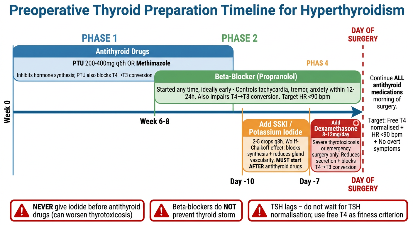
- Phase 1 (Weeks 0-8): Antithyroid drugs (PTU/Methimazole) - the backbone of preparation
- Phase 2 (ongoing): Propranolol - for heart rate and symptom control, with the bonus of T4→T3 blockade
- Phase 3 (Day -7 to -10): SSKI added for rapid gland preparation before surgery
- Phase 4 (emergency only): Dexamethasone for severe or urgent cases
- Day of Surgery: Continue all drugs; fitness criterion is free T4 normalised + HR <90
Anaesthesia in hyperthyroidism for md theory exam and viva
Anaesthesia in Hyperthyroidism
MD Theory Exam & Viva Answer
THEORY ANSWER FORMAT (Long Essay / Short Note)
Introduction
A. PREOPERATIVE MANAGEMENT
1. Assessment
- Cardiovascular: palpitations, AF, heart failure, systolic hypertension, widened pulse pressure
- Airway: stridor, dyspnoea, dysphagia (tracheal/oesophageal compression from goitre)
- Neuromuscular: proximal muscle weakness, tremor
- Metabolic: weight loss, heat intolerance, diarrhoea
- Free T4, T3, TSH (free T4 is the operative fitness criterion - TSH lags)
- ECG (AF, sinus tachycardia, LVH)
- Echocardiography if heart failure suspected
- CT/MRI neck and chest - mandatory for large goitres to assess tracheal deviation, substernal extension
- Indirect laryngoscopy (baseline vocal cord function)
- U&E (hypokalaemia, hypercalcaemia)
- Blood glucose (risk of unmasking DM)
2. Preoperative Preparation - The Timeline
Primary goal: Achieve euthyroidism before elective surgery (Miller's, Stoelting's)
Step 1 - Antithyroid Drugs (weeks 0 to 6-8)
| Drug | Dose | Mechanism |
|---|---|---|
| PTU | 200-400 mg q6-8h | Blocks iodide organification (synthesis) + blocks T4→T3 conversion |
| Methimazole | 10-30 mg/day | Blocks synthesis only |
- Takes 6-8 weeks to achieve euthyroidism
- Toxic effects: rash, agranulocytosis, hepatitis, arthralgias
- Continue on morning of surgery
Step 2 - Beta-Blockers (start early; can be added at any stage)
| Drug | Why preferred | Dose |
|---|---|---|
| Propranolol | Drug of choice - also blocks T4→T3 conversion | Titrated to effect; onset 12-24h |
- Controls: tachycardia, tremor, anxiety, heat intolerance
- Any beta-blocker acceptable; long-acting agents more convenient
- Combination: propranolol + SSKI standard pre-op regimen (7-14 days)
- Does NOT prevent thyroid storm
- Does NOT inhibit hormone synthesis
Step 3 - Potassium Iodide/SSKI (day -7 to -10 before surgery)
- Wolff-Chaikoff effect: blocks iodide organification → inhibits synthesis AND release
- Also reduces gland size and vascularity (important for thyroidectomy)
- Faster control: 7-10 days vs 6-8 weeks with antithyroid drugs alone
- Dose: 2-5 drops every 8 hours (or sodium iodide 250 mg q6h)
- ⚠️ MUST start antithyroid drugs first - iodide alone can worsen thyrotoxicosis
Step 4 - Glucocorticoids (severe disease / emergency surgery)
- Dexamethasone 8-12 mg/day or hydrocortisone 50-100 mg q6h
- Reduces thyroid hormone secretion + blocks T4→T3 conversion
- Reserved for: severe thyrotoxicosis, emergency surgery, thyroid storm
Emergency Surgery Protocol
- PTU/methimazole immediately
- SSKI after antithyroid drug
- Propranolol → HR < 90 bpm
- Dexamethasone
- Proceed when haemodynamically stable
B. AIRWAY MANAGEMENT
| Situation | Approach |
|---|---|
| Routine goitre | Standard laryngoscopy |
| Difficult intubation incidence | 5-8% in goitre surgery |
| Risk predictor | Thyroid cancer (not goitre size) |
| Tracheal deviation/compression | Awake fibreoptic intubation |
| Large substernal goitre | Review CT/MRI; risk of intrathoracic airway collapse post-induction; awake technique |
| Long-standing goitre | Risk of tracheomalacia - have plan for reintubation at extubation |
| Transoral thyroidectomy | Nasal intubation required |
- General endotracheal anaesthesia (standard)
- LMA - allows real-time vocal cord visualisation with spontaneous breathing (Stoelting's)
- Bilateral superficial cervical plexus block - for limited thyroidectomy
C. INTRAOPERATIVE MANAGEMENT
1. What to AVOID
| Drug | Reason |
|---|---|
| Ketamine | Sympathomimetic → aggravates tachycardia and hypertension - avoid even when clinically euthyroid (Stoelting's) |
| Pancuronium | Sympathomimetic → worsens tachycardia |
| Anticholinergics (atropine, glycopyrrolate) | Worsen tachycardia |
| Ephedrine | Indirect vasopressor - releases catecholamines in sensitised patient |
| Epinephrine in LA solutions | Increased catecholamine sensitivity |
| Aspirin (if fever) | Displaces T4 from binding proteins → worsens thyrotoxicosis |
2. Safe / Preferred Drugs
| Drug | Notes |
|---|---|
| Propofol | Preferred induction agent; clearance ↑ and Vd ↑ in hyperthyroidism → increase TIVA infusion rates |
| All volatile agents | Acceptable; MAC is NOT altered by hyperthyroidism |
| Succinylcholine / Rocuronium | Safe for intubation |
| Phenylephrine | Direct-acting vasopressor of choice for hypotension |
| Esmolol infusion | First-line for intraoperative tachycardia (50-350 mcg/kg/h) |
| Regional anaesthesia | Excellent alternative; avoid adrenaline-containing solutions |
3. Key Intraoperative Principles
- Induction may be slower - elevated cardiac output increases drug distribution; higher agent concentrations required
- MAC unchanged - do not alter volatile agent targets
- Maintain adequate depth - prevent exaggerated sympathetic response to surgical stimulation
- Muscle relaxants - reduce initial dose - increased incidence of myasthenia gravis in hyperthyroidism
- Use nerve stimulator (twitch monitor) for all subsequent NMB dosing (Stoelting's)
- Direct vasopressors only for hypotension (phenylephrine)
- Invasive monitoring individualised - especially if significant LV dysfunction
- NIM tube for RLN monitoring: avoid NMB and topical LA if used
4. Expected Haemodynamic Profile
- Systolic hypertension + widened pulse pressure
- ↓ SVR with ↑ cardiac output
- Sinus tachycardia / AF
- Increased catecholamine sensitivity (↑ β-receptor density)
D. THYROID STORM
Triggers
Clinical Features
Thyroid Storm vs MH - The Critical Distinction
| Feature | Thyroid Storm | Malignant Hyperthermia |
|---|---|---|
| Timing | Postoperative (usually) | Intraoperative |
| ETCO2 | Normal | ↑ (early sign) |
| CK | Normal | Massively elevated |
| Rigidity | Absent | Present |
| K+ | Hypokalemia | Hyperkalemia |
| Metabolic acidosis | Mild | Severe |
| Trigger | Surgery in hyperthyroid pt | Volatiles/succinylcholine |
| Treatment | Antithyroid drugs + support | Dantrolene |
Management of Thyroid Storm (Stoelting's Table 47-3)
| Step | Drug | Dose |
|---|---|---|
| Inhibit synthesis | PTU | 200-400 mg PO/NGT q6h |
| Block release (after PTU) | Sodium iodide | 250 mg PO/IV q6h |
| Reduce secretion + T4→T3 | Hydrocortisone | 50-100 mg IV q6h |
| Control HR | Propranolol | 10-40 mg PO q4-6h |
| Or | Esmolol infusion | Titrate |
| Antipyretic | Paracetamol + cooling blankets | Avoid aspirin |
| Prevent shivering | Pethidine | 25-50 mg IV q4-6h |
| Adjunct | Cholestyramine | 4g q6h (interrupts enterohepatic T4 recirculation) |
| Refractory | Plasmapheresis | |
| Fluids | IV resuscitation | Correct electrolytes |
E. POSTOPERATIVE COMPLICATIONS (after Thyroidectomy)
| Complication | Timing | Signs | Action |
|---|---|---|---|
| Bilateral RLN injury | Immediate | Stridor, aphonia, glottic closure | Immediate reintubation → tracheostomy |
| Unilateral RLN injury | Immediate | Hoarseness (often transient) | Laryngoscopy; observe |
| Hypocalcaemia | 24-96 hours | Perioral tingling, Chvostek's sign, laryngeal stridor → laryngospasm | IV calcium chloride/gluconate; check Mg |
| Tracheomalacia | On extubation | Airway collapse | Reintubate; tracheostomy |
| Thyroid storm | During or first 18 hours | See above | See above |
| Haematoma | First few hours | Neck swelling, tracheal compression | Urgent surgical decompression |
| Pneumothorax | Intraoperative | Desaturation, ↓ breath sounds | Chest drain |
VIVA QUESTIONS & MODEL ANSWERS
To achieve euthyroidism before elective surgery. The operative fitness criterion is normalisation of free T4 with absence of overt symptoms - not TSH, which lags weeks behind clinical response.
Propranolol is unique among beta-blockers because it not only controls hyperadrenergic symptoms (tachycardia, tremor, anxiety, heat intolerance) but also inhibits the peripheral conversion of T4 to the more active T3 - an effect seen over 1-2 weeks. Other beta-blockers lack this additional antithyroid mechanism.
Iodide (SSKI) works via the Wolff-Chaikoff effect to block hormone synthesis and release. However, if given alone initially, it can paradoxically worsen thyrotoxicosis (by transiently increasing hormone availability or through Jod-Basedow effect). Starting antithyroid drugs first ensures that any hormone released by iodide cannot contribute to toxicity.
No. MAC is not altered by hyperthyroidism (Miller's, Stoelting's). However, induction may be slower because the elevated cardiac output increases drug distribution volume and clearance, requiring higher concentrations. Propofol TIVA infusion rates must be increased for the same reason.
Hyperthyroidism is associated with an increased incidence of myasthenia gravis (an autoimmune overlap). Myasthenic patients are highly sensitive to non-depolarising NMBs. The initial dose should be reduced and a nerve stimulator (twitch monitor) used to guide all subsequent dosing.
The key distinguishing features are: thyroid storm typically presents postoperatively, with hypokalemia, no rise in ETCO2, no muscle rigidity, and no elevation of CK. MH presents intraoperatively during volatile/succinylcholine exposure with early rise in ETCO2, hyperkalemia, rigidity, massively elevated CK, and severe metabolic acidosis. MH responds to dantrolene; thyroid storm does not.
Aspirin displaces T4 from thyroid-binding globulin, increasing the concentration of free (active) T4 in circulation - this worsens the thyrotoxic state. Paracetamol (acetaminophen) is the safe antipyretic of choice.
A substernal goitre behaves like an anterior mediastinal mass and can cause intrathoracic airway obstruction after induction of general anaesthesia due to loss of negative intrathoracic pressure and muscle tone supporting the airway. CT/MRI must be reviewed preoperatively. An awake fibreoptic approach should be considered.
Hypocalcaemia due to inadvertent parathyroid removal (hypoparathyroidism). Symptoms develop 24-96 hours after total thyroidectomy. Laryngeal stridor progressing to laryngospasm is one of the first signs of hypocalcaemic tetany. Treatment: IV calcium chloride or calcium gluconate; also check and correct magnesium.
Hyperthyroidism upregulates β-adrenergic receptor density in the heart, skeletal muscle, and adipose tissue, making the patient highly sensitive to catecholamines. Indirect vasopressors (ephedrine) work by releasing endogenous catecholamines, which in this sensitised state can precipitate severe hypertension, tachyarrhythmias, and myocardial ischaemia. Direct-acting vasopressors (phenylephrine) bypass this mechanism and are safe.
No. Beta-blockers only provide symptomatic control by blunting the hyperadrenergic manifestations. They do not inhibit thyroid hormone synthesis or secretion and cannot prevent thyroid storm. Only achieving euthyroidism (via antithyroid drugs) reduces that risk.
Apathetic hyperthyroidism occurs in elderly patients who present with depression, withdrawal, and cardiac features (AF, heart failure) without the classic sympathomimetic features (anxiety, tremor, weight loss). It is relevant because the diagnosis may be missed preoperatively, and these patients are at high risk of haemodynamic instability and thyroid storm under anaesthesia without appropriate preparation.
One-Line Revision Points
| Point | Answer |
|---|---|
| MAC in hyperthyroidism | Unchanged |
| Induction speed | Slower (high CO) |
| Propofol TIVA | Increase infusion rate |
| Best vasopressor | Phenylephrine (direct-acting) |
| Avoid | Ketamine, pancuronium, ephedrine, anticholinergics, adrenaline in LA |
| Pre-op beta-blocker | Propranolol (also blocks T4→T3) |
| Iodide rule | Always after antithyroid drugs |
| Fitness criterion | Free T4 normal (not TSH) |
| MH vs thyroid storm K+ | MH = hyperK; Storm = hypoK |
| Post-thyroidectomy laryngospasm | Hypocalcaemia |
| Thyroid storm antipyretic | Paracetamol (not aspirin) |
| Storm timing | Usually postoperative (first 18h) |
| NMB in hyperthyroidism | Reduce dose + use nerve stimulator |
Extubation strategy
Extubation Strategy After Thyroid Surgery
From Miller's Anesthesia (10e) & Barash/Stoelting's Clinical Anesthesia (9e)
Why Extubation is High-Risk After Thyroid Surgery
| Risk | Mechanism |
|---|---|
| Bilateral RLN injury | Unopposed cord adduction → complete glottic closure on extubation |
| Unilateral RLN injury | Partial airway compromise; may be clinically silent initially |
| Haematoma | Venous engorgement from coughing/bucking on ETT → haematoma compresses airway |
| Tracheomalacia | Weakened tracheal rings in long-standing goitre collapse inward once ETT removed |
| Hypocalcaemia | Laryngospasm 24-96h post-op (late complication, not immediate) |
| Airway oedema | From surgical manipulation and prolonged retraction |
The Core Principle
"Gentle emergence from anaesthesia is necessary to avoid coughing on the ETT and the possibility of haematoma formation from venous engorgement." — Miller's Anesthesia 10e
Step-by-Step Extubation Strategy
Step 1 - Pre-extubation Checklist (Before Any Attempt)
| Criterion | Detail |
|---|---|
| Fully awake and cooperative | Not drowsy; follows commands |
| Adequate spontaneous ventilation | Respiratory rate, tidal volume, SpO2 acceptable |
| Complete NMB reversal | Train-of-four ratio ≥0.9; confirm with nerve stimulator |
| Haemodynamically stable | HR <90 bpm (especially in hyperthyroid patient) |
| Normothermic | Hypothermia worsens muscle weakness |
| Surgical field checked | No active bleeding; wound closed and dry |
| Vocal cord function documented | Ask patient to phonate "E" on the table, or do laryngoscopy - confirm pre-extubation cord function |
Step 2 - Smooth Emergence (Prevent Coughing)
| Technique | Mechanism | Notes |
|---|---|---|
| Remifentanil infusion (0.01-0.05 mcg/kg/min) (Miller's) | Blunts cough reflex and sympathetic response during extubation | Most practical and effective method; titrate infusion down slowly as patient wakes |
| Lignocaine IV (1-1.5 mg/kg) 2-3 min before extubation | Suppresses cough reflex | Simple adjunct |
| Dexmedetomidine infusion | Smooth awake sedation; reduces emergence agitation and coughing | Useful in anxious/agitated patients |
| Deep extubation | Removes tube before cough reflex returns | ⚠️ Avoid in thyroid surgery - risk of airway obstruction; Barash/Stoelting explicitly states many clinicians avoid this technique wherever possible due to airway obstruction risk |
Key point from Miller's: Deep extubation is NOT recommended after thyroid surgery despite reducing coughing - the risk of airway obstruction outweighs the benefit.
Step 3 - Assess for Tracheomalacia Before Extubation
| Test | Method |
|---|---|
| Cuff-deflation test | Deflate cuff while tube still in situ; positive if patient can breathe around tube (adequate tracheal lumen) |
| Direct fibreoptic bronchoscopy | Through ETT before extubation - visualise tracheal wall integrity and any dynamic collapse |
| Surgeon's intraoperative assessment | Palpation of tracheal rings during surgery to assess rigidity |
- Do not extubate in the operating theatre - transfer to ICU with ETT in situ
- Consider planned tracheostomy intraoperatively (discuss with surgeon)
- If extubation attempted - use Airway Exchange Catheter (AEC) strategy (see Step 5)
Step 4 - RLN Assessment at Extubation
- Ask patient to phonate: say the sound "E" (Stoelting's)
- Normal phonation = bilateral cord function likely intact
- Hoarse/weak voice = suspect unilateral injury (usually tolerated; observe)
- No voice / aphonia + stridor = suspect bilateral injury → do NOT extubate; reintubate or proceed to tracheostomy
- Intraoperative EMG monitoring provides real-time RLN data
- Surgeon should communicate nerve integrity status before extubation
Step 5 - The "Staged / Bailey Manoeuvre" Extubation
- Long-standing/substernal goitre (tracheomalacia risk)
- Difficult original intubation
- Significant intraoperative airway oedema
- Bilateral RLN at risk (e.g. completion thyroidectomy, central neck dissection)
- Any doubt about post-extubation airway safety
- Preoxygenate fully (FiO2 1.0) before extubation
- Lubricate AEC; select AEC with external diameter closest to internal diameter of the reintubation ETT
- Insert AEC to depth of ETT bevel (match depth markings on AEC with markings on ETT)
- Aim for mid-tracheal positioning of AEC tip to avoid endobronchial placement
- Remove ETT carefully while holding AEC position
- Leave AEC in situ as a guide rail for reintubation if needed
- AEC has a central lumen - can provide supplemental O2 (jet ventilation in emergency)
- Keep AEC in place for 30-60 minutes post-extubation while monitoring in theatre or recovery
- If reintubation required: advance ETT over AEC; if hang-up at glottis - lift tongue with laryngoscope + rotate ETT 90° anticlockwise
Step 6 - Post-Extubation Position and Monitoring
| Measure | Rationale |
|---|---|
| Head-up / 30-45° elevation | Reduces venous engorgement; minimises haematoma risk and oedema |
| Avoid tight neck dressings | Can compress venous drainage |
| Continuous SpO2 and capnography in recovery | Detect early desaturation from cord oedema, haematoma, tracheomalacia |
| Wound inspection every 15 min (first hour) | Early haematoma detection |
| Voice check before discharge from recovery | Confirm cord function |
Post-Extubation Emergencies - Recognition and Response
1. Haematoma (Most Time-Critical)
| Sign | Action |
|---|---|
| Neck swelling, tracheal deviation, stridor, dyspnoea, rising distress | Call surgeon immediately |
| Open wound at bedside | Release skin clips/sutures to decompress haematoma (this alone may relieve compression enough to allow reintubation) |
| Reintubate awake if possible | Haematoma distorts anatomy - use videolaryngoscopy / fibreoptic |
| If cannot intubate | Surgical airway (scalpel-bougie-tube) |
2. Bilateral RLN Palsy
| Sign | Action |
|---|---|
| Sudden stridor and aphonia on extubation | Immediate reintubation |
| If reintubation fails | Emergency tracheostomy |
3. Tracheomalacia
| Sign | Action |
|---|---|
| Airway collapse immediately after extubation | Reintubate; CPAP/PEEP may temporarily stent the airway; definitive = tracheostomy |
4. Hypocalcaemia / Laryngospasm
| Sign | Timing | Action |
|---|---|---|
| Laryngeal stridor → laryngospasm | 24-96 hours post-op | IV calcium chloride (10 mL of 10%) or calcium gluconate; check and correct Mg |
Summary - Extubation Decision Framework
Before extubation:
    ├─ Long-standing goitre? → Test for tracheomalacia (cuff test / bronchoscopy)
    ├─ Difficult original intubation? → Use AEC strategy
    ├─ RLN at high risk? → Phonate "E" + laryngoscopy before extubation
    └─ All clear?
           ↓
    Smooth emergence:
    → Remifentanil infusion 0.01-0.05 mcg/kg/min
    → Avoid deep extubation
    → Fully awake, cooperative, NMB reversed
           ↓
    Extubate in 30-45° head-up position
           ↓
    AEC left in situ if ANY doubt (staged extubation)
           ↓
    Recovery: SpO2 monitoring + wound checks q15 min
           ↓
    Voice check before discharge from recovery
Viva Points on Extubation
Although deep extubation prevents coughing and reduces haematoma risk, it is avoided because there is a significant risk of airway obstruction - both from cord oedema/palsy and tracheomalacia. Recovery of airway reflexes is needed to protect against obstruction. (Barash/Stoelting)
A low-dose remifentanil infusion (0.01-0.05 mcg/kg/min) is a popular technique to allow smooth, awake extubation without coughing - preserving airway reflexes while blunting the cough reflex. This prevents venous engorgement and haematoma formation. (Miller's)
With the ETT still in situ, the cuff is deflated. If the patient can breathe around the ETT, there is adequate tracheal lumen and tracheomalacia/oedema is less likely to cause obstruction after extubation. Failure suggests critical narrowing.
Haematoma typically presents within the first few hours with visible neck swelling and progressive stridor - relieved partially by wound opening. Tracheomalacia presents immediately on extubation as sudden airway collapse without external swelling; requires reintubation and tracheostomy.
Bailey manouvre
The Bailey Manoeuvre
From Miller's Anesthesia (10e) & Barash/Stoelting's Clinical Anesthesia (9e)
Definition
"An alternative extubation technique, known as the Bailey maneuver, involves exchanging an ETT for an SGA while the patient is under deep anaesthesia." — Miller's Anesthesia, 10e
The Problem It Solves
| Technique | Advantage | Disadvantage |
|---|---|---|
| Awake extubation | Patent airway maintained; reflexes intact | Coughing, straining, haemodynamic surge → haematoma, raised ICP, wound dehiscence |
| Deep extubation | Smooth, no coughing | Risk of airway obstruction and hypoventilation before reflexes return |
- Smooth like deep extubation (no coughing on ETT)
- Protected like awake (SGA maintains airway until patient wakes fully)
Indications
| Clinical Situation | Risk Avoided |
|---|---|
| Patients at high risk of bronchospasm (asthma, reactive airways) (Barash/Stoelting) | ETT triggers bronchospasm on emergence |
| Thyroid / neck surgery | Coughing → venous engorgement → haematoma |
| Intracranial / neurosurgery | Coughing → ↑ ICP |
| Intraocular surgery | Coughing → ↑ intraocular pressure |
| Maxillofacial / ENT surgery | Surgical site disruption |
| Known or suspected difficult airway | Deep extubation too risky; Bailey gives airway bridge |
| Vascular / aortic surgery | Coughing → haemodynamic surge |
How It Is Performed - Step by Step
Prerequisites Before Starting
- Patient is deeply anaesthetised (surgical stage III)
- Neuromuscular blockade fully reversed
- Oropharynx suctioned under deep anaesthesia
- Bite block placed (rolled gauze between molars - not OPA, which can cause dental damage) (Miller's)
- Patient preoxygenated (FiO2 1.0)
- Size-appropriate LMA selected and checked
The Technique (Barash/Stoelting, Ch. 28)
Step 1:
Patient deeply anaesthetised; ETT still in situ
              ↓
Step 2:
Deflate LMA cuff completely
              ↓
Step 3:
Insert the deflated LMA into the oropharynx
BEHIND the in-situ ETT
(slide LMA posteriorly into hypopharynx around the ETT)
              ↓
Step 4:
Confirm LMA tip seated in hypopharynx
              ↓
Step 5:
Remove the ETT (withdraw over the LMA)
              ↓
Step 6:
Inflate the LMA cuff
              ↓
Step 7:
Confirm airway patency via capnography / chest rise / SpO2
              ↓
Step 8:
Allow patient to emerge via LMA
(smooth, no laryngeal stimulation, no coughing)
              ↓
Step 9:
Remove LMA when fully awake - either:
  (a) Deeply anaesthetised, OR
  (b) After full return of protective reflexes + patient opens mouth on command
Variant - When SGA Was Used as Intubation Conduit
- At end of surgery, deflate the SGA cuff and leave device in situ
- Remove the ETT, leaving the SGA as the primary airway
- Conduct emergence via SGA
"This technique can be of great benefit in cases in which an ETT is needed intraoperatively but coughing or hypertension during emergence would be deleterious." (Barash/Stoelting)
Advantages of the Bailey Manoeuvre
| Advantage | Mechanism |
|---|---|
| No coughing on ETT | ETT removed under deep anaesthesia |
| Smooth haemodynamics | No sympathetic surge from laryngeal stimulation |
| Airway maintained | LMA provides reliable conduit until full consciousness |
| Reduced bronchospasm | LMA far less stimulating than ETT (Barash/Stoelting) |
| Reduced laryngospasm | LMA removal associated with less laryngospasm than ETT (Barash/Stoelting Table 28-9) |
| Improved SpO2 after device removal | Patient breathing via LMA until fully awake |
| Oxygen until reflexes return | LMA permits ventilation and oxygenation through emergence |
Disadvantages / Limitations
| Limitation | Detail |
|---|---|
| Aspiration risk | LMA does not protect against regurgitation; contraindicated if full stomach |
| Not for restricted access | Cannot be used if mouth opening is severely limited |
| Requires skill | Inserting deflated LMA behind in-situ ETT is technically demanding |
| Not a safety net for reintubation | Unlike AEC, LMA does not provide a guide rail for rapid reintubation if airway is lost |
| Not first-line for difficult airway | ASA recommends AEC as preferred strategy for difficult airway extubation (Miller's) |
Bailey Manoeuvre vs. AEC vs. Deep Extubation
| Feature | Bailey Manoeuvre | AEC | Deep Extubation |
|---|---|---|---|
| Mechanism | ETT → LMA exchange (deep) | Guide rail left in trachea | ETT removed under deep anaesthesia |
| Airway maintained by | LMA | Patient's own airway + AEC guide | Patient's own airway |
| Smooth emergence | ✓ Yes | ✓ Yes | ✓ Yes |
| Reintubation route | Via laryngoscopy | Over AEC directly | Via laryngoscopy |
| Best for | Bronchospasm risk; cough prevention | Difficult airway (ASA preferred) | Low-risk routine cases |
| Aspiration risk | Higher than ETT | Lower | Moderate |
| Requires skill | High | Moderate | Low |
| ASA recommendation | Alternative technique | First-line for difficult airway | Routine cases only |
LMA Removal During/After Bailey Manoeuvre
- While deeply anaesthetised - smooth, no stimulation
- After protective reflexes fully returned - patient can open mouth on command
Avoid removal during stage II excitation - associated with coughing and laryngospasm.
Relevance to Thyroid Surgery Specifically
- Need for ETT intraoperatively: surgical access, RLN monitoring (NIM tube), airway protection
- Need to avoid coughing at extubation: prevents venous engorgement and haematoma
- Full intraoperative ETT + NIM monitoring
- Smooth exchange to LMA under deep anaesthesia at end of surgery
- Patient waking on LMA without ever coughing on the tracheal tube
Viva Questions
Exchanging an endotracheal tube for a supraglottic airway device (typically LMA) while the patient is under deep anaesthesia, allowing smooth cough-free emergence while maintaining airway control.
The LMA cuff is deflated and the LMA is inserted into the oropharynx behind the in-situ ETT. The ETT is then withdrawn, the LMA cuff is inflated, and the patient emerges through the LMA.
Patients at high risk of bronchospasm in whom tracheal intubation was required for surgery - the LMA causes far less bronchospasm than an ETT on emergence.
The AEC is recommended by the ASA as first-line for difficult airway extubation because it provides a direct guide rail for immediate reintubation if the airway is lost after extubation. The Bailey manoeuvre does not provide this safety feature - if the LMA fails, standard laryngoscopy is required with no pre-positioned guide. (Miller's)
Full stomach / aspiration risk - the LMA does not protect against regurgitation. Also avoid with very limited mouth opening.
Is the pre op preparation according to latest guidelines
hyperthyroidism preoperative preparation guidelines
ATA ETA 2024 2025 guidelines hyperthyroidism preoperative preparation thyroidectomy antithyroid drugs
hyperthyroidism thyroidectomy preoperative preparation potassium iodide beta blocker
ATA 2016 Graves disease guidelines preoperative preparation methimazole iodide beta blocker update 2024
https://pmc.ncbi.nlm.nih.gov/articles/PMC11472198
preoperative preparation hyperthyroidism thyroid surgery methimazole iodide
Is the Preoperative Preparation According to Latest Guidelines?
Side-by-Side Comparison
1. Antithyroid Drugs - UPDATED GUIDANCE
| Aspect | Textbooks (Miller's/Stoelting's) | Current Guidelines (ATA 2016 / AAES 2020) | Verdict |
|---|---|---|---|
| First-line drug | PTU or methimazole mentioned equally | Methimazole is the preferred first-line drug (ATA 2016) | ⚠️ Textbooks underemphasise this |
| When PTU is preferred | Not specified clearly | PTU only in: first trimester of pregnancy, thyroid storm, patients with methimazole side effects | ⚠️ Important nuance missed |
| Why methimazole preferred | Not stated | More effective, longer half-life (once-daily dosing), fewer side effects; PTU has serious hepatotoxicity risk (ETA 2022: "should not be used" outside exceptions) | ⚠️ PTU hepatotoxicity risk not highlighted |
| Methimazole dosing | Not dose-specified | Dose titrated to fT4 level: 5-10 mg/day if fT4 1-1.5x ULN; 10-20 mg/day if 1.5-2x ULN; 30-40 mg/day if 2-3x ULN | ⚠️ New - textbooks do not include this dosing scheme |
| Monitoring | Continue morning of surgery | Check fT4 every 4-8 weeks; monitor T3 separately (may remain elevated even when T4 normalises) | ⚠️ T3 monitoring emphasis is new |
| Time to euthyroidism | 6-8 weeks | 3-8 weeks | ✅ Consistent |
Key update: Methimazole is the first-line drug. PTU is second-line due to hepatotoxicity risk. For the MD exam, always state methimazole as preferred and know the dose-titration scheme.
2. Beta-Blockers - CONSISTENT WITH UPDATES
| Aspect | Textbooks | Guidelines | Verdict |
|---|---|---|---|
| Propranolol as drug of choice | ✅ Stated | ✅ Confirmed - propranolol preferred (also blocks T4→T3) | ✅ Correct |
| Any beta-blocker acceptable | ✅ Stated | ✅ Confirmed - long-acting agents more convenient | ✅ Correct |
| Target heart rate | <90 bpm | <80 bpm in current protocols for rapid preparation (2024 review) | ⚠️ Minor discrepancy - guidelines suggest tighter control |
| Beta-blockers do not prevent storm | ✅ Stated | ✅ Confirmed | ✅ Correct |
| Used in all hyperthyroid patients | ✅ Stated | Recommended especially in: elderly, severe thyrotoxicosis, cardiovascular disease | ✅ Consistent |
3. Potassium Iodide / Lugol's Solution - UPDATED GUIDANCE
| Aspect | Textbooks | Guidelines | Verdict |
|---|---|---|---|
| Wolff-Chaikoff mechanism | ✅ Stated | ✅ Confirmed | ✅ Correct |
| Antithyroid drug must precede iodide | ✅ Stated | ✅ Confirmed - essential rule | ✅ Correct |
| Duration | Up to 10 days | Not more than 10 days - risk of escape from Wolff-Chaikoff effect after 10 days; hormone synthesis resumes | ⚠️ Textbooks do not mention escape phenomenon |
| Exception to "ATD first" rule | Not mentioned | In Graves' patients who cannot tolerate thionamides: iodide + beta-blocker alone may still be effective (ATA 2016) | ⚠️ New exception not in textbooks |
| Additional benefit | Not fully stated | Iodide reduces thyroid blood flow, vascularity, and VEGF levels - benefits observed even in already euthyroid patients pre-thyroidectomy | ⚠️ Surgical benefit worth knowing |
| Iodide timing | Mentioned broadly | Give iodine 1 hour after thionamide administration for optimal sequencing | ⚠️ Specific timing not in textbooks |
| Universal pre-thyroidectomy iodide | Not addressed | Some guidelines (Huang et al. 2024) recommend iodide preoperatively in all Graves' thyroidectomy patients (even euthyroid) for haemostasis benefits | ⚠️ Evolving evidence |
4. Glucocorticoids - CONSISTENT
| Aspect | Textbooks | Guidelines | Verdict |
|---|---|---|---|
| Dexamethasone 8-12 mg/day | ✅ Stated | ✅ Confirmed for severe thyrotoxicosis | ✅ Correct |
| Blocks T4→T3 conversion | ✅ Stated | ✅ Confirmed | ✅ Correct |
| Hydrocortisone 100 mg IV q8h on day of surgery (then taper) | Partially mentioned | Hydrocortisone 100 mg IV q8h on day of surgery, tapered over 3 days | ⚠️ Specific intraoperative hydrocortisone protocol not in textbooks |
5. Rapid Preparation Regimen - NEW IN GUIDELINES (Not in Textbooks)
Option A - Iodide-based rapid preparation (ATA / current reviews)
| Drug | Dose | Duration |
|---|---|---|
| Lugol's 5% solution | 13 drops three times daily (~81 mg iodine/day) | 10 days max |
| Dexamethasone | 2 mg twice daily | Same period |
| Beta-blocker | Titrate to HR <80 bpm | Same period |
Option B - Iodinated contrast agent (iopanoic acid / iodide)
| Drug | Dose |
|---|---|
| Iopanoic acid (IOP) | 500 mg every 6 hours |
| Betamethasone | 0.5 mg every 6 hours |
| Propranolol | 40 mg every 8 hours |
These rapid protocols are increasingly used in real clinical practice but are not in Miller's or Stoelting's - important for viva.
6. Cholestyramine - MENTIONED IN TEXTBOOKS, CONFIRMED IN GUIDELINES
- 4g q6h used as adjunct (bile acid sequestrant)
- Interrupts enterohepatic recirculation of thyroid hormones
- Confirmed as useful adjunct in rapid preparation regimens
- ✅ Consistent between textbooks and guidelines
7. Fitness Criterion for Surgery - UPDATED
| Aspect | Textbooks | Guidelines | Verdict |
|---|---|---|---|
| Free T4 normalisation | ✅ Stated | ✅ Confirmed | ✅ Correct |
| TSH lags - do not wait for TSH | ✅ Stated | ✅ Confirmed | ✅ Correct |
| Monitor T3 separately | Not stated | Monitor serum T3 - may remain elevated even when fT4 normalised; euthyroidism not confirmed until T3 also normal | ⚠️ Important gap in textbooks |
What the Textbooks Get Right
✅ Propranolol as preferred beta-blocker (T4→T3 blockade)
✅ "Give antithyroid drug before iodide" rule
✅ Target HR <90 bpm for emergency surgery
✅ Dexamethasone in severe disease
✅ Continue all drugs on morning of surgery
✅ Free T4 as fitness criterion (not TSH)
✅ 7-14 day preparation timeline with full regimen
What the Textbooks Miss or Understate
| Gap | Current Guideline Position |
|---|---|
| Methimazole > PTU | Methimazole is clearly first-line; PTU reserved for specific situations only |
| PTU hepatotoxicity | Significant risk; ETA 2022 says "should not be used" routinely |
| Methimazole dose by fT4 level | Dose-titration scheme based on degree of elevation |
| Monitor T3 as well as fT4 | T3 may remain elevated when fT4 normalises |
| 10-day max for iodide | Wolff-Chaikoff escape occurs after 10 days |
| Iodide 1 hour after thionamide | Optimal timing for sequencing |
| Rapid preparation protocols (iodide + steroid + beta-blocker) | Achieves euthyroidism in 7-10 days without thionamides |
| Iodide even in euthyroid Graves' pre-op | Reduces intraoperative blood loss and vascularity |
| Hydrocortisone taper post-operative | 100 mg IV q8h day of surgery, taper over 3 days |
For Your MD Exam and Viva - What to Say
"The preoperative preparation taught in Miller's and Stoelting's is broadly consistent with ATA 2016 guidelines. However, current guidelines clarify that methimazole is the preferred first-line antithyroid drug over PTU due to PTU's hepatotoxicity risk. PTU is now reserved for first-trimester pregnancy, thyroid storm, and methimazole intolerance. The methimazole dose should be titrated to the degree of fT4 elevation. Iodide should not be continued beyond 10 days due to the risk of Wolff-Chaikoff escape. In patients intolerant of thionamides, a rapid preparation regimen using Lugol's solution, dexamethasone, and beta-blockers can achieve clinical euthyroidism within 7-10 days. T3 levels should be monitored separately from fT4 as the criterion for euthyroidism, as T3 may remain elevated despite fT4 normalisation."
Give it systematically to write in exam
Preoperative Preparation for Hyperthyroidism Before Surgery
Exam Answer (ATA 2016 + AAES 2020 + Current Evidence)
Introduction
- Achieve euthyroidism before elective surgery
- Minimise risk of thyroid storm perioperatively
- Reduce intraoperative blood loss (for thyroidectomy)
- Control cardiovascular manifestations
Fitness criterion: Normalisation of free T4 AND T3 + absence of overt symptoms + HR <80 bpm (Do NOT wait for TSH - it lags weeks behind clinical response)
STEP 1 - Antithyroid Drugs (Weeks 0 to 6-8)
Drug of Choice: Methimazole (ATA 2016 - First Line)
| fT4 level | Methimazole dose |
|---|---|
| 1 - 1.5× upper limit of normal | 5-10 mg/day (once daily) |
| 1.5 - 2× upper limit of normal | 10-20 mg/day |
| 2 - 3× upper limit of normal | 30-40 mg/day |
- Monitor TFTs every 4-8 weeks
- Monitor T3 separately - may remain elevated even when fT4 normalises
- Reduce to maintenance (5-10 mg/day) as thyroid function improves
- Continue on the morning of surgery
When to Use PTU Instead (ATA 2016 - Second Line Only)
- First trimester of pregnancy
- Thyroid storm
- Methimazole intolerance or side effects
⚠️ PTU is NOT first-line due to risk of hepatotoxicity (hepatic failure, agranulocytosis). ETA 2022 states it "should not be used" routinely.
STEP 2 - Beta-Blockers (Start Early; Can Add at Any Stage)
Drug of Choice: Propranolol
- Controls hyperadrenergic symptoms within 12-24 hours
- Uniquely also inhibits peripheral T4→T3 conversion (over 1-2 weeks)
- No other beta-blocker has this additional antithyroid effect
- Does NOT inhibit hormone synthesis
- Does NOT prevent thyroid storm
- Use cautiously in heart failure - test with small dose of esmolol (0.1-0.2 mg/kg) first
STEP 3 - Potassium Iodide / Lugol's Solution (Day -10 to Day -1 Before Surgery)
Mechanism - Wolff-Chaikoff Effect
Preparation
| Preparation | Dose |
|---|---|
| SSKI (saturated solution of potassium iodide) | 2-5 drops every 8 hours |
| Lugol's solution 5% | 13 drops three times daily (~81 mg iodine/day) |
| Sodium iodide (IV - for thyroid storm) | 250 mg every 6 hours |
Key Rules (ATA 2016)
-
ALWAYS start antithyroid drug BEFORE iodide
- Iodide given alone can initially worsen thyrotoxicosis
- Optimal: give iodide 1 hour after thionamide dose
- Exception: In patients intolerant of thionamides, iodide + beta-blocker alone may still be used
-
Do NOT continue iodide beyond 10 days
- Risk of Wolff-Chaikoff escape after 10 days: gland adapts, resumes hormone synthesis
- Textbooks do not mention this - important exam point
-
Benefits for thyroid surgery specifically:
- Reduces thyroid blood flow and vascularity
- Reduces intraoperative blood loss
- Some guidelines recommend iodide in all Graves' thyroidectomy patients (even if euthyroid) for this haemostatic benefit
STEP 4 - Glucocorticoids (Severe Disease / Emergency Surgery)
Indications
- Severe thyrotoxicosis
- Emergency surgery in uncontrolled hyperthyroid patient
- Thyroid storm
Regimen
| Drug | Dose | Benefit |
|---|---|---|
| Dexamethasone | 2 mg twice daily OR 8-12 mg/day | Inhibits T4→T3 conversion + reduces hormone secretion |
| Hydrocortisone | 100 mg IV every 8 hours on day of surgery | Stress cover + same antithyroid effects |
| Taper | Over 3 days post-surgery | Gradual withdrawal |
Glucocorticoids block T4→T3 conversion - the same mechanism as propranolol and PTU. Triple blockade of T4→T3 conversion (propranolol + PTU + dexamethasone) is used in thyroid storm.
STEP 5 - Cholestyramine (Adjunct)
- Dose: 4 g every 6 hours
- Mechanism: Bile acid sequestrant - interrupts enterohepatic recirculation of thyroid hormones
- Reduces circulating T4/T3 load faster
- Used as adjunct in rapid preparation and thyroid storm
Standard Preoperative Timeline
WEEKS 0-6                WEEK 6-8               DAY -10 to -1         DAY OF SURGERY
─────────────────────────────────────────────────────────────────────────►
│ Methimazole           │ Confirm euthyroid    │ Add SSKI/Lugol's    │ Continue ALL drugs
│ (dose per fT4)        │ Free T4 + T3 normal  │ 2-5 drops q8h       │ in morning
│                       │                      │ Max 10 days only     │
│ Propranolol           │ HR <80 bpm           │                     │ Check HR, fT4
│ (titrate to HR)       │                      │                     │
└───────────────────────┴──────────────────────┴─────────────────────┴──────────────────
Emergency / Rapid Preparation (When Surgery Cannot Wait)
For patients who can take thionamides:
- Methimazole (high dose) or PTU
- SSKI / Lugol's (1 hour after thionamide)
- Propranolol (HR <80 bpm)
- Dexamethasone
- Cholestyramine (adjunct) → Target: 7-10 days to clinical euthyroidism
For patients intolerant of thionamides (ATA 2016 exception):
| Drug | Dose |
|---|---|
| Lugol's 5% | 13 drops three times daily |
| Dexamethasone | 2 mg twice daily |
| Beta-blocker | Titrate to HR <80 bpm |
Iopanoic acid (iodinated contrast) based regimen:
| Drug | Dose |
|---|---|
| Iopanoic acid | 500 mg every 6 hours |
| Betamethasone | 0.5 mg every 6 hours |
| Propranolol | 40 mg every 8 hours |
Fitness for Surgery - Checklist
| Criterion | Standard |
|---|---|
| Free T4 | Normalised |
| T3 | Normalised (check separately - may lag fT4) |
| TSH | May still be low - do NOT delay surgery waiting for TSH |
| Heart rate | <80 bpm at rest |
| Symptoms | No overt hyperthyroid features |
| Cardiovascular | AF/failure controlled |
| Vitamin D | Replete (reduces post-op hypocalcaemia risk) |
Summary - Key Exam Points
| Point | Answer |
|---|---|
| First-line antithyroid drug | Methimazole (not PTU) |
| PTU reserved for | Pregnancy (1st trimester), thyroid storm, methimazole intolerance |
| Why methimazole preferred | Once daily, more effective, safer (PTU: hepatotoxicity) |
| Methimazole dose | Titrated to fT4 level (5 → 40 mg/day) |
| Beta-blocker of choice | Propranolol (also blocks T4→T3) |
| Iodide rule 1 | Always after antithyroid drug |
| Iodide rule 2 | Not beyond 10 days (Wolff-Chaikoff escape) |
| Iodide timing | 1 hour after thionamide dose |
| Fitness criterion | Free T4 AND T3 normal; NOT TSH |
| Emergency HR target | <80 bpm (current) / <90 bpm (textbook) |
| Steroid role | T4→T3 blockade + reduces hormone secretion |
| Cholestyramine role | Interrupts enterohepatic recirculation |
| Continue drugs on day of surgery | Yes - all of them |
Intraoperative management exam type
Intraoperative Management of Hyperthyroidism
MD Exam Answer (Miller's 10e + Stoelting's/Barash 9e)
Introduction
"To achieve a depth of anaesthesia that prevents an exaggerated sympathetic response to surgical stimulation, while avoiding drugs that stimulate the sympathetic nervous system." — Stoelting's (Barash), Ch. 47
A. MONITORING
Standard Monitoring (All Cases)
- ECG (continuous - detect AF, tachyarrhythmias)
- SpO2
- ETCO2 (capnography - also helps distinguish thyroid storm from MH)
- Temperature (continuous - detect early hyperthermia)
- Non-invasive blood pressure
Enhanced / Invasive Monitoring (Individualised)
- Haemodynamic instability preoperatively
- Significant LV dysfunction / heart failure
- Uncontrolled AF or high-output failure
| Monitor | Indication |
|---|---|
| Arterial line | Continuous BP monitoring; beat-to-beat in cardiovascularly compromised patients |
| Central venous catheter | Fluid guidance in unstable patients |
| PA catheter / TOE | Significant LV dysfunction; especially useful in guiding management of patients with low EF |
"Invasive haemodynamic monitoring is especially useful in guiding treatment of patients with significant left ventricular dysfunction." - Stoelting's
B. AIRWAY MANAGEMENT
Preoperative Airway Assessment (Review Before Induction)
- Review CT/MRI for tracheal deviation, substernal extension, airway narrowing
- Indirect laryngoscopy - document baseline vocal cord function
- Assess mouth opening, neck extension, Mallampati
Expected Difficulties
| Risk | Detail |
|---|---|
| Difficult intubation | Incidence 5-8% in goitre surgery (Stoelting's) |
| Predictor of difficulty | Thyroid cancer (not goitre size) |
| Tracheal deviation | Lateral displacement from goitre |
| Substernal goitre | Behaves like anterior mediastinal mass; risk of intrathoracic airway collapse after induction |
| Tracheomalacia | Weakened rings from chronic compression |
Airway Plan
| Situation | Strategy |
|---|---|
| Routine goitre, no deviation | Standard laryngoscopy; videolaryngoscope available |
| Tracheal deviation / compression | Awake fibreoptic intubation |
| Substernal goitre | CT/MRI reviewed; consider awake fibreoptic technique; avoid muscle relaxation until airway secured |
| RLN monitoring required (NIM tube) | Avoid muscle relaxants post-intubation and avoid topical laryngeal LA |
| Tracheomalacia suspected | Plan for staged extubation / AEC |
Tube Size and Position
- Standard ETT unless nasal intubation needed (transoral thyroidectomy)
- NIM (Nerve Integrity Monitor) ETT if surgeon requires RLN monitoring intraoperatively
- Confirm tube position after head and neck repositioning for surgical exposure
C. INDUCTION
Agent of Choice: Propofol
| Feature | Detail |
|---|---|
| Safe and preferred | Smooth induction; no sympathomimetic effect |
| ↑ Clearance in hyperthyroidism | Due to elevated cardiac output and hepatic blood flow |
| ↑ Volume of distribution | Increased due to altered drug kinetics |
| Action required | Increase propofol induction dose and TIVA infusion rates compared to euthyroid patient |
Avoid at Induction
| Drug | Reason |
|---|---|
| Ketamine | Direct sympathomimetic → aggravates tachycardia and hypertension; avoid even when clinically euthyroid (Stoelting's) |
| Anticholinergics (atropine, glycopyrrolate) | Worsen tachycardia; avoid unless essential |
Induction Speed
- Induction may be slower than expected - elevated CO distributes drug rapidly
- Higher inhalational concentrations or higher propofol doses may be required
D. MAINTENANCE OF ANAESTHESIA
Inhalational Agents
| Feature | Detail |
|---|---|
| All volatile agents acceptable | Sevoflurane, desflurane, isoflurane all safe |
| MAC is NOT altered by hyperthyroidism | Do not adjust MAC targets (Miller's, Stoelting's) |
| Sympathomimetic volatile agents | Historically desflurane avoided in thyroid surgery as it can cause sympathetic surge on airway manipulation |
TIVA (Total Intravenous Anaesthesia)
| Feature | Detail |
|---|---|
| Propofol safe | Preferred for TIVA |
| Increase infusion rate | Clearance and Vd both increased; standard weight-based rates will be sub-therapeutic |
| Remifentanil | Useful adjunct; low-dose infusion (0.01-0.05 mcg/kg/min) provides smooth anaesthesia and blunts sympathetic responses |
Depth of Anaesthesia - Critical Principle
- Maintain adequate anaesthetic depth throughout surgery
- Inadequate depth → sympathetic surge → tachycardia, hypertension, risk of storm
- Use BIS/entropy monitoring if available to guide depth
- Deepen anaesthesia before skin incision and during stimulating surgical steps
E. DRUGS TO AVOID - COMPLETE LIST
| Drug | Category | Reason |
|---|---|---|
| Ketamine | Induction agent | Sympathomimetic → tachycardia, hypertension |
| Pancuronium | NMB | Sympathomimetic (vagolytic + catecholamine release) |
| Anticholinergics (atropine, glycopyrrolate) | Reversal agents | Worsen tachycardia |
| Ephedrine | Vasopressor | Indirect acting → releases catecholamines |
| Epinephrine/adrenaline in LA | Local anaesthetic additive | Increased catecholamine sensitivity; risk of arrhythmia |
| Aspirin | Antipyretic (if fever) | Displaces T4 from binding proteins → worsens thyrotoxicosis |
F. SAFE / PREFERRED DRUG CHOICES
| Drug | Category | Notes |
|---|---|---|
| Propofol | Induction + TIVA | Preferred; increase dose for hyperthyroid kinetics |
| Sevoflurane / Isoflurane | Maintenance | All volatile agents acceptable; MAC unchanged |
| Succinylcholine | Intubation NMB | Safe; standard dose |
| Rocuronium (small dose) | Intubation NMB | Safe; use nerve stimulator for all subsequent doses |
| Phenylephrine | Vasopressor | Direct-acting - drug of choice for hypotension |
| Esmolol | Rate control | First-line for intraoperative tachycardia; 50-350 mcg/kg/h infusion |
| Propranolol | Rate control | 10-40 mg IV; also blocks T4→T3 |
| Paracetamol | Antipyretic | Safe; do NOT use aspirin |
| Regional anaesthesia | Alternative technique | Excellent choice; avoid adrenaline-containing solutions |
G. MUSCLE RELAXANTS - SPECIAL CONSIDERATION
"The incidence of myasthenia gravis is increased in hyperthyroid patients; thus, the initial dose of muscle relaxant should be reduced, and a twitch monitor should be used to titrate subsequent doses." — Stoelting's (Barash)
| Principle | Detail |
|---|---|
| Reduce initial NMB dose | Myasthenia gravis association (autoimmune overlap) |
| Use nerve stimulator for ALL subsequent doses | TOF monitoring mandatory |
| NIM tube: avoid NMB post-intubation | NMB abolishes EMG signal; gives false-negative RLN monitoring |
| Reversal: avoid atropine | Use glycopyrrolate cautiously or neostigmine + glycopyrrolate only if essential; prefer sugammadex (no anticholinergic needed) |
H. CARDIOVASCULAR MANAGEMENT
Expected Haemodynamic Profile
| Parameter | Finding in Hyperthyroidism |
|---|---|
| Heart rate | ↑ (sinus tachycardia or AF) |
| Systolic BP | ↑ |
| Diastolic BP | ↓ (↑ pulse pressure) |
| SVR | ↓ |
| Cardiac output | ↑↑ (2-3× normal) |
| Catecholamine sensitivity | ↑ (upregulated β-receptors) |
Intraoperative Haemodynamic Targets
| Target | Value |
|---|---|
| Heart rate | <80 bpm (current) / <90 bpm (emergency) |
| Systolic BP | Maintain within 20% of baseline |
| Avoid | Sympathetic surges |
Rate Control Intraoperatively
- Test dose: 0.1-0.2 mg/kg IV bolus
- If HR slows without worsening heart failure → start infusion
- Infusion: 50-350 mcg/kg/h, titrate to HR
- Beta-blockers used cautiously
- Controlled HR improves diastolic filling and cardiac function
- Short-acting esmolol titrated by clinical response
Vasopressors
| Drug | Type | Use |
|---|---|---|
| Phenylephrine | Direct α-agonist | First-line for hypotension |
| Vasopressin | Direct | Alternative direct-acting agent |
| Ephedrine | Indirect | AVOID - releases catecholamines |
| Adrenaline | Direct + indirect | Avoid if possible; use only for cardiac arrest |
I. FLUID MANAGEMENT
| Consideration | Detail |
|---|---|
| Pre-existing dehydration | Diarrhoea in thyrotoxicosis → correct preoperatively |
| Electrolyte imbalance | Hypokalaemia common → check and correct pre-op and intraoperatively |
| Intraoperative fluids | Standard; guided by invasive monitoring if haemodynamically compromised |
| Avoid | Fluid overload in high-output heart failure |
J. TEMPERATURE MANAGEMENT
| Measure | Reason |
|---|---|
| Continuous temperature monitoring | Detect early hyperthermia (first sign of thyroid storm) |
| Active warming | NOT routine - hyperthyroid patients already hyperthermic tendency |
| Have cooling ready | Ice packs, cooling blankets on standby for all cases |
| If temp rises intraoperatively | First think thyroid storm (not MH - different profile) |
K. THYROID STORM - INTRAOPERATIVE RECOGNITION AND RESPONSE
Intraoperative Signs (occur during or within 18 hours post-op)
| Feature | Thyroid Storm | Malignant Hyperthermia |
|---|---|---|
| Timing | Usually postoperative | Intraoperative |
| ETCO2 | Normal | ↑ (early) |
| Temperature | ≥40°C | ≥40°C |
| CK | Normal | ↑↑ (massive) |
| Rigidity | Absent | Present |
| K+ | Hypokalemia | Hyperkalemia |
| Metabolic acidosis | Mild | Severe |
| Treatment | Antithyroid drugs + supportive | Dantrolene |
Immediate Management of Thyroid Storm (Stoelting's Table 47-3)
| Priority | Drug | Dose |
|---|---|---|
| Inhibit synthesis | PTU | 200-400 mg via NGT q6h |
| Block release (after PTU) | Sodium iodide | 250 mg IV/PO q6h |
| Reduce T4→T3 + secretion | Hydrocortisone | 50-100 mg IV q6h |
| Control HR | Esmolol infusion OR propranolol 10-40 mg | Titrate |
| Cool | Cooling blankets + ice packs | Active cooling |
| Antipyretic | Paracetamol only (NOT aspirin) | Standard dose |
| Prevent shivering | Pethidine | 25-50 mg IV q4-6h |
| Adjunct | Cholestyramine | 4g q6h |
| Refractory | Plasmapheresis | ICU |
| Fluids | IV resuscitation + electrolytes | Correct K+, glucose |
L. POSITIONING
- Neck extension for thyroid surgery - can cause haemodynamic changes and stress on cervical vessels
- Ensure adequate padding under neck/shoulders
- Recheck ETT/NIM tube position after final positioning
- Head-up tilt (15-20°) reduces venous engorgement and surgical bleeding
M. RLN MONITORING (When NIM Tube Used)
| Requirement | Detail |
|---|---|
| NIM tube electrodes on vocal cords | Confirm position after repositioning |
| No NMB post-intubation | Abolishes EMG signal; surgeon gets no feedback |
| No topical laryngeal LA | Same reason |
| Succinylcholine/small rocuronium | Acceptable for intubation only |
| Surgeon uses nerve stimulator | Tests suspicious structures; EMG response confirms RLN |
Summary Table for Exam
| Aspect | Key Point |
|---|---|
| Induction agent | Propofol (↑ dose - increased kinetics) |
| Avoid induction | Ketamine, anticholinergics |
| MAC | Unchanged |
| TIVA rate | Increase (↑ clearance + Vd) |
| Vasopressor | Phenylephrine (direct-acting only) |
| Rate control | Esmolol infusion (50-350 mcg/kg/h) |
| NMB | Reduce dose + nerve stimulator (MG association) |
| NIM tube | No NMB, no topical LA post-intubation |
| Avoid | Ketamine, pancuronium, ephedrine, anticholinergics, adrenaline in LA, aspirin |
| Temperature | Continuous monitoring; cooling on standby |
| Thyroid storm vs MH | Storm = post-op, hypoK, no ↑ETCO2; MH = intraop, hyperK, ↑ETCO2, rigidity |
| Storm antipyretic | Paracetamol only (NOT aspirin) |
Anaesthetic management in hypothyroidism
Anaesthetic Management in Hypothyroidism
MD Exam Answer — Miller's 10e + Barash/Stoelting 9e
Introduction
Causes
- Autoimmune: Hashimoto thyroiditis (most common worldwide)
- Previous radioiodine (131I) therapy
- Surgical removal of thyroid
- Drugs: PTU, methimazole, amiodarone, lithium
- Severe iodine depletion
- Infiltrative disease (amyloidosis, sarcoidosis)
Distinguish from Euthyroid Sick Syndrome (critical illness): low T3/T4 with near-normal TSH; no treatment required - resolves with underlying illness (Miller's)
Pathophysiology - Multi-system Effects
| System | Effect | Anaesthetic Relevance |
|---|---|---|
| Cardiovascular | Bradycardia, ↓ cardiac output, ↑ SVR, pericardial effusion (low voltage ECG), hypercholesterolaemia, cardiomyopathy | Haemodynamic instability; exaggerated hypotension under anaesthesia |
| Respiratory | Hypoventilation, ↓ response to hypoxia and hypercapnia, muscle weakness, OSA | Prolonged ventilatory depression from anaesthesia; avoid sedatives/opioids |
| Airway | Large tongue (macroglossia), periorbital oedema, vocal cord oedema, hoarse voice | Difficult intubation; aspiration risk |
| Neurological | Cognitive dysfunction, peripheral neuropathy, slow reflexes, myopathy | Slow emergence; increased sensitivity to CNS depressants |
| Gastrointestinal | Ileus, gastric atony, constipation | Full stomach risk; aspiration |
| Haematological | Anaemia, acquired von Willebrand syndrome → coagulopathy | Increased bleeding risk; check coagulation |
| Metabolic | Hypothermia, hyponatraemia, hypoglycaemia | Temperature management critical |
| Adrenal | Blunted stress response, adrenal depression in longstanding disease | Risk of Addisonian crisis perioperatively |
A. PREOPERATIVE MANAGEMENT
1. Assessment
- Duration and severity of hypothyroidism
- Current thyroid replacement therapy and compliance
- Symptoms: fatigue, cold intolerance, constipation, cognitive slowing, voice change, OSA symptoms
- Cardiovascular symptoms: chest pain, dyspnoea, ankle swelling
- Macroglossia, goitre, periorbital oedema, hoarse voice
- Bradycardia, BP (narrow pulse pressure)
- Signs of pericardial effusion (muffled heart sounds, raised JVP)
- Signs of heart failure
- TSH, free T4, T3
- ECG: bradycardia, low voltage, prolonged QTc, flat/inverted T waves
- Echocardiography: pericardial effusion, LV dysfunction
- Haemoglobin (anaemia common)
- Electrolytes: hyponatraemia, hypoglycaemia
- Coagulation screen (von Willebrand factor deficiency)
- Cholesterol (often elevated)
- Cortisol (if adrenal insufficiency suspected)
2. When to Operate - Severity Guide (Miller's + Stoelting's)
| Severity | Approach |
|---|---|
| Mild/Moderate hypothyroidism | Proceed with surgery - no compelling reason to postpone (Stoelting's); no increase in serious complications |
| Severe hypothyroidism | Postpone elective surgery until at least partially treated; severe disease risks cardiac and respiratory decompensation |
| Myxedema coma | Only lifesaving surgery - treat aggressively first |
"Anesthesia management has few implications in patients with mild to moderate hypothyroidism." - Miller's
3. Preoperative Thyroid Replacement
| Situation | Management |
|---|---|
| Already on levothyroxine | Continue on morning of surgery (long half-life of T4 = ~7 days; missing one dose inconsequential) |
| Rapid control needed (e.g. severe/urgent) | IV T4 (levothyroxine) 200-500 mcg loading dose, then 50-100 mcg/day IV (Miller's) |
| Even faster control | Liothyronine (T3) IV - faster onset than T4 |
| Ischaemic heart disease | Reduce and carefully titrate T4 replacement dose - rapid replacement can precipitate myocardial ischaemia/angina (both textbooks) |
| Myxedema coma | IV T4 + IV T3 combination + hydrocortisone (see below) |
Special rule for CAD patients: Thyroid replacement can unmask or worsen angina - initiate at low dose and titrate slowly. In symptomatic/unstable CAD: delay thyroid replacement until after coronary revascularisation (Stoelting's)
4. Adrenal Insufficiency Consideration
- Longstanding or severe hypothyroidism → blunted stress response → risk of adrenal insufficiency
- Give stress-dose hydrocortisone perioperatively if suspected
- Hydrocortisone 100 mg IV at induction; continue 25 mg q6h for 24 hours
B. AIRWAY MANAGEMENT
| Risk | Detail |
|---|---|
| Macroglossia | Large tongue → difficult laryngoscopy |
| Vocal cord oedema | Hoarse voice preoperatively is a warning |
| Periorbital and airway oedema | Myxedematous infiltration |
| OSA (obstructive sleep apnoea) | Common in hypothyroid patients |
| Aspiration risk | Gastric dysmotility + obtunded airway reflexes in severe disease |
Airway Strategy
| Situation | Plan |
|---|---|
| Mild/moderate disease | Standard airway assessment; routine intubation |
| Severe disease with macroglossia/cord oedema | Consider awake fibreoptic intubation |
| Aspiration risk (severe disease, myxedema) | Rapid sequence induction (Miller's) |
| Goitre with tracheal deviation | CT/MRI review; awake technique if deviation significant |
| Post-thyroidectomy patient | Be aware of tracheomalacia, RLN injury, and hypocalcaemia history |
C. INDUCTION
Preferred Agent: Ketamine (Stoelting's - unique to hypothyroidism)
"Ketamine has been proposed as the ideal induction agent because it stimulates the sympathetic nervous system." - Stoelting's
- Counteracts the bradycardia, ↓ CO, and ↓ SVR of hypothyroidism
- Maintains heart rate and blood pressure
- Contrast with hyperthyroidism (where ketamine is absolutely contraindicated)
Propofol / Other Agents
- Can be used, but:
- Vasodilatory and cardiodepressant effects exaggerated in hypothyroidism
- Use reduced doses - severe disease has ↓ cardiac output, ↓ drug clearance, ↑ sensitivity
- Profound hypotension can result from standard doses
Key Principle
- All induction agents have exaggerated haemodynamic effects in severe hypothyroidism due to:
- Already reduced cardiac output
- Fixed bradycardia
- Decreased intravascular volume (narrow pulse pressure)
D. MAINTENANCE
Inhalational Agents
- All acceptable
- MAC: little if any decrease in hypothyroidism (Stoelting's)
- However, vasodilatory + cardiodepressant effects can exaggerate hypotension - use lower concentrations in severe disease
TIVA
- Propofol safe at reduced doses
- Drug clearance decreased (↓ hepatic blood flow, ↓ cardiac output) → drugs last longer
- Avoid high infusion rates; titrate carefully
Regional Anaesthesia
- Excellent choice in hypothyroid patients (Stoelting's)
- Avoids respiratory depression, prolonged sedation
- Caveat: maintain intravascular volume - sympathetic blockade from neuraxial anaesthesia + already reduced SVR → profound hypotension
E. DRUGS TO USE CAREFULLY OR AVOID
| Drug | Issue | Action |
|---|---|---|
| Opioids / sedatives | ↓ ventilatory response to hypoxia and hypercapnia potentiated | Use reduced doses; titrate carefully |
| Long-acting sedating agents | Prolonged recovery; ↑ OSA risk | Dose judiciously (Miller's) |
| Volatile agents | Exaggerate hypotension and cardiodepression | Reduce concentration in severe disease |
| Anticholinergics | May worsen pre-existing tachyarrhythmias or ileus | Use with caution |
| Warfarin | Increased anticoagulant effect in hypothyroidism | Monitor INR carefully |
| Digoxin | Increased toxicity (reduced clearance + electrolyte disturbances) | Reduce dose |
Safe/Preferred
| Drug | Reason |
|---|---|
| Ketamine | Sympathomimetic - supports BP and HR |
| Vasopressors (direct-acting) | Phenylephrine, vasopressin - treat hypotension |
| Regional anaesthesia | Avoids CNS/respiratory depressants |
F. CARDIOVASCULAR MANAGEMENT
Expected Intraoperative Haemodynamic Profile
| Parameter | Change |
|---|---|
| Heart rate | ↓ (bradycardia, slow AF) |
| Cardiac output | ↓↓ |
| SVR | ↑ |
| Pulse pressure | Narrow |
| Intravascular volume | Reduced |
| ECG | Low voltage, prolonged QTc, flat/inverted T waves, possible pericardial effusion pattern |
Key Considerations
- Monitor for hypotension - cardinal intraoperative risk; direct monitoring (arterial line) in severe cases
- Bradycardia - treat with atropine or glycopyrrolate; chronotropic agents may be needed
- Pericardial effusion - tamponade risk; echocardiography preoperatively if suspected
- Prolonged QTc - risk of ventricular arrhythmias; avoid QT-prolonging drugs
- Congestive heart failure - rarely occurs without coexisting heart disease; but monitor for it
G. RESPIRATORY MANAGEMENT
| Risk | Detail |
|---|---|
| ↓ Hypoxic ventilatory response | Cannot increase RR/TV appropriately in response to ↓ SpO2 |
| ↓ Hypercapnic ventilatory response | Cannot respond to rising CO2 |
| Exacerbated by opioids, sedatives, GA | Potentiates all these defects |
| Ventilatory muscle weakness | May need prolonged ventilatory support |
Implications
- Consider controlled ventilation intraoperatively (do not rely on spontaneous breathing in severe cases)
- Postoperative: high risk for respiratory depression and prolonged ventilation
- Extubate only when fully awake, with good tidal volumes and reversal confirmed
- Monitor closely for OSA in recovery
H. TEMPERATURE MANAGEMENT
- Hypothyroid patients are already hypothermic (reduced basal metabolic rate)
- Active warming mandatory: forced-air warming blanket, warm IV fluids, warm theatre
- However, Stoelting's states: "Cover to conserve body heat; no warming blankets" in myxedema coma (risk of vasodilation causing cardiovascular collapse)
- Monitor temperature continuously
I. METABOLIC AND ELECTROLYTE MANAGEMENT
| Abnormality | Action |
|---|---|
| Hyponatraemia | Correct preoperatively (avoid acute correction - risk of central pontine myelinolysis) |
| Hypoglycaemia | Monitor blood glucose; dextrose infusion if needed |
| Anaemia | Transfuse if severe; also increases myocardial ischaemia risk |
| Coagulopathy (acquired vWS) | FFP or desmopressin (DDAVP) if significant bleeding |
J. POSTOPERATIVE MANAGEMENT
| Risk | Management |
|---|---|
| Prolonged sedation | Extended recovery monitoring; delayed discharge from PACU |
| Respiratory depression | High-flow O2; be ready for reintubation; OSA monitoring |
| Hypothermia | Active warming in recovery |
| GI ileus | Early mobilisation; laxatives; NG tube if needed |
| Adrenal insufficiency | Continue stress-dose steroids if started |
| Delayed wound healing | Meticulous surgical and nursing care |
| Resume thyroid replacement | As soon as oral intake possible |
K. MYXEDEMA COMA - THE EMERGENCY
Definition
Precipitants
Features
Anaesthetic Rule
"Only lifesaving surgery should proceed in the face of myxedema coma." - Stoelting's
Management (Stoelting's Table 47-5)
| Step | Drug/Action | Dose |
|---|---|---|
| Airway | Tracheal intubation + controlled ventilation | As needed |
| Thyroid replacement | Levothyroxine (T4) IV - loading dose | 200-300 mcg IV over 5-10 min, then 100 mcg IV q24h |
| Or | Liothyronine (T3) IV | More rapid onset - use if available |
| Severe/coma | Combination IV T4 + IV T3 | Rapid restoration (Miller's) |
| Adrenal cover | Hydrocortisone IV | 100 mg IV, then 25 mg q6h |
| Fluids + electrolytes | Guided by serum electrolytes | Correct Na+, glucose |
| Temperature | Conserve body heat | Blankets - NO active warming (vasodilation → cardiovascular collapse) |
| Treat precipitant | Antibiotics, treat MI etc. | Essential - without this, mortality remains high |
Expected response: improvements in HR, BP, and temperature within 24 hours of thyroid replacement
⚠️ Thyroid replacement can precipitate myocardial ischaemia - especially in patients with CAD
Summary Tables for Exam
Hypothyroidism vs Hyperthyroidism - Anaesthetic Contrast
| Feature | Hypothyroidism | Hyperthyroidism |
|---|---|---|
| Induction agent preference | Ketamine (sympathomimetic) | Propofol (avoid ketamine) |
| Drug doses | Reduce (↓ clearance) | Increase (↑ clearance) |
| MAC | Unchanged (or minimal ↓) | Unchanged |
| TIVA rate | Decrease | Increase |
| Vasopressor | Phenylephrine | Phenylephrine |
| Temperature | Hypothermia risk; warm | Hyperthermia risk; cool |
| Bradycardia | Common; treat with atropine | Tachycardia; treat with esmolol |
| Postoperative risk | Prolonged sedation, respiratory depression, OSA | Thyroid storm (18h) |
| Cardiac output | ↓↓ | ↑↑ |
| Drug sensitivity | ↑ (increased CNS/respiratory depression) | ↑ (catecholamine sensitivity) |
High-Yield Exam Points
- Mild/moderate hypothyroidism: no compelling reason to postpone surgery (Stoelting's)
- Severe hypothyroidism: postpone elective surgery until partially treated
- Ketamine is the preferred induction agent (sympathomimetic) - contrast with hyperthyroidism
- MAC is not significantly reduced (Stoelting's)
- Drug clearance is decreased - all drugs last longer; use reduced doses
- Opioids and sedatives exaggerate already-impaired ventilatory responses - use judiciously
- Regional anaesthesia is excellent - but maintain volume (hypotension risk)
- Coagulopathy from acquired vWS - check coagulation; DDAVP available
- T4 replacement can cause angina in CAD patients - titrate slowly
- In CAD: delay T4 replacement until after coronary revascularisation (Stoelting's)
- Myxedema coma: only lifesaving surgery; IV T4 200-300 mcg loading; IV hydrocortisone; NO active warming
- Myxedema mortality: 25-50%
- Euthyroid sick syndrome: do not treat - resolves with underlying illness
Eye signs for graves ophthalmopathy
Eye Signs in Graves' Ophthalmopathy
MD Exam Answer
Introduction
Key distinction: Lid retraction and lid lag from sympathetic overactivity occur in ALL causes of thyrotoxicosis. True Graves' ophthalmopathy (proptosis, ophthalmoplegia, chemosis) is unique to Graves' disease and is autoimmune in origin - it can occur even without hyperthyroidism (10% of cases).
Pathophysiology
- TSH receptor antibodies cross-react with antigens on orbital fibroblasts and extraocular muscle cells
- This triggers lymphocytic infiltration and deposition of glycosaminoglycans in retro-orbital and orbital tissues
- Results in: swelling of extraocular muscles + proliferation of orbital fat → increased orbital pressure → proptosis
- Late stage: fibrosis → muscle tethering → restrictive ophthalmoplegia
Classification of Eye Signs
Group A - Non-specific (Sympathomimetic - Any Thyrotoxicosis)
| Sign | Eponym | Description | How to Test |
|---|---|---|---|
| Lid retraction | Dalrymple's sign | Upper lid margin at or above superior limbus → sclera visible above cornea ("scleral show"); widened palpebral fissure | Observe in primary gaze |
| Staring expression | Kocher's sign | Frightened, staring appearance particularly on attentive fixation | Observe during focused attention |
| Lid lag | Von Graefe's sign | Upper eyelid lags behind the eyeball when patient looks downward - white sclera appears above iris on downgaze | Ask patient to follow finger slowly downward |
| Infrequent blinking | Stellwag's sign | Staring look with reduced blink rate; widened palpebral fissure; due to toxic contraction of striated fibres of levator palpebrae | Count blinks per minute; observe |
| Absent forehead wrinkling on upward gaze | Joffroy's sign | No forehead wrinkling when patient looks upward with face inclined downward | Ask patient to look up with head slightly down |
| Failure of convergence | Möbius' sign | Inability or failure to converge eyeballs on near object | Ask patient to follow finger approaching nose |
| Conjunctival injection | Goldzeiher's sign | Injection of conjunctiva; redness | Observe conjunctiva |
Group B - Specific to Graves' Ophthalmopathy (Infiltrative/Autoimmune)
| Feature | Detail |
|---|---|
| Exophthalmos / Proptosis | Axial forward displacement of eyeball (>22 mm on exophthalmometer). Both lids retracted - sclera visible above AND below iris. Caused by ↑ retro-orbital fat and muscle swelling. Unilateral in up to 10% |
| Periorbital oedema | Puffiness and oedema around the orbit from inflammatory infiltration |
| Chemosis | Oedema of conjunctiva - conjunctiva becomes oedematous, thickened, and crinkled; caused by obstruction of venous/lymphatic drainage by raised retro-orbital pressure |
| Epibulbar hyperaemia | Redness overlying horizontal rectus muscle insertion |
| Restrictive ophthalmoplegia / diplopia | Occurs in 30-50% of TED patients; inflammatory oedema then fibrosis of extraocular muscles causes restricted movement; most commonly inferior rectus (→ defect in elevation, mimics superior rectus palsy); diplopia on upward and lateral gaze |
| Corneal exposure / keratopathy | Incomplete lid closure (lagophthalmos) → exposure keratitis → corneal ulceration and infection |
| Superior limbic keratoconjunctivitis | Specific inflammatory keratopathy at superior limbus |
| Compressive optic neuropathy | Most serious complication - swollen extraocular muscles compress optic nerve at orbital apex → papilloedema → visual field defects → permanent vision loss if untreated |
Clinical Images (Kanski's Clinical Ophthalmology, 10th Ed.)
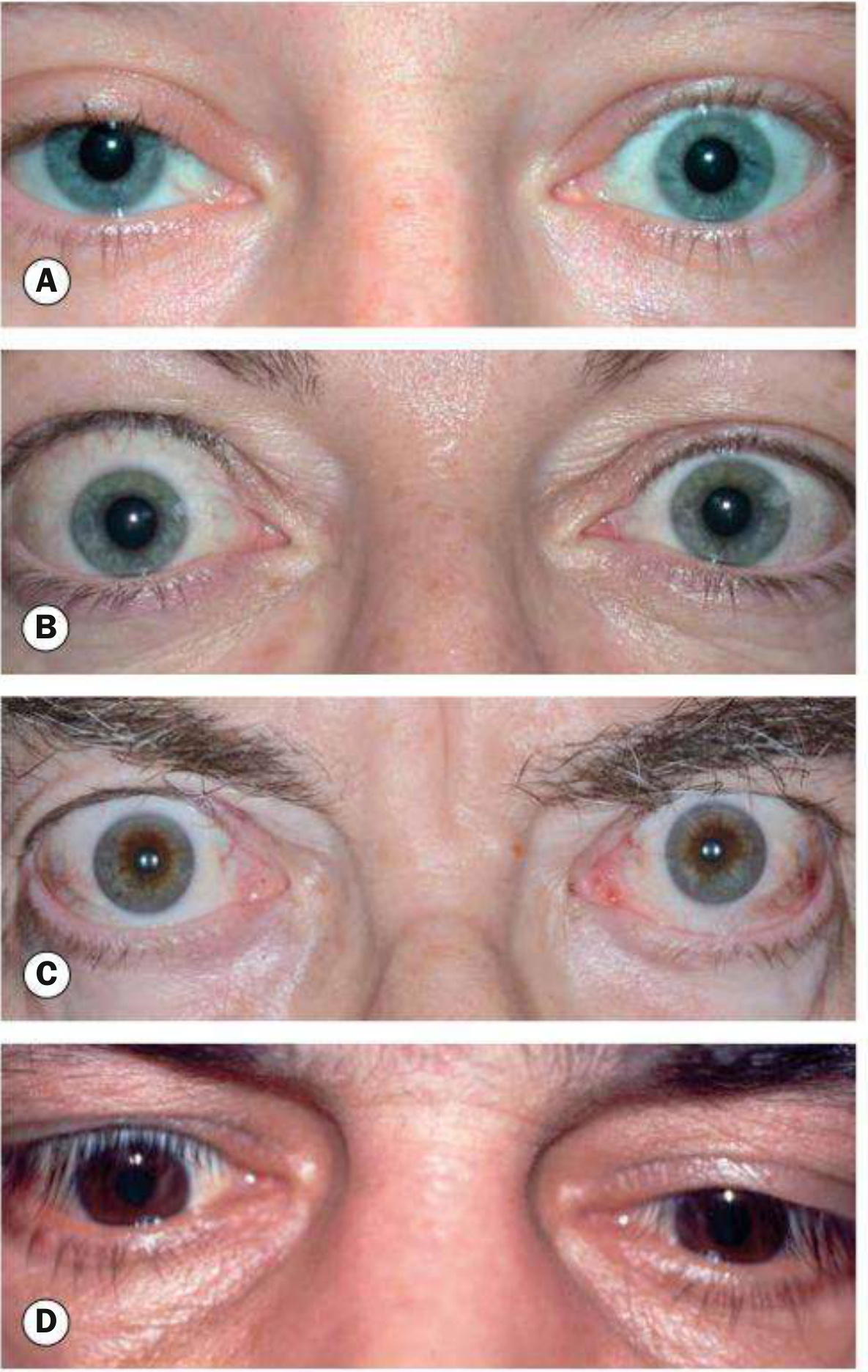
NO SPECS Classification (Harrison's 22e)
| Class | Letter | Features |
|---|---|---|
| 0 | No signs or symptoms | Normal |
| 1 | Only signs | Lid retraction or lag only; no symptoms |
| 2 | Soft tissue involvement | Periorbital oedema, chemosis, conjunctival injection |
| 3 | Proptosis | >22 mm on exophthalmometer |
| 4 | Extraocular muscle involvement | Diplopia; restrictive ophthalmoplegia |
| 5 | Corneal involvement | Exposure keratopathy, ulceration |
| 6 | Sight loss | Compressive optic neuropathy; visual field defects |
⚠️ Patients do not necessarily progress sequentially through classes - class 6 (optic neuropathy) can occur without severe proptosis. EUGOGO (European Group on Graves' Orbitopathy) scoring is now preferred in clinical practice as it assesses disease activity separately from severity.
Summary Table - All Named Eye Signs
| Eponym | Sign | Mechanism | Specific to Graves'? |
|---|---|---|---|
| Dalrymple's sign | Upper lid retraction in primary gaze; scleral show above | Sympathetic - levator overaction | No (all thyrotoxicosis) |
| Von Graefe's sign | Lid lag on downgaze | Sympathetic - levator fails to relax | No (all thyrotoxicosis) |
| Kocher's sign | Staring, frightened appearance on fixation | Sympathetic overactivity | No |
| Stellwag's sign | Infrequent blinking + wide fissure | Toxic levator contraction | No |
| Joffroy's sign | No forehead wrinkling on upward gaze | Frontalis muscle involvement | No |
| Möbius' sign | Failure of convergence | Extraocular muscle weakness | Partly infiltrative |
| Goldzeiher's sign | Conjunctival injection | Venous obstruction / inflammation | More specific |
| Exophthalmos | Axial proptosis >22 mm | Retro-orbital fat + muscle swelling | Yes - Graves' specific |
| Chemosis | Conjunctival oedema | Venous/lymphatic obstruction from raised orbital pressure | Yes |
| Restrictive ophthalmoplegia | Diplopia; most common: ↓ elevation | Inferior rectus fibrosis (most common) | Yes |
| Compressive optic neuropathy | Visual field loss, ↓ acuity, colour vision loss | Apex compression of optic nerve | Yes - most dangerous |
Clinical Course (Harrison's 22e)
- Typically worsens over 3-6 months, then plateaus 12-18 months, then partial improvement
- Thyroid dysfunction can worsen eye signs; but ophthalmopathy follows its own independent course from thyroid disease
- Can precede or follow thyrotoxicosis by years
- Radioiodine therapy (especially in smokers) can worsen ophthalmopathy - use steroids cover if radioiodine given in active disease
- Smoking is the single most important modifiable risk factor
Urgent Referral Criteria (Scott-Brown's)
- Visual acuity deteriorating
- Colour vision affected
- Corneal exposure / ulceration
- Papilloedema
- Severe periorbital oedema with inability to close eye
High-Yield Exam Points
- Graves' ophthalmopathy occurs in ~1/3 clinically; most have subclinical orbital changes on imaging
- 10% have ophthalmopathy without hyperthyroidism (euthyroid ophthalmopathy)
- Von Graefe, Dalrymple, Stellwag, Kocher = sympathomimetic (non-specific)
- Proptosis, chemosis, ophthalmoplegia = infiltrative (Graves' specific)
- Most serious complication = compressive optic neuropathy (→ permanent blindness)
- Most common motility deficit = inferior rectus fibrosis → defect in elevation (mimics SR palsy)
- NO SPECS: 0-1 (lid signs) → 2 (soft tissue) → 3 (proptosis) → 4 (EOM) → 5 (cornea) → 6 (sight)
- Smoking = biggest modifiable risk; radioiodine can worsen active disease
- Course of ophthalmopathy is independent of thyroid disease
- Thyroid dermopathy (pretibial myxoedema) almost always accompanies moderate-severe ophthalmopathy
Blood conservation strategies