
Tell me about overin cyst. Size and treatment in detail, research base
"ovarian cyst" AND management
ovarian cyst size treatment guidelines when to remove surgery
ovarian cyst ultrasound simple complex
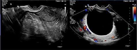
This diagnostic image consists of two side-by-side transvaginal ultrasound views of the pelvic region. The left panel shows the 'Rt Adnexa' with a heterogeneous, mixed echogenicity appearance, suggesting complex pathology such as a tubo-ovarian complex or hydrosalpinx. The right panel displays the 'Rt Ovary' using Color Doppler imaging, highlighting a large, unilocular, simple cyst. The cyst is characterized by its uniformly anechoic (black) internal fluid, well-defined smooth walls, and thin peripheral vascularization indicated by the red and blue Doppler signals. The comparison illustrates the visual difference between a simple ovarian cyst and complex adnexal structures. This material is suitable for medical education in obstetrics and gynecology, specifically for teaching sonographic identification of pelvic masses, ovarian cysts, and adnexal inflammation.
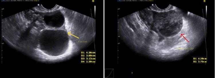
This diagnostic image consists of two side-by-side transvaginal ultrasound frames demonstrating ovarian pathology in a 27-year-old female. The left panel shows a simple ovarian cyst (indicated by a yellow arrow). It is characterized by a well-defined, thin-walled, anechoic (black) fluid-filled sac with acoustic enhancement posteriorly, representing a classic simple cyst morphology. The right panel demonstrates a complex ovarian cyst (indicated by a red arrow). This structure is characterized by a heterogeneous and disorganized internal architecture, containing low-level internal echoes and thickened, irregular borders, which contrast with the clear fluid of the simple cyst. Both images include linear measurement callouts in the bottom-right corners (e.g., D1, D2) for volumetric assessment. These findings are clinically significant in the differential diagnosis of adnexal masses, particularly in cases of suspected ectopic pregnancy or hemorrhagic cysts, where distinguishing between simple fluid and complex internal debris is critical for determining management pathways.
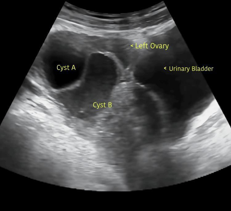
This diagnostic image is a transabdominal pelvic ultrasound demonstrating bilateral ovarian enlargement due to multicystic masses, specifically focused on the left ovary. Two distinct cystic structures are identified and labeled. Cyst A is a smaller, well-defined, simple anechoic cyst with smooth, thin borders and no internal septations or solid components. Cyst B is significantly larger, predominantly anechoic with some irregular internal echoes or artifacts, and appears less circumscribed than Cyst A. Anatomical landmarks labeled include the 'Left Ovary' and the 'Urinary Bladder' situated at the periphery. The ultrasound findings are consistent with ovarian hyperstimulation or multicystic ovaries, demonstrating significant volume increase in the adnexal structures. This visual is intended for educational use in obstetrics and gynecology to illustrate sonographic characteristics of simple versus complex ovarian cysts and pelvic anatomy under pathological conditions.
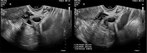
This diagnostic image consists of two side-by-side transvaginal pelvic ultrasound views of the right adnexa. The scan reveals a unilocular simple cyst within the right ovary. Visually, the cyst is characterized by a well-defined, thin, smooth wall and a purely anechoic (black) internal lumen, signifying clear fluid content without internal septations, papillary projections, or solid mural components. Acoustic enhancement is visible deep to the cyst, a typical finding for fluid-filled structures. The surrounding ovarian stroma and adjacent pelvic soft tissues exhibit heterogeneous, intermediate echogenicity. Measurement markers on the second frame assist in determining the volume and dimensions of the cyst. This image serves as a clinical example of a benign-appearing ovarian cyst, used in gynecological education to differentiate simple cysts from complex tubo-ovarian masses or hydrosalpinx.
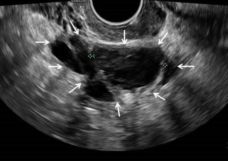
This diagnostic image is a transvaginal ultrasound scan of the female pelvis focusing on the right adnexa. It demonstrates a right ovarian cyst, approximately 3.5 cm in size, highlighted by multiple white arrows. The lesion presents as a complex cystic structure with mixed echogenicity. Key visual features include well-defined but irregular borders and a heterogeneous internal composition characterized by low-level internal echoes and thin, stringy internal septations. These fine, hyperechoic strands cross the primarily hypoechoic fluid-filled space, creating a multi-loculated appearance typical of complex cysts. In the provided clinical context, these findings are consistent with an evolving ovarian dermoid cyst (mature cystic teratoma) or a hemorrhagic cyst. The image serves as an educational example of pelvic sonography used to differentiate simple fluid-filled follicles from complex adnexal masses based on internal architecture and echotexture.
PMID: 36787526
Ovarian Cysts: Size Criteria and Treatment
What it is
Size thresholds guiding management
- < 1 cm: normal follicles
- 1-3 cm: dominant/functional follicles, physiological
- > 3 cm: termed a "cyst," warrants characterization (wall thickness, septations, solid components, Doppler flow)
| Size / feature | Typical management |
|---|---|
| Simple, unilocular, thin-walled, < 5 cm, asymptomatic, normal CA-125 | Reassurance; often no follow-up needed (very likely physiological) |
| Simple, unilocular, < 10 cm, normal CA-125 (any age) | Can be followed conservatively with serial ultrasound; malignancy risk is extremely low - Berek & Novak's Gynecology |
| Postmenopausal, predominantly cystic, up to 8 cm | May be observed or given a short course of oral contraceptives; repeat imaging in 2 menstrual cycles - Berek & Novak's Gynecology |
| Simple cyst > 4-5 cm | Generally considered for resection as spontaneous resolution is unlikely - Schwartz's Principles of Surgery |
| Persistent or symptomatic simple cyst 5-10 cm | Surgical evaluation favored, especially if symptomatic |
| Any complex cyst (septations, solid parts, papillary projections, ascites) regardless of size | Surgical evaluation warranted, work-up for malignancy |
| > 10 cm, or solid component, or abnormal Doppler/CA-125 | Higher malignancy suspicion; surgical staging capability should be available |
| Up to ~20 cm | Can usually still be removed laparoscopically |
| > 20 cm | Often requires open laparotomy for safe removal |
Treatment approach by scenario
- < 5-8 cm, asymptomatic: expectant management, repeat transvaginal ultrasound in 6-8 weeks (many are corpus luteum cysts that resolve within 1-3 cycles). Oral contraceptives do not shrink existing cysts but can reduce new functional cyst formation.
- Persistent, enlarging, symptomatic, or > 5-10 cm: laparoscopic cystectomy (ovary-sparing) preferred over oophorectomy when fertility is a priority.
- Simple, unilocular cysts up to 8-10 cm with normal CA-125: can be safely observed. A 2023 systematic review/meta-analysis of 1,672 postmenopausal women found simple cysts resolved spontaneously in ~34% and remained stable in ~39%; only ~19% went to surgery (usually by patient preference), and the malignancy rate was about 1 in 10,000 (PMID: 36787526). This supports conservative management for truly simple cysts even after menopause.
- Complex morphology, elevated CA-125, solid components, or rapid growth: warrants gynecologic oncology referral and surgical excision (usually oophorectomy given lower fertility priority and higher cancer suspicion in this group).
- Symptomatic or those affecting fertility are typically managed surgically (cystectomy). Medical therapy (GnRH agonists, danazol, progestins, aromatase inhibitors) can suppress growth/symptoms but does not eliminate the cyst - a 2024 meta-analysis found medical therapy reduces cyst size and pain but recurrence is common after stopping treatment (PMID: 37944155).
- Caution: laparoscopic electrocoagulation of the cyst bed to control bleeding after endometrioma cystectomy is associated with measurable damage to ovarian reserve (lower AMH) compared with suturing - a 2024 systematic review/meta-analysis (PMID: 39145875).
- Most regress spontaneously. A 2024 European Paediatric Surgeons' Association consensus statement recommends observation for simple cysts, with intervention (aspiration or laparoscopic surgery) reserved for large (typically > 4-5 cm), symptomatic, or complex/torsed cysts to preserve ovarian tissue (PMID: 37557903).
- Larger cysts (generally > 5 cm) carry higher torsion risk. Acute severe pain, peritoneal signs, or hemodynamic instability from rupture/hemorrhage requires urgent surgical evaluation, sometimes emergent laparoscopy.
- Intraoperative cyst spillage (e.g., dermoid, endometrioma) during cystectomy has a well-documented but generally low risk of chemical peritonitis or local recurrence, per a 2021 systematic review/meta-analysis (PMID: 33279627); careful surgical technique to avoid spillage is still recommended.
Surgical options
- Cystectomy (ovary-sparing removal of the cyst wall): preferred when fertility preservation matters and the cyst appears benign.
- Oophorectomy: considered for postmenopausal women, very large cysts, or when malignancy is suspected.
- Laparoscopy: first-line for most benign-appearing cysts, feasible for masses up to ~15-20 cm depending on surgeon experience and cyst characteristics.
- Laparotomy: reserved for very large cysts, suspected malignancy needing full staging, or when laparoscopic removal isn't safe.
Bottom line
35 mm in size unilateral adrenal
"adrenal incidentaloma" AND management
adrenal incidentaloma 35mm size cutoff surgery guidelines
PMID: 37318239
35 mm Unilateral Adrenal Mass (Adrenal Incidentaloma)
Step 1: Is it functional? (biochemical work-up - do this regardless of size)
- 1 mg overnight dexamethasone suppression test (cortisol > 1.8 µg/dL [>50 nmol/L] suggests autonomous cortisol secretion - now termed "mild autonomous cortisol secretion," MACS)
- Plasma or 24-hour urinary fractionated metanephrines (rule out pheochromocytoma)
- Plasma aldosterone/renin ratio if hypertensive and/or hypokalemic (rule out primary aldosteronism)
- Clinical screen for Cushingoid features, catecholamine excess symptoms (headache, palpitations, diaphoresis), and hypokalemia/refractory hypertension
Step 2: Malignancy risk by imaging + size
- Homogeneous and ≤10 Hounsfield units (HU) -> classic benign adenoma appearance -> no further imaging needed, regardless of size (this is a key change in the 2023 ESE guideline)
- If not clearly benign (HU >10, heterogeneous, irregular margins) -> needs further characterization (washout CT, MRI, or multidisciplinary discussion)
| Size | Approx. ACC risk | Typical recommendation |
|---|---|---|
| < 4 cm | ~2-3% | Observation if benign imaging features and non-functional |
| 4-6 cm | ~6-7% | Consider adrenalectomy if surgically fit |
| > 6 cm | ~25% | Adrenalectomy generally recommended |
Step 3: Decision
- Non-functional + clearly benign imaging (homogeneous, ≤10 HU): no surgery, no further imaging needed at all - this is a genuinely benign adenoma.
- Non-functional + indeterminate imaging: multidisciplinary discussion; options are additional imaging (washout CT or MRI), interval follow-up imaging at 6-12 months, or (individualized) surgery. A significant interval growth (ESE: >20% and ≥5 mm; older AACE/AAES criteria: ≥0.5-1 cm) raises suspicion and favors resection.
- Functional (even mild/subclinical cortisol excess with comorbidities like hypertension or type 2 diabetes): surgery should be considered on an individualized basis even without malignant features.
- Suspicious features regardless of size (irregular margins, rapid growth, necrosis, calcification, local invasion): resection.
If surgery is chosen
Bottom line for a 35 mm unilateral adrenal incidentaloma
- Rule out hormone excess (cortisol, metanephrines, aldosterone/renin if hypertensive/hypokalemic).
- Characterize on unenhanced CT: if homogeneous and ≤10 HU, this is a benign adenoma - no surgery, no further imaging.
- If indeterminate imaging or any functionality, individualize between interval imaging (6-12 months) and laparoscopic adrenalectomy - size alone (3.5 cm) is not an automatic indication for surgery, since it's below the ~4-6 cm thresholds most guidelines use to trigger resection based on size.
Fluid Field
What are you asking about with 'Fluid Field'?
Ovarian Cysts: Size Criteria and Treatment
What it is
Size thresholds guiding management
- < 1 cm: normal follicles
- 1-3 cm: dominant/functional follicles, physiological
- > 3 cm: termed a "cyst," warrants characterization (wall thickness, septations, solid components, Doppler flow)
| Size / feature | Typical management |
|---|---|
| Simple, unilocular, thin-walled, < 5 cm, asymptomatic, normal CA-125 | Reassurance; often no follow-up needed (very likely physiological) |
| Simple, unilocular, < 10 cm, normal CA-125 (any age) | Can be followed conservatively with serial ultrasound; malignancy risk is extremely low - Berek & Novak's Gynecology |
| Postmenopausal, predominantly cystic, up to 8 cm | May be observed or given a short course of oral contraceptives; repeat imaging in 2 menstrual cycles - Berek & Novak's Gynecology |
| Simple cyst > 4-5 cm | Generally considered for resection as spontaneous resolution is unlikely - Schwartz's Principles of Surgery |
| Persistent or symptomatic simple cyst 5-10 cm | Surgical evaluation favored, especially if symptomatic |
| Any complex cyst (septations, solid parts, papillary projections, ascites) regardless of size | Surgical evaluation warranted, work-up for malignancy |
| > 10 cm, or solid component, or abnormal Doppler/CA-125 | Higher malignancy suspicion; surgical staging capability should be available |
| Up to ~20 cm | Can usually still be removed laparoscopically |
| > 20 cm | Often requires open laparotomy for safe removal |
Treatment approach by scenario
- < 5-8 cm, asymptomatic: expectant management, repeat transvaginal ultrasound in 6-8 weeks (many are corpus luteum cysts that resolve within 1-3 cycles). Oral contraceptives do not shrink existing cysts but can reduce new functional cyst formation.
- Persistent, enlarging, symptomatic, or > 5-10 cm: laparoscopic cystectomy (ovary-sparing) preferred over oophorectomy when fertility is a priority.
- Simple, unilocular cysts up to 8-10 cm with normal CA-125: can be safely observed. A 2023 systematic review/meta-analysis of 1,672 postmenopausal women found simple cysts resolved spontaneously in ~34% and remained stable in ~39%; only ~19% went to surgery (usually by patient preference), and the malignancy rate was about 1 in 10,000 (PMID: 36787526). This supports conservative management for truly simple cysts even after menopause.
- Complex morphology, elevated CA-125, solid components, or rapid growth: warrants gynecologic oncology referral and surgical excision (usually oophorectomy given lower fertility priority and higher cancer suspicion in this group).
- Symptomatic or those affecting fertility are typically managed surgically (cystectomy). Medical therapy (GnRH agonists, danazol, progestins, aromatase inhibitors) can suppress growth/symptoms but does not eliminate the cyst - a 2024 meta-analysis found medical therapy reduces cyst size and pain but recurrence is common after stopping treatment (PMID: 37944155).
- Caution: laparoscopic electrocoagulation of the cyst bed to control bleeding after endometrioma cystectomy is associated with measurable damage to ovarian reserve (lower AMH) compared with suturing - a 2024 systematic review/meta-analysis (PMID: 39145875).
- Most regress spontaneously. A 2024 European Paediatric Surgeons' Association consensus statement recommends observation for simple cysts, with intervention (aspiration or laparoscopic surgery) reserved for large (typically > 4-5 cm), symptomatic, or complex/torsed cysts to preserve ovarian tissue (PMID: 37557903).
- Larger cysts (generally > 5 cm) carry higher torsion risk. Acute severe pain, peritoneal signs, or hemodynamic instability from rupture/hemorrhage requires urgent surgical evaluation, sometimes emergent laparoscopy.
- Intraoperative cyst spillage (e.g., dermoid, endometrioma) during cystectomy has a well-documented but generally low risk of chemical peritonitis or local recurrence, per a 2021 systematic review/meta-analysis (PMID: 33279627); careful surgical technique to avoid spillage is still recommended.
Surgical options
- Cystectomy (ovary-sparing removal of the cyst wall): preferred when fertility preservation matters and the cyst appears benign.
- Oophorectomy: considered for postmenopausal women, very large cysts, or when malignancy is suspected.
- Laparoscopy: first-line for most benign-appearing cysts, feasible for masses up to ~15-20 cm depending on surgeon experience and cyst characteristics.
- Laparotomy: reserved for very large cysts, suspected malignancy needing full staging, or when laparoscopic removal isn't safe.
Bottom line
Step 1: Is it functional? (biochemical work-up - do this regardless of size)
- 1 mg overnight dexamethasone suppression test (cortisol > 1.8 µg/dL [>50 nmol/L] suggests autonomous cortisol secretion - now termed "mild autonomous cortisol secretion," MACS)
- Plasma or 24-hour urinary fractionated metanephrines (rule out pheochromocytoma)
- Plasma aldosterone/renin ratio if hypertensive and/or hypokalemic (rule out primary aldosteronism)
- Clinical screen for Cushingoid features, catecholamine excess symptoms (headache, palpitations, diaphoresis), and hypokalemia/refractory hypertension
Step 2: Malignancy risk by imaging + size
- Homogeneous and ≤10 Hounsfield units (HU) -> classic benign adenoma appearance -> no further imaging needed, regardless of size (this is a key change in the 2023 ESE guideline)
- If not clearly benign (HU >10, heterogeneous, irregular margins) -> needs further characterization (washout CT, MRI, or multidisciplinary discussion)
| Size | Approx. ACC risk | Typical recommendation |
|---|---|---|
| < 4 cm | ~2-3% | Observation if benign imaging features and non-functional |
| 4-6 cm | ~6-7% | Consider adrenalectomy if surgically fit |
| > 6 cm | ~25% | Adrenalectomy generally recommended |
Step 3: Decision
- Non-functional + clearly benign imaging (homogeneous, ≤10 HU): no surgery, no further imaging needed at all - this is a genuinely benign adenoma.
- Non-functional + indeterminate imaging: multidisciplinary discussion; options are additional imaging (washout CT or MRI), interval follow-up imaging at 6-12 months, or (individualized) surgery. A significant interval growth (ESE: >20% and ≥5 mm; older AACE/AAES criteria: ≥0.5-1 cm) raises suspicion and favors resection.
- Functional (even mild/subclinical cortisol excess with comorbidities like hypertension or type 2 diabetes): surgery should be considered on an individualized basis even without malignant features.
- Suspicious features regardless of size (irregular margins, rapid growth, necrosis, calcification, local invasion): resection.
If surgery is chosen
Bottom line for a 35 mm unilateral adrenal incidentaloma
- Rule out hormone excess (cortisol, metanephrines, aldosterone/renin if hypertensive/hypokalemic).
- Characterize on unenhanced CT: if homogeneous and ≤10 HU, this is a benign adenoma - no surgery, no further imaging.
- If indeterminate imaging or any functionality, individualize between interval imaging (6-12 months) and laparoscopic adrenalectomy - size alone (3.5 cm) is not an automatic indication for surgery, since it's below the ~4-6 cm thresholds most guidelines use to trigger resection based on size.
- A fluid-filled cyst/lesion on imaging (e.g., ovarian, adrenal, or another site)
- The field of view in an ultrasound or CT scan showing a fluid collection
- Something else entirely (a different term or topic)