
Ileitis causes
ileitis etiology causes
terminal ileitis endoscopy Crohn's disease
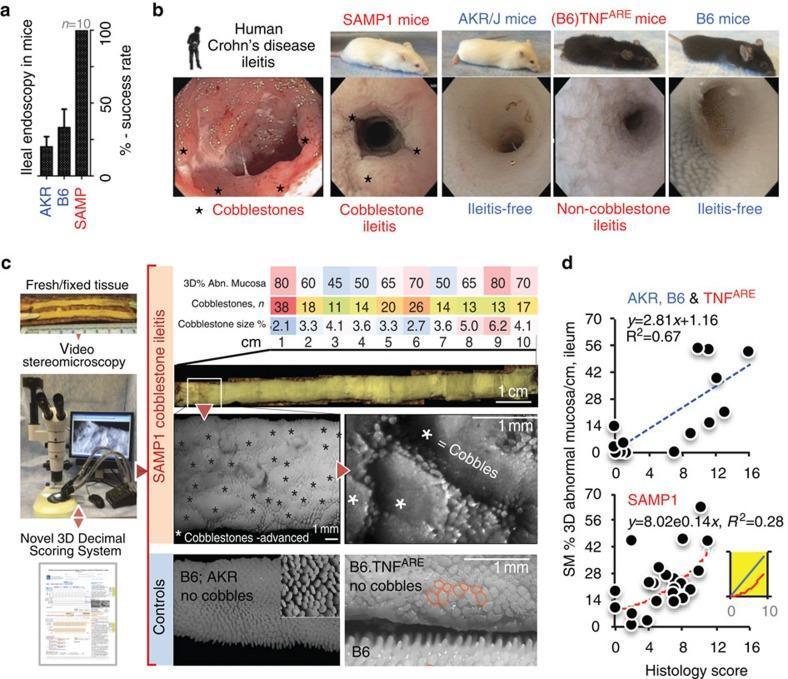
Educational panel illustrating murine models of Crohn’s disease-like ileitis through endoscopy and 3D stereomicroscopy (SM). Panel (a) compares ileal endoscopy success rates across AKR, B6, and SAMP mouse strains. Panel (b) provides comparative endoscopic views of the terminal ileum. Human Crohn’s disease and SAMP1 mice both display characteristic 'cobblestone' ileitis with raised, nodular mucosal lesions. AKR/J and B6 mice serve as healthy, ileitis-free controls, while (B6)TNFARE mice exhibit a distinct non-cobblestone inflammatory pattern. Panel (c) details a novel 3D-SMAPgut assessment protocol for topographical mucosal analysis. It includes high-resolution stereomicroscopy images showing advanced cobblestone lesions in SAMP1 ileum versus regular villous architecture in B6 and AKR controls. B6.TNFARE mice show abnormal mucosal architecture without cobblestone formation. Quantitative data tables track the percentage of abnormal mucosa and lesion counts per centimeter. Panel (d) features scatter plots correlating 3D-SM abnormal mucosa percentages with traditional histological scores. AKR, B6, and TNFARE show a linear correlation (R²=0.67), whereas SAMP1 exhibits an exponential relationship (R²=0.28), indicating the superior sensitivity of 3D-SM in capturing complex structural disease progression compared to 2D histology.
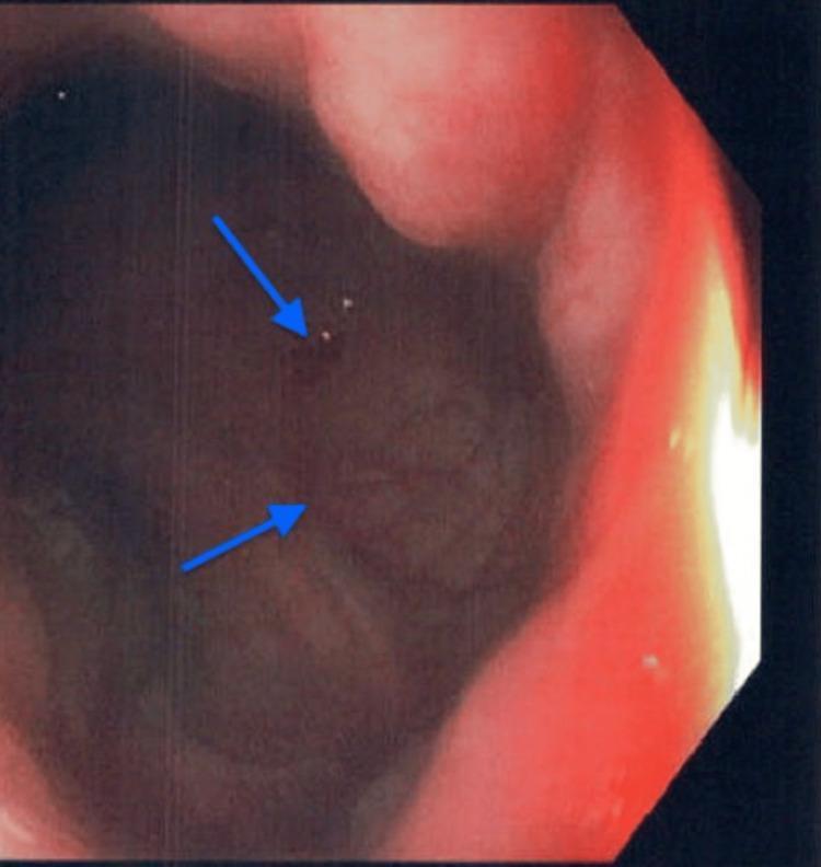
This diagnostic endoscopic image captures the terminal ileum during a colonoscopy, demonstrating active terminal ileitis. The visual field shows a dark red, erythematous mucosal lining with significant congestion. Key pathological features include uneven mucosal texture and patchy distribution of surface ulcerations, indicated by blue arrows pointing to disruptions in the tissue and small bright spots of reflecting light. The image illustrates signs of acute inflammation within the distal small bowel, a hallmark finding in conditions such as Crohn's disease or drug-induced enteropathy. The surrounding luminal walls appear thickened and edematous, while the loss of the typical vascular pattern suggests underlying mucosal damage and inflammation. This educational visual serves as a representative example of inflammatory bowel disease (IBD) manifestations during lower gastrointestinal endoscopy.
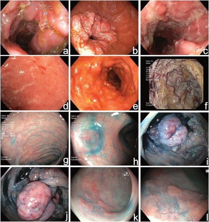
This composite of twelve endoscopic images (a-l) demonstrates high-resolution video endoscopy findings in Inflammatory Bowel Disease (IBD). Panels a-f utilize standard white-light endoscopy for initial diagnosis, showcasing various disease manifestations: (a) acute terminal ileitis in Crohn's disease with mucosal edema; (b) duodenal Crohn's stenosis with significant luminal narrowing; (c) segmental fissural ulcerations in the colon; and (d-f) progressive severity of ulcerative colitis (UC) ranging from mild erythema (UCEIS 3) to severe mucosal architectural distortion and deep ulceration (UCEIS 8). Panels g-l demonstrate surveillance colonoscopy using chromoendoscopy with 0.1% indigocarmine dye. The dye highlights surface topography: (g) normal mucosal pit patterns; (h) a small flat hyperplastic polyp; (i-j) elevated pseudopolyps; and (k-l) a flat, inhomogeneous polypoid lesion. Frame (l) employs near-focus imaging to reveal subtle mucosal irregularities characteristic of high-grade intraepithelial neoplasia. This comparison illustrates the utility of chromoendoscopy in enhancing contrast for better detection of dysplastic lesions during IBD surveillance compared to conventional white-light imaging.
Causes of Ileitis
1. Inflammatory Bowel Disease (most common)
- Crohn's disease - the ileum (especially terminal ileum) is involved in the majority of cases. Tintinalli's Emergency Medicine notes that the terms "regional enteritis," "terminal ileitis," and "granulomatous ileocolitis" all describe the same process. It is a transmural, discontinuous, granulomatous inflammation with skip lesions.
- Backwash ileitis in Ulcerative Colitis - retrograde inflammation of the terminal ileum due to reflux through an incompetent ileocecal valve in pancolitis.
2. Infectious Causes
- Yersinia enterocolitica and Y. pseudotuberculosis - the classic infectious mimic of Crohn's; causes fever, gastroenteritis, and terminal ileitis that can even perforate
- Mycobacterium tuberculosis - intestinal TB commonly affects the ileocecal region
- Mycobacterium avium complex (MAC) - in immunocompromised patients
- Salmonella spp.
- Campylobacter jejuni
- Aeromonas
- Shigella
- Enterohemorrhagic E. coli
- Clostridioides difficile
- Actinomyces israelii
- Cytomegalovirus (CMV) - especially in immunosuppressed patients
- HIV
- Histoplasma capsulatum
- Cryptococcus neoformans
- Entamoeba histolytica
- Anisakis simplex (from undercooked fish)
- Helminths
3. Drug-Related
- NSAIDs - cause ileal ulcers, strictures, and a "diaphragm disease" pattern; a well-recognized cause of non-specific ileitis
- Immune checkpoint inhibitors
- Medications causing neutropenic enterocolitis (chemotherapy)
4. Vascular Disorders
- Intestinal ischemia (focal segmental ischemia - acute or chronic enteritis, stricture; chronic mesenteric ischemia)
- Drug-induced ischemic ileitis: amphetamines, ergotamine, cocaine, phenylephrine
- Vasculitis: Henoch-Schonlein purpura (IgA vasculitis), polyarteritis nodosa, eosinophilic granulomatosis with polyangiitis (Churg-Strauss), SLE, Takayasu arteritis, granulomatosis with polyangiitis (Wegener's), giant cell arteritis, rheumatoid vasculitis, thromboangiitis obliterans, lymphomatoid granulomatosis
- Behcet disease - well-recognized cause of ileitis and/or colitis
5. Infiltrative Disorders
- Eosinophilic gastroenteritis - eosinophilic infiltration of the ileal wall
- Amyloidosis - ileal deposition
- Lymphoid nodular hyperplasia
6. Neoplasms (masquerading as ileitis)
- Carcinoid tumor (ileum is the most common GI site)
- Cecal or ileal adenocarcinoma
- Lymphoma
- Metastatic cancer
7. Radiation Enteritis
8. Spondyloarthropathy-Associated Ileitis
- Subclinical gut inflammation is found in a significant proportion of patients with ankylosing spondylitis and reactive arthritis; overt ileitis can be a manifestation.
9. Gynecologic / Adjacent Organ Disorders
- Endometriosis involving the ileum
- Ectopic pregnancy
- Ovarian cyst or tumor
- Ovarian torsion
- Pelvic inflammatory disease (PID)
- Tubo-ovarian abscess
- Appendicitis / appendiceal abscess
- Cecal diverticulitis
- Torsion of the appendiceal epiploica
Summary Table
| Category | Key Examples |
|---|---|
| IBD | Crohn's disease, backwash ileitis (UC) |
| Infection | Yersinia, TB, CMV, Salmonella, MAC |
| Drug-induced | NSAIDs, checkpoint inhibitors |
| Vascular | Ischemia, vasculitis, Behcet |
| Infiltrative | Eosinophilic gastroenteritis, amyloid |
| Neoplasm | Lymphoma, carcinoid, adenocarcinoma |
| Radiation | Radiation enteritis |
| Rheumatologic | Spondyloarthropathy |
| Gynecologic/adjacent | Endometriosis, appendicitis, PID |