
cholelithiasis and cancer of gallbladder
cholelithiasis AND gallbladder carcinoma
NCI gallbladder cancer treatment PDQ cholelithiasis gallstones guidance
https://www.cancer.gov/types/gallbladder/hp/gallbladder-trea…
ultrasonography gallstones cholelithiasis acoustic shadowing gallbladder
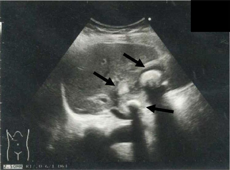
This abdominal ultrasonography image demonstrates the gallbladder in a patient with cholelithiasis. The primary findings consist of multiple hyperechoic structures (indicated by black arrows) within the gallbladder lumen, consistent with cholelithiasis (gallstones). These stones exhibit high echogenicity and are accompanied by prominent posterior acoustic shadowing, a hallmark diagnostic feature of dense calcified structures on ultrasound. The gallbladder wall appears thickened, suggesting underlying inflammation or cholecystitis. The surrounding liver parenchyma shows a relatively homogeneous echotexture, providing a clear acoustic window for the gallbladder. This diagnostic image is used to assess biliary pathology and plan clinical interventions such as percutaneous transhepatic gallbladder drainage (PTGBD). The orientation is shown via the body habitus icon in the bottom-left corner, indicating a right upper quadrant scan. This visual serves as a primary tool for medical students and clinicians to identify the classic sonographic presentation of gallstones and associated pericholecystic changes.
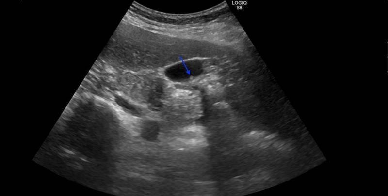
This diagnostic image is an abdominal ultrasound in a transverse or oblique plane focusing on the gallbladder. The gallbladder appears as a well-defined, anechoic (black) fluid-filled structure. A blue arrow identifies a small, hyperechoic, non-shadowing intraluminal lesion measuring approximately 7 mm, consistent with a gallbladder polyp. The polyp is attached to the dependent wall of the gallbladder and does not demonstrate posterior acoustic shadowing, which helps differentiate it from cholelithiasis (gallstones). The surrounding hepatic parenchyma exhibits a homogeneous, medium-level echogenicity. The gallbladder wall appears slightly thickened, suggesting possible chronic cholecystitis. This image is used to demonstrate the sonographic appearance of gallbladder polyps, which in this clinical context was pathologically confirmed as heterotopic pancreatic tissue. The educational focus is on the detection and characterization of intraluminal gallbladder pathologies using ultrasonography.
gallbladder carcinoma CT imaging mass gallbladder
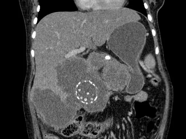
Contrast-enhanced computed tomography (CT) of the abdomen, analyzed in this coronal view, demonstrates porcelain gallbladder with calcifications in the gallbladder wall and multiple gallstones. An irregular, bulky soft-tissue mass arises from the gallbladder fossa and penetrates the adjacent hepatic parenchyma, creating an infiltrative interface with liver tissue. The mass shows heterogenous attenuation with possible focal necrosis and associated pericholecystic inflammation. There is evidence of hepatic parenchymal infiltration by the tumor, consistent with locally advanced gallbladder carcinoma. Biliary dilation is not prominently featured in this single view, but the extent of hepatic invasion indicates advanced disease. The imaging pattern - gallbladder wall calcification (porcelain gallbladder), gallstones, and an irregular mass with liver invasion - supports a diagnosis of gallbladder carcinoma arising in porcelain gallbladder. Differential considerations include chronic cholecystitis with calcification and less likely cholangiocarcinoma or metastatic disease; however, location and invasion favor primary gallbladder malignancy. Clinically, the patient presents with an abdominal mass and diffuse abdominal pain, aligning with oncologic etiology. Management implications include accurate staging to determine resectability; when feasible, radical cholecystectomy with partial hepatectomy may be curative, whereas unresectable disease requires systemic therapy and palliative care. This case highlights porcelain gallbladder as a potential malignant precursor and the necessity of surgical consultation for optimal treatment planning.
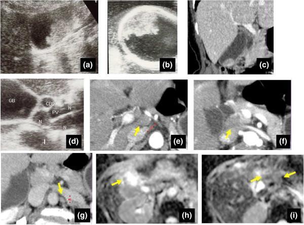
Multi-modal diagnostic imaging of a gallbladder mass and associated lymphadenopathy. (a-b) Ultrasonography (US) images reveal a highly echoic, sessile mass measuring approximately 30 mm in the gallbladder fundus. Intraoperative US (b) demonstrates preservation of the hyperechoic outer layer, suggesting a 'Type B' tumor depth limited to the muscularis propria. (c) Sagittal CT reconstruction confirms the intraluminal gallbladder mass without gross invasion into the hepatic parenchyma. (d-g) Abdominal US and axial CT scans identify multiple swollen lymph nodes (labeled 'N' or indicated by arrows) located within the hepatoduodenal ligament and para-aortic regions. (h-i) Diffusion-Weighted Imaging (DWI) highlights a significant signal intensity discordance: the gallbladder mass exhibits marked hyperintensity, while the enlarged lymph nodes appear hypointense. These images illustrate the diagnostic workflow for staging gallbladder carcinoma and differentiating between metastatic involvement and benign lymphadenopathy (e.g., sarcoid-like reactions) using signal intensity patterns on MRI.
PMID: 39370088
Cholelithiasis and cancer of the gallbladder
1. Cholelithiasis
- Female sex, increasing age, obesity, pregnancy, family history
- Rapid weight loss, bariatric surgery, prolonged fasting or parenteral nutrition
- Cirrhosis
- Hemolytic disorders, for example sickle-cell disease or hereditary spherocytosis, causing black pigment stones
- Biliary infection or parasites, associated with brown pigment stones
- Many patients are asymptomatic and stones are detected incidentally.
- Biliary colic: episodic steady right-upper-quadrant or epigastric pain, often after a fatty meal, sometimes radiating to the right shoulder or back, with nausea/vomiting.
- In uncomplicated biliary colic, the patient is usually afebrile and laboratory tests may be normal.
- Acute or chronic cholecystitis
- Choledocholithiasis and obstructive jaundice
- Ascending cholangitis
- Acute biliary pancreatitis
- Mirizzi syndrome
- Gallstone ileus
- Increased association with gallbladder carcinoma
- Right-upper-quadrant ultrasound is first line. Stones are mobile echogenic foci with posterior acoustic shadowing. It also assesses gallbladder-wall thickening, pericholecystic fluid, and bile-duct dilatation.
- Liver tests and bilirubin help assess possible common-bile-duct obstruction.
- MRCP, EUS, ERCP, CT, or a HIDA scan are selected based on whether duct stones, acute cholecystitis, or an alternative diagnosis is suspected.
- Asymptomatic gallstones: usually reassurance and observation, not prophylactic surgery.
- Symptomatic biliary colic: elective laparoscopic cholecystectomy is the usual definitive treatment.
- Acute cholecystitis: analgesia, fluids, antibiotics when indicated, and early laparoscopic cholecystectomy when appropriate.
- Common-bile-duct stone/cholangitis: endoscopic clearance or drainage, commonly with ERCP, followed by cholecystectomy where suitable.
2. Gallbladder cancer
Relationship with gallstones
- Large gallstones, especially >3 cm, and long duration of stone disease
- Gallbladder polyps, particularly ≥10 mm, sessile lesions, or enlarging polyps
- Porcelain gallbladder, especially incomplete or selective mucosal calcification
- Chronic Salmonella carriage and certain biliary infections
- Choledochal cysts, abnormal pancreaticobiliary duct junction, biliary cysts
- Obesity and smoking
- Higher-incidence geographic/ethnic populations
Presentation
- Persistent right-upper-quadrant pain
- Anorexia, weight loss, fatigue
- Jaundice and pruritus from biliary obstruction
- Palpable mass, hepatomegaly, ascites
- Fever or recurrent cholecystitis-like episodes
Diagnosis and staging
- Ultrasound: may show irregular wall thickening, a sessile mass, a large polyp, calcification, or liver invasion.
- Contrast CT and MRI/MRCP: assess local hepatic invasion, nodes, vascular/biliary involvement, and metastases.
- Diagnosis is confirmed histologically, often from the cholecystectomy specimen or through targeted biopsy when disease is unresectable.
Harrison’s Principles of Internal Medicine, 22nd ed., p. 713.
Treatment principles
| Disease setting | Main approach |
|---|---|
| T1a disease incidentally found after cholecystectomy | Simple cholecystectomy may be adequate |
| T1b or T2 and selected resectable tumors | Re-resection or radical cholecystectomy, often including liver segments IVb/V wedge resection and regional lymph-node dissection |
| Resected disease | Consider adjuvant capecitabine, guided by oncology/pathology review |
| Unresectable, recurrent, or metastatic disease | Systemic therapy, often gemcitabine-cisplatin plus durvalumab for fit patients; biliary drainage and symptom-directed care as needed |
Key distinction
- Gallstones are common and usually harmless if asymptomatic.
- Gallbladder cancer is rare, although gallstones are found in many affected patients.
- Prophylactic cholecystectomy is not automatically needed for every patient with asymptomatic stones. It is considered when symptoms, complications, or high-risk features are present.
Relationship between both conditions
cholelithiasis AND gallbladder cancer
NCI gallbladder cancer cholelithiasis less than 1 percent patients gallstones
Relationship
-
Gallstones are the most important risk factor
- Gallstones are present in roughly 65% to 90% of people diagnosed with gallbladder carcinoma.
- Populations with a high prevalence of cholelithiasis also tend to have higher rates of gallbladder cancer.
Sleisenger and Fordtran's Gastrointestinal and Liver Disease, Etiology section.
-
Proposed mechanism: chronic inflammation
- Recurrent mechanical irritation from stones, biliary stasis, and repeated episodes of chronic cholecystitis can cause persistent mucosal inflammation.
- Over years, inflammation promotes epithelial injury, regeneration, dysplasia, and potentially carcinoma.
- Large stones, particularly >3 cm, and a long duration of stone exposure are associated with higher risk.
-
Association is not the same as inevitable progression
- Gallstones are common, while gallbladder cancer is rare.
- Only about 0.5% to 3% of people with cholelithiasis develop gallbladder cancer.
- Conversely, about 10% to 35% of gallbladder cancer cases occur without demonstrable gallstones.
Sleisenger and Fordtran's Gastrointestinal and Liver Disease, Etiology section.
Maingot's Abdominal Operations, Epidemiology section.
-
Shared high-risk settings The cancer risk is greater when gallstones coexist with:
- Large or longstanding stones
- Porcelain gallbladder, especially selective mucosal calcification
- Gallbladder polyp ≥10 mm, sessile or growing polyp
- Chronic Salmonella carriage or biliary infection
- Choledochal cyst or anomalous pancreaticobiliary duct junction
- Obesity and certain high-incidence geographic regions
Clinical implication
Gallstones increase the risk of gallbladder carcinoma mainly through long-term chronic inflammation, but only a small minority of patients with gallstones ever develop cancer.