I now have all the content needed from authoritative textbooks. Let me compile the complete answers.Here are all 6 Tier 2 Short Essays written as complete, exam-ready 5-mark answers for RGUHS Karnataka:
TIER 2 SHORT ESSAYS - Complete 5-Mark Answers
Genetics & Molecular Biology | RGUHS Karnataka MBBS 1st Year
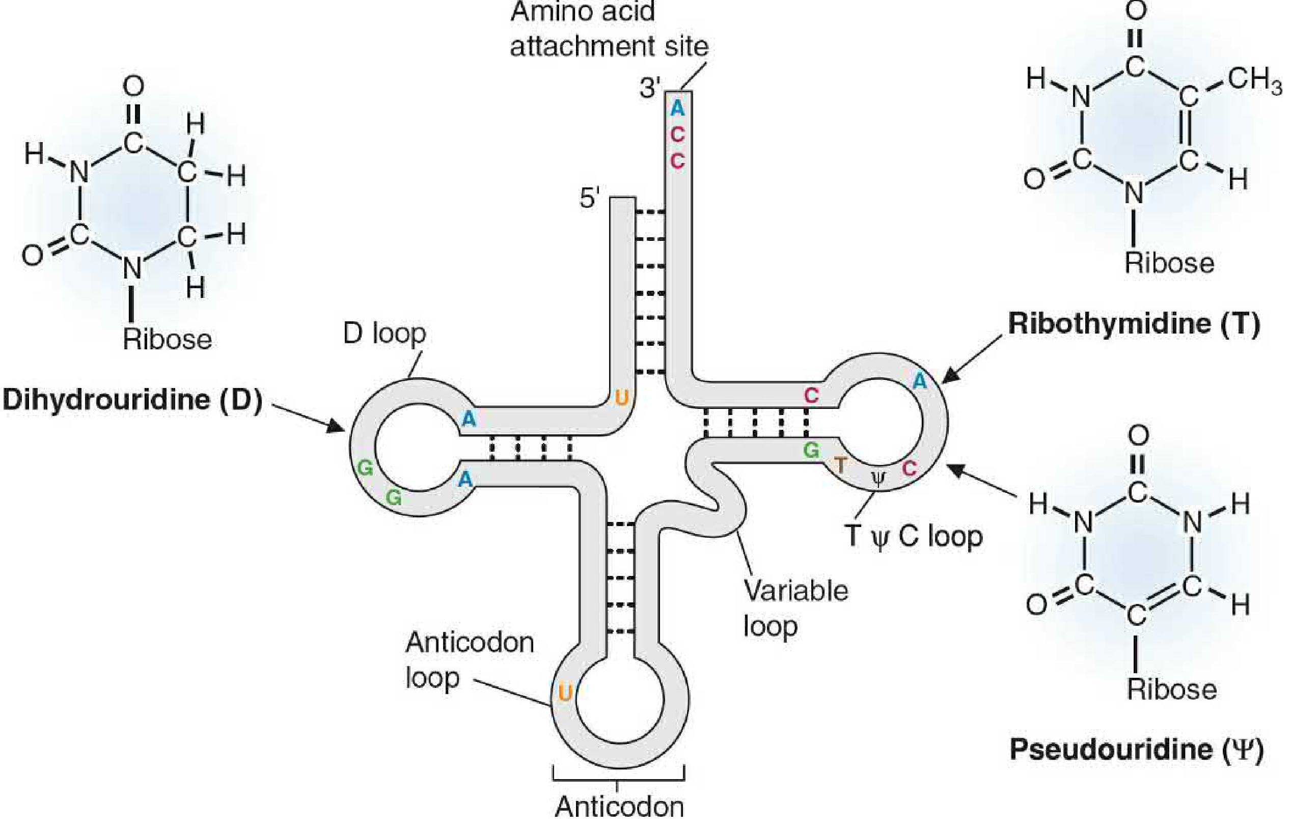
1. Genetic Code and Wobble Hypothesis
Definition
The genetic code is the set of rules by which the nucleotide sequence of mRNA is read in triplets (codons) and translated into the amino acid sequence of a protein.
Features of Genetic Code (9 Features - Must Know All)
| Feature | Meaning | Example |
|---|
| 1. Triplet | Each codon = 3 nucleotides | AUG, GGU, UAA |
| 2. Non-overlapping | Each nucleotide belongs to only one codon | AUGCAU is read AUG-CAU, not AUG-UGC-GCA |
| 3. Comma-free | No punctuation marks between codons | Read continuously without gaps |
| 4. Degenerate (redundant) | Most amino acids have MORE than one codon | Leucine has 6 codons; Serine has 6 |
| 5. Unambiguous | Each codon specifies only ONE amino acid | AUG = Met only (never anything else) |
| 6. Universal | Same in almost all organisms | AUG = Met in bacteria, humans, plants |
| 7. Ordered | Related codons code for related amino acids | Reduces impact of mutation |
| 8. Initiator codon | AUG = start codon (codes for Met / fMet) | Signals start of translation |
| 9. Terminator codons | UAA, UAG, UGA = stop codons | Do NOT code for any amino acid |
Note for exam: There are 64 total codons (4³). 61 code for amino acids, 3 are stop codons.
Wobble Hypothesis (Crick, 1966)
Problem it solves: If 61 codons exist but most amino acids have multiple codons, why don't cells need 61 different tRNAs?
Explanation:
- Strict base pairing (A-U, G-C) applies only at the 1st and 2nd positions of the codon
- At the 3rd position (called the "wobble position"), the base pairing rules are relaxed
- A single tRNA anticodon can base-pair with more than one codon by wobbling at the 3rd codon position
Wobble base pairing rules:
| Anticodon 5'-base (wobble position) | Can pair with codon 3'-base |
|---|
| C | G only |
| A | U only |
| U | A or G |
| G | C or U |
| Inosine (I) | A, C, or U (most versatile) |
Example: The tRNA with anticodon 5'-IGC-3' can recognize three alanine codons: GCU, GCC, and GCA - because inosine (I) at the wobble position pairs with U, C, or A.
Result: Fewer than 61 tRNAs are needed to read all 61 codons. Humans have approximately 45 different tRNA molecules.
Exception: Mitochondria use a slightly different code:
- UGA codes for Tryptophan (not stop)
- AUA codes for Methionine (not Isoleucine)
Source: Basic Medical Biochemistry - A Clinical Approach, 6e, p. 475
2. PCR - Polymerase Chain Reaction
Definition
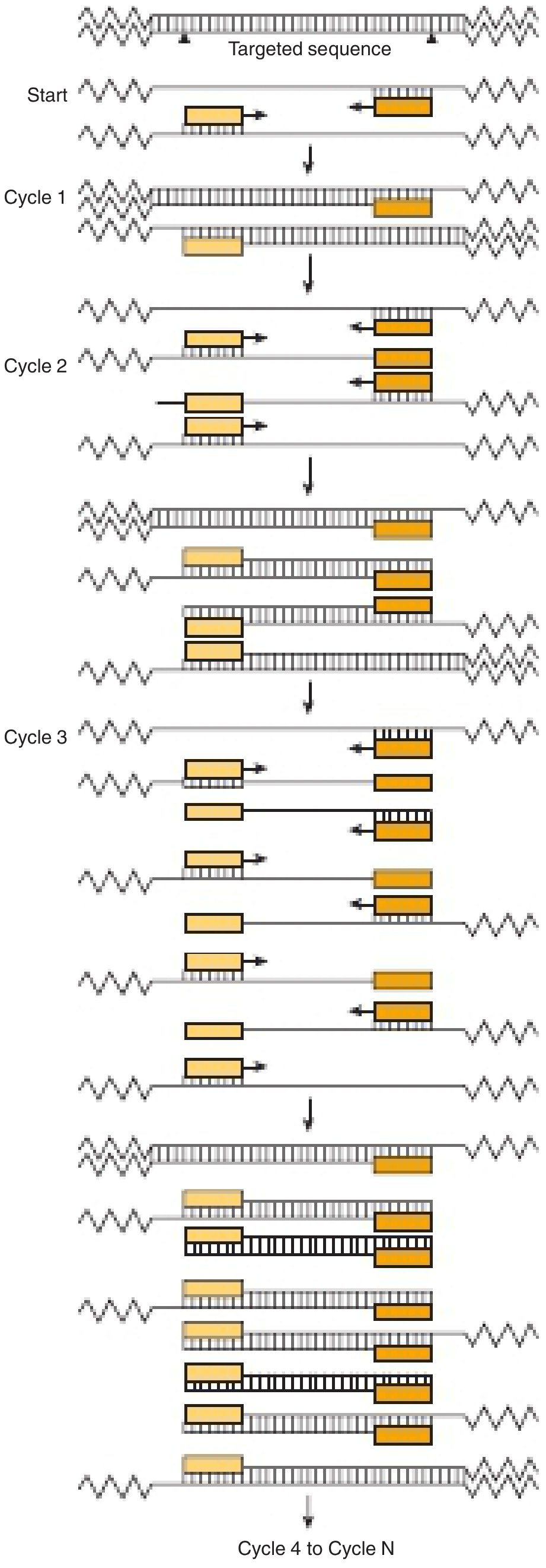
PCR is an in vitro method of exponentially amplifying a specific target DNA sequence from a minute amount of starting material using repeated cycles of denaturation, annealing, and extension.
Components Required
- Template DNA - DNA containing the target sequence
- Two primers - short oligonucleotides (~18-25 bp), one complementary to each strand, flanking the target
- Taq DNA polymerase - heat-stable DNA polymerase from Thermus aquaticus (withstands 94°C)
- dNTPs (dATP, dTTP, dCTP, dGTP) - building blocks
- MgCl₂ - cofactor for Taq polymerase
- Buffer - maintains optimal pH
- Thermocycler - machine that automatically cycles temperatures
Three Steps of Each PCR Cycle
| Step | Temperature | Duration | What Happens |
|---|
| 1. Denaturation | 94-96°C | 30 sec | dsDNA strands separate (H-bonds broken) |
| 2. Annealing | 50-65°C | 30 sec | Primers bind to complementary sequences on each strand |
| 3. Extension | 72°C | 1 min/kb | Taq polymerase extends primers using dNTPs (5'→3' direction) |
Exponential Amplification
- After 1 cycle: 2 copies
- After 20 cycles: 2²⁰ = ~10⁶ copies
- After 30 cycles: 2³⁰ = ~10⁹ copies
- Product doubles with each cycle
Medical Applications of PCR
| Application | Example |
|---|
| Diagnosis of infectious disease | HIV, TB (M. tuberculosis), COVID-19 (RT-PCR), Hepatitis B/C |
| Genetic disease diagnosis | Sickle cell anemia, cystic fibrosis, thalassemia (prenatal diagnosis) |
| Forensic medicine | DNA profiling from single hair, bloodstain, or spermatozoon |
| Paternity testing | DNA comparison |
| HLA typing | Before organ transplantation |
| Cancer diagnosis | Detecting gene amplifications, translocations (e.g., BCR-ABL in CML) |
| Quantitative RNA analysis | RT-PCR for gene expression studies |
Important Variant - RT-PCR
- First, viral RNA is converted to cDNA using Reverse Transcriptase
- Then cDNA is amplified by PCR
- Used for: COVID-19 diagnosis, HIV quantification (viral load), gene expression studies
Source: Harper's Illustrated Biochemistry, 32e, p. 464
3. Regulation of Gene Expression - Lac Operon
What is an Operon?
An operon is a unit of prokaryotic gene expression consisting of:
- Structural genes - encode the actual enzymes/proteins
- Operator - binding site for repressor protein
- Promoter - binding site for RNA polymerase
- Regulator gene - encodes the repressor protein (located separately)
The Lac Operon (Jacob and Monod, 1961)
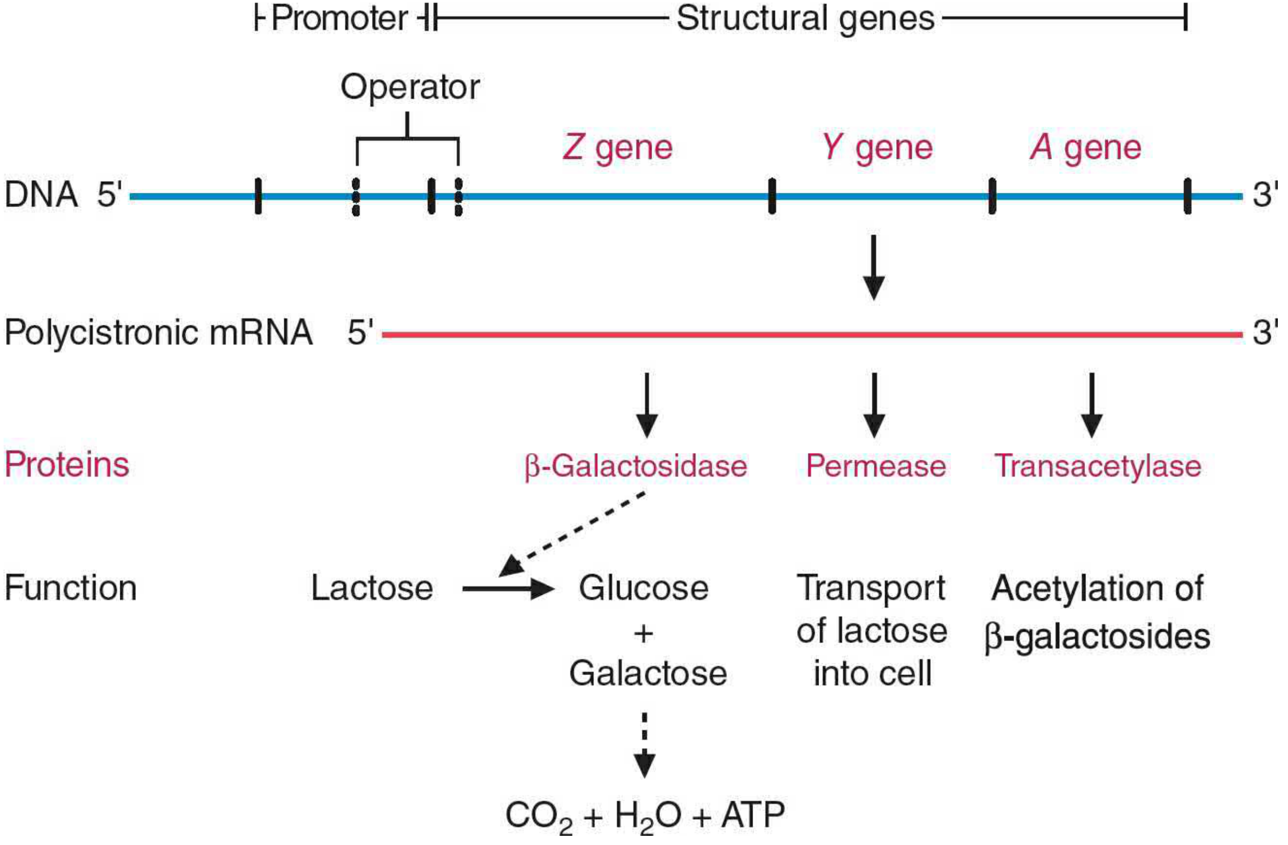
The lac operon encodes enzymes for lactose metabolism in E. coli. It is an inducible operon - normally OFF, switched ON only when lactose is present and glucose is absent.
Structural Genes and Their Products:
| Gene | Protein Product | Function |
|---|
| lacZ | β-Galactosidase | Hydrolyzes lactose → glucose + galactose |
| lacY | Permease | Transports lactose into the cell |
| lacA | Transacetylase | Acetylates β-galactosides |
Regulation of the Lac Operon
A. Negative Control (Repressor-Operator System):
When NO lactose is present (operon OFF):
- LacI repressor protein is active
- Repressor binds to operator region
- RNA polymerase CANNOT bind to promoter
- No transcription → no enzymes made
When LACTOSE is present (operon ON):
- Lactose → converted to allolactose (the true inducer) by small amounts of existing β-galactosidase
- Allolactose binds to the LacI repressor → repressor changes shape → CANNOT bind to operator
- RNA polymerase binds to promoter and transcribes lacZ, lacY, lacA as a polycistronic mRNA
- Enzymes produced → lactose metabolized
B. Positive Control (CAP-cAMP System):
Even when lactose is present, the lac operon is ONLY fully expressed when glucose is absent.
- No glucose → high cAMP levels → cAMP binds to CAP (Catabolite Activator Protein / CRP)
- cAMP-CAP complex binds to the promoter → stimulates RNA polymerase binding → maximum transcription
- Glucose present → low cAMP → CAP inactive → reduced transcription (even if lactose is present)
Summary Table:
| Glucose | Lactose | cAMP | Repressor | Transcription |
|---|
| Present | Absent | Low | Active | None |
| Present | Present | Low | Inactive | Low (basal) |
| Absent | Absent | High | Active | None |
| Absent | Present | High | Inactive | Maximum |
Trp Operon (Repressible Operon - Briefly)
- Normally ON (tryptophan synthesis occurs constitutively)
- When tryptophan is available, it acts as a corepressor - binds inactive repressor → active repressor → binds operator → transcription stops
- This saves energy when tryptophan is already available
Source: Basic Medical Biochemistry - A Clinical Approach, 6e, p. 507-509
4. DNA Repair Mechanisms
Why DNA Repair is Needed
DNA is constantly damaged by UV radiation, chemicals, reactive oxygen species, and replication errors. If unrepaired, mutations accumulate leading to cancer or cell death.
Four Major DNA Repair Mechanisms
A. Mismatch Repair (MMR)
When used: Corrects base-pair mismatches that escape proofreading during replication
Mechanism (E. coli):
- MutS protein recognizes the mismatched base pair
- MutL is recruited by MutS; complex activates MutH
- MutH cleaves the unmethylated daughter strand (discrimination from methylated parental strand via GATC methylation)
- Exonuclease removes the mismatched stretch
- DNA pol III fills the gap; DNA ligase seals
Rate improvement: Reduces replication error from 1 in 10⁷ to 1 in 10⁹ nucleotides
Clinical Significance: Defects in human MMR genes (MSH2, MLH1) → Lynch syndrome (HNPCC - Hereditary Non-Polyposis Colorectal Cancer)
B. Nucleotide Excision Repair (NER)
When used: Removes bulky DNA lesions - UV-induced thymine dimers, chemical adducts (e.g., benzo[a]pyrene from cigarette smoke)
What is a thymine dimer? UV radiation causes covalent bonding between two adjacent thymine bases on the same strand → blocks DNA replication
Mechanism (Prokaryotes):
- UvrABC excinuclease recognizes the bulky dimer
- Cuts the damaged strand on both sides (5' and 3') of the lesion
- A short oligonucleotide (~12-13 bp in prokaryotes, ~25-30 bp in eukaryotes) is excised
- DNA pol I fills the gap
- DNA ligase seals the nick
Clinical Significance: Defect in NER genes (XPA-XPG) → Xeroderma Pigmentosum (XP)
- Cannot repair UV-induced pyrimidine dimers
- Extreme sensitivity to sunlight
- Multiple skin cancers in early childhood
C. Base Excision Repair (BER)
When used: Corrects individual altered or damaged bases (spontaneous deamination, oxidation, alkylation)
Common example: Cytosine slowly deaminates to Uracil (C → U). This must be repaired because U base-pairs with A instead of G, causing a transition mutation.
Mechanism:
- DNA glycosylase removes the abnormal base by cleaving the N-glycosidic bond → creates an AP site (apurinic/apyrimidinic site)
- AP endonuclease cleaves the phosphodiester backbone at the AP site
- Deoxyribophosphodiesterase removes the deoxyribose remnant
- DNA pol β (in eukaryotes) inserts the correct nucleotide
- DNA ligase seals the nick
Examples of damage repaired by BER:
- Deamination: C → U, A → Hypoxanthine
- Oxidation: Guanine → 8-oxoguanine (can cause G-C to T-A transversion)
- Alkylation: N7-methylguanine
D. Direct Repair
Photolyase (Light repair):
- Directly reverses UV-induced thymine dimers in the presence of visible light (photoactivation)
- Not present in placental mammals (present in bacteria, plants, lower organisms)
O⁶-methylguanine methyltransferase:
- Directly removes methyl groups from O⁶-methylguanine
- Suicide enzyme - permanently inactivated after one repair event
Summary Table
| Type | Damage Repaired | Key Enzymes | Disease if Defective |
|---|
| Mismatch Repair | Replication errors | MutS, MutL, MutH | Lynch syndrome (HNPCC) |
| Nucleotide Excision | UV dimers, bulky adducts | UvrABC, DNA pol I | Xeroderma Pigmentosum |
| Base Excision | Altered/lost bases | DNA glycosylase, AP endonuclease, pol β | Various cancers |
| Direct Repair | Alkylated bases | Methyltransferase, Photolyase | - |
Source: Biochemistry, Lippincott 8e, p. 1179-1185
5. Mutations - Types and Clinical Examples
Definition
A mutation is a permanent heritable change in the nucleotide sequence of DNA.
Classification of Mutations
A. Based on Size / Extent
1. Point Mutation (Single Base Change)
i) Substitution mutations:
| Type | Definition | Example |
|---|
| Transition | Purine → Purine OR Pyrimidine → Pyrimidine | A→G, C→T |
| Transversion | Purine → Pyrimidine OR Pyrimidine → Purine | A→C, G→T |
ii) Effects of substitution mutations:
| Effect | Definition | Example |
|---|
| Silent (synonymous) | Codon changes but SAME amino acid (due to degeneracy) | GAA → GAG (both Glu) |
| Missense | Codon changes → DIFFERENT amino acid | GAG → GTG (Glu → Val) = Sickle cell anemia |
| Nonsense | Codon changes → STOP codon | Premature termination → truncated protein = Beta-thalassemia |
2. Frameshift Mutation
- Insertion or deletion of 1-2 nucleotides (not multiples of 3)
- Shifts the reading frame of all codons downstream
- Results in completely different amino acid sequence from point of mutation onwards
- Often creates a premature stop codon
Example: Duchenne Muscular Dystrophy (DMD) - deletion in dystrophin gene causes frameshift
B. Based on Cause
| Cause | Examples |
|---|
| Spontaneous | Tautomeric shifts, deamination (C→U), depurination |
| Physical mutagens | UV radiation (thymine dimers), ionizing radiation (γ-rays, X-rays) |
| Chemical mutagens | Base analogs (5-bromouracil, 2-aminopurine), deaminating agents (nitrous acid), alkylating agents (nitrogen mustard, EMS), intercalating agents (acridine dyes → frameshift) |
C. Based on Location
| Type | Effect on Inheritance |
|---|
| Somatic mutation | Affects only the individual (not inherited) - can cause cancer |
| Germline mutation | Present in germ cells - passed to offspring |
Important Clinical Examples
| Disease | Mutation Type | Gene | Specific Change |
|---|
| Sickle Cell Anemia | Missense point mutation (transition) | β-globin | GAG → GTG (Glu₆ → Val₆) |
| Beta-thalassemia | Various (frameshift, nonsense, splice site) | β-globin | Multiple - reduces/abolishes β-globin |
| Phenylketonuria | Missense | PAH (phenylalanine hydroxylase) | Point mutation |
| Huntington's disease | Trinucleotide repeat expansion | HTT | CAG repeat expansion |
| Xeroderma Pigmentosum | NER defect | XP genes | UV-induced dimer accumulation |
| Duchenne MD | Frameshift deletion | Dystrophin | Large deletion shifts reading frame |
Carcinogenesis and Mutations
- Most cancers result from acquired somatic mutations in proto-oncogenes (gain-of-function) and tumor suppressor genes (loss-of-function)
- Mutagens are therefore also called carcinogens
- Ames test: identifies potential carcinogens by their mutagenicity in bacteria
6. Restriction Endonucleases
Definition
Restriction endonucleases (restriction enzymes) are bacterial enzymes that recognize specific short DNA sequences and cleave the DNA at or near that sequence. They are part of the bacterial defense system (restriction-modification system) that protects against foreign DNA (bacteriophages).
Discovery
- Arber, Smith, and Nathans (1978 Nobel Prize) - discovered restriction endonucleases
Types of Restriction Enzymes
| Type | Characteristics | Use in Biotechnology |
|---|
| Type I | Cuts far from recognition site, requires ATP | Not used (non-specific cleavage) |
| Type II | Cuts at or within recognition site, simple cofactor | Most widely used in recombinant DNA technology |
| Type III | Cuts downstream of recognition site | Not commonly used |
Recognition Sequences - Palindromic Nature
Type II restriction enzymes recognize palindromic sequences - short sequences (4-8 bp) that read the same on both strands in the 5'→3' direction.
Important Examples:
| Enzyme | Source Organism | Recognition Sequence | Cut Site |
|---|
| EcoRI | E. coli | 5'...G↓AATTC...3' | Produces 4-base 5' overhang (sticky ends) |
| BamHI | Bacillus | 5'...G↓GATCC...3' | Produces 4-base 5' overhang (sticky ends) |
| HindIII | H. influenzae | 5'...A↓AGCTT...3' | Produces 4-base 5' overhang (sticky ends) |
| EcoRV | E. coli | 5'...GAT↓ATC...3' | Produces blunt ends |
| SmaI | Serratia marcescens | 5'...CCC↓GGG...3' | Produces blunt ends |
Types of Cut Ends
Sticky Ends (Cohesive Ends):
- Staggered cuts leave short single-stranded overhangs
- Can anneal with complementary sticky ends from any other DNA cut with the same enzyme
- More efficient for ligation (DNA joining)
5'...G     AATTC...3'    →    5'...G           3' + 5' AATTC...3'
3'...CTTAA     G...5'         3'...CTTAA  5'         3'     G...5'
Blunt Ends:
- Cut straight through both strands at the same position
- No single-stranded overhang
- Can be ligated but less efficiently
Role in Recombinant DNA Technology
- Both the vector (plasmid) and foreign DNA are cut with the same restriction enzyme → compatible sticky ends
- Foreign DNA fragment anneals to the cut vector by hydrogen bonding
- DNA ligase covalently seals the nicks → recombinant DNA (chimeric DNA)
- Inserted into host bacterium (transformation) for cloning and expression
Other Applications
| Application | How Restriction Enzymes Are Used |
|---|
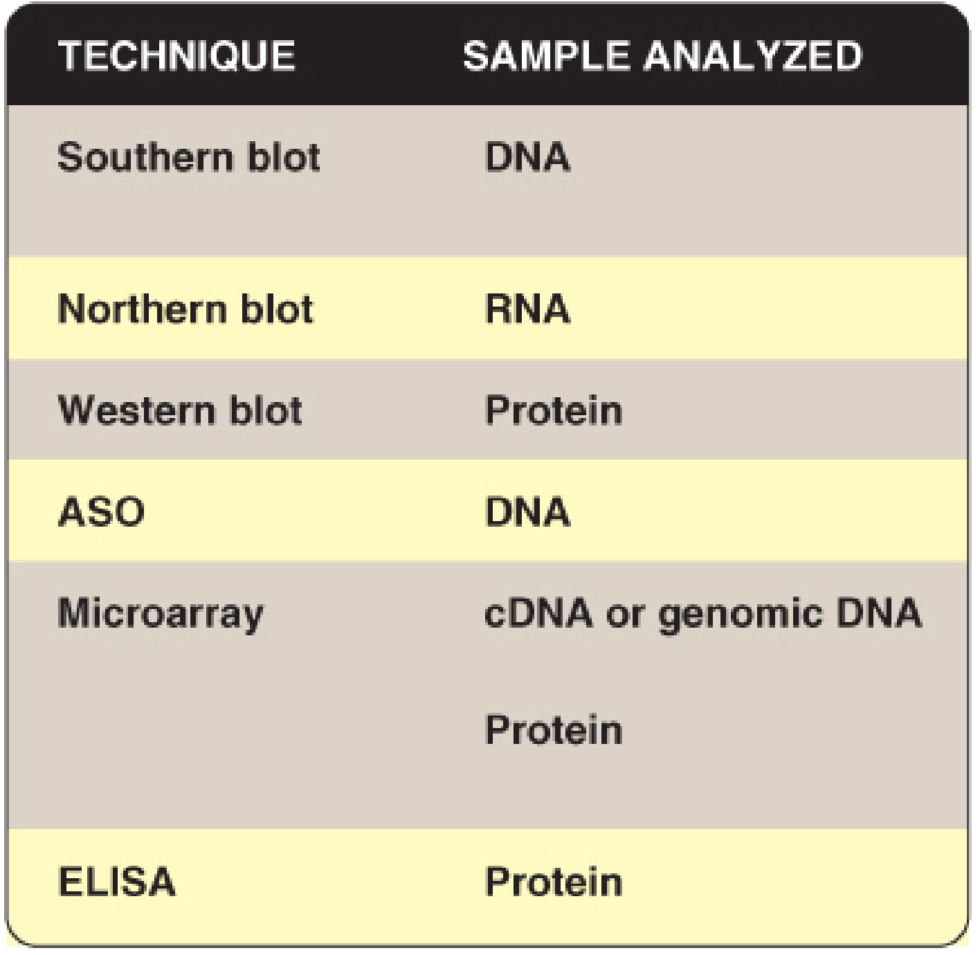
| RFLP / DNA Fingerprinting | Digest genomic DNA → different fragment sizes between individuals → Southern blot |
| Gene mapping | Restriction maps show positions of cut sites along a chromosome |
| Diagnosis of genetic diseases | Sickle cell anemia - the mutation in β-globin destroys an MstII recognition site (GAG→GTG) - loss of this band on Southern blot confirms diagnosis |
| Prenatal diagnosis | Restriction digestion of fetal DNA from amniocentesis or CVS |
Bacterial Protection Against Own Restriction Enzymes
- Bacteria methylate their own DNA at the same recognition sequences (using methyltransferases)
- Methylated DNA is NOT cut by the restriction enzyme
- Foreign DNA (unmethylated) IS cut
Source: Biochemistry, Lippincott 8e; Harper's Illustrated Biochemistry, 32e
Quick Revision - All 6 Tier 2 Topics at a Glance
| Topic | Critical Lines to Remember |
|---|
| Genetic Code | 64 codons; 61 amino acid + 3 stop; AUG = start; Wobble = 3rd position flexibility by Inosine |
| PCR | Denaturation (94°C) → Annealing (55-65°C) → Extension (72°C/Taq); 30 cycles = 10⁹ copies |
| Lac Operon | Inducible; Negative control (allolactose inactivates repressor); Positive control (cAMP-CAP needed); Maximum expression when lactose present + glucose absent |
| DNA Repair | MMR = mismatch → Lynch; NER = thymine dimers → Xeroderma Pigmentosum; BER = altered bases |
| Mutations | Missense: same position, different AA (sickle cell); Nonsense: premature stop; Frameshift: insertion/deletion → reading frame shift |
| Restriction Enzymes | Type II used in biotech; Palindromic sequences; EcoRI (GAATTC); Sticky vs blunt ends; Used in recombinant DNA + RFLP |
Exam strategy: For Lac Operon - always draw the ON/OFF diagram showing RNA polymerase, repressor, operator, and structural genes. For Mutations - always give the sickle cell anemia example (GAG→GTG). For PCR - write the 3 steps with temperatures. These details are what separate a 4-mark answer from a 5-mark answer in RGUHS exams.