I now have comprehensive information to write a full, exam-ready answer. Let me compile this into a detailed response.
Transverse Section at the Level of Pyramidal Decussation - Clinically Applied Anatomy (MBBS Exam Answer)
INTRODUCTION
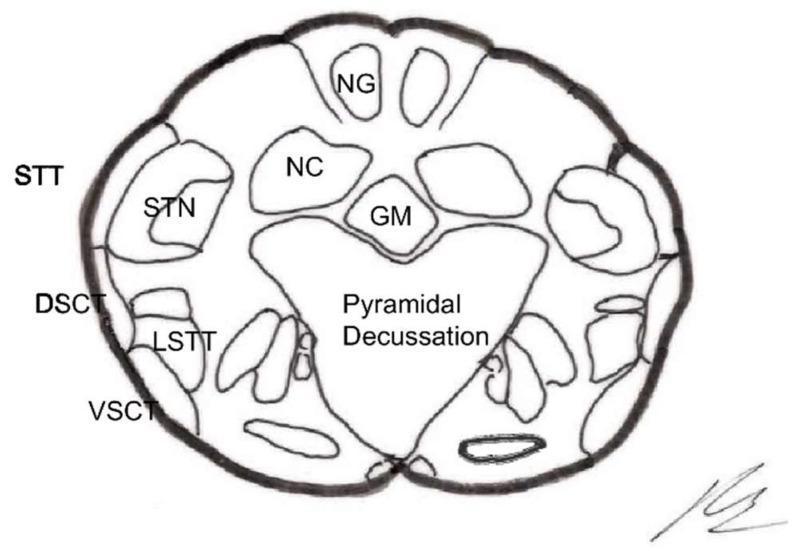
The transverse section at the level of the pyramidal decussation (also called the great motor decussation or Mistichelli crossing) is the lowest level of the medulla oblongata, located just above the foramen magnum where the medulla transitions into the spinal cord. This is one of the three classically described transverse sections of the medulla and is the most important for MBBS exams because of its rich clinical correlations.
LEVEL / LOCATION
- Situated at the caudal (lower) end of the medulla oblongata
- Just superior to the foramen magnum
- Corresponds to the junction of medulla and spinal cord
- Also called the level of the decussation of the pyramids
STRUCTURES SEEN IN TRANSVERSE SECTION
1. ANTERIOR (VENTRAL) REGION
| Structure | Details |
|---|
| Decussating corticospinal (pyramidal) fibers | The dominant feature - bundles of fibers crossing the midline in an interdigitating pattern, obliterating the anterior median fissure |
| Anterior median fissure | Interrupted/obliterated by decussating fiber bundles |
| Anterior corticospinal tract (uncrossed ~10-25%) | Descends ipsilaterally in the anterior funiculus |
| Lateral corticospinal tract (crossed ~75-90%) | Newly formed on the contralateral side |
Key fact: Approximately 75-90% of corticospinal fibers decussate here to form the lateral corticospinal tract (LCST). The remaining 10-25% descend uncrossed as the anterior corticospinal tract (ACST).
- Localization in Clinical Neurology, 8e, p. 917
- Adams and Victor's Principles of Neurology, 12th Ed.
2. POSTERIOR (DORSAL) REGION
| Structure | Position | Function |
|---|
| Nucleus gracilis | Posteromedial | Receives proprioception, vibration, fine touch from lower limbs (below T6) |
| Nucleus cuneatus | Posterolateral | Receives proprioception, vibration, fine touch from upper limbs (above T6) |
| Fasciculus gracilis | Medial column | Ascending sensory fibers to nucleus gracilis |
| Fasciculus cuneatus | Lateral column | Ascending sensory fibers to nucleus cuneatus |
| Central grey matter | Surrounds central canal | |
Note: At this level, the nuclei gracilis and cuneatus are just beginning to emerge - their full development appears more rostrally at the level of internal arcuate fiber decussation.
3. LATERAL REGION
| Structure | Note |
|---|
| Spinal nucleus of trigeminal nerve (Spinal trigeminal nucleus) | Receives pain and temperature from ipsilateral face; extends down to C3 spinal cord level |
| Spinal tract of trigeminal nerve | Lies lateral to the nucleus |
| Dorsal spinocerebellar tract (DSCT) | Lateral periphery |
| Ventral spinocerebellar tract (VSCT) | Anterolateral periphery |
| Lateral spinothalamic tract (LST) | Pain and temperature from contralateral body |
| Descending sympathetic fibers | Run in the lateral tegmentum |
KEY ANATOMICAL FACTS FOR EXAMINATIONS
The Decussation of the Pyramids - Mechanism
- At the caudal medulla, the corticospinal fibers in the medullary pyramid obliquely cross the midline, interdigitating with each other
- The crossing fibers pass dorsolaterally to reach the opposite lateral funiculus of the spinal cord
- The fiber bundles cross alternately - upper limb fibers cross before lower limb fibers (rostral-to-caudal sequence)
- This decussation interrupts the anterior median fissure
Somatotopic Organization
- Within the pyramids (before decussation): lower limb fibers are more lateral, upper limb fibers more medial
- This somatotopy is clinically significant for partial lesions
- Localization in Clinical Neurology, 8e, p. 917
VASCULAR SUPPLY AT THIS LEVEL
| Territory | Artery | Structures supplied |
|---|
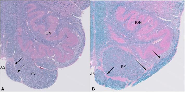
| Paramedian (medial) | Anterior spinal artery / Vertebral artery paramedian branches | Pyramid, medial lemniscus, hypoglossal nucleus/fibers |
| Lateral | Posterior inferior cerebellar artery (PICA) / vertebral artery | Lateral medulla, spinothalamic tract, spinal trigeminal nucleus/tract, nucleus ambiguus, inferior cerebellar peduncle, descending sympathetics |
CLINICAL CORRELATIONS (HIGH-YIELD FOR EXAMS)
1. Hemiplegia Cruciata (Cruciate Hemiplegia) - MOST IMPORTANT
This is the pathognomonic clinical syndrome of a lesion at the pyramidal decussation level.
Mechanism: Because upper limb fibers cross BEFORE lower limb fibers during decussation, a unilateral lesion at the exact level of the pyramidal decussation affects:
- Crossed fibers to the arm (already decussated) → ipsilateral arm paresis
- Uncrossed fibers to the leg (not yet decussated) → contralateral leg paresis
Result: Ipsilateral arm paresis + Contralateral leg paresis - a "diagonal" or "cruciate" pattern
This is called hemiplegia cruciata (also written hemiplegia cruciata or alternating hemiplegia).
- Localization in Clinical Neurology, 8e, p. 920
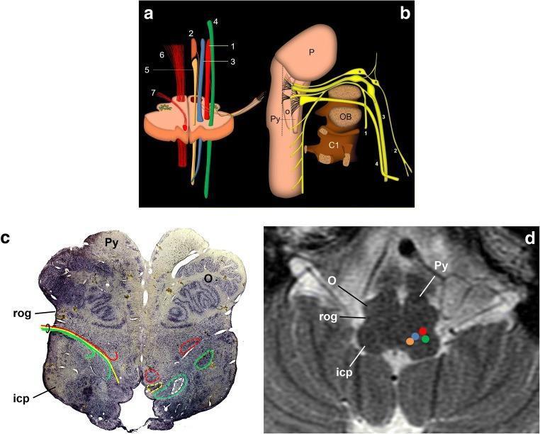
2. Medial Medullary Syndrome (Dejerine Syndrome / Anterior Bulbar Syndrome)
Caused by occlusion of the anterior spinal artery or vertebral artery paramedian branches
Classic triad:
| Feature | Structure damaged | Side |
|---|
| Tongue paralysis (LMN-type), tongue deviates to lesion side | CN XII nucleus/fibers | Ipsilateral |
| Hemiplegia (UMN, face spared) | Pyramid | Contralateral |
| Loss of vibration, proprioception, fine touch | Medial lemniscus | Contralateral |
Pain and temperature sensation are SPARED (spinothalamic tract is in the lateral medulla, not affected here).
Bilateral occlusion results in: flaccid quadriplegia (face spared), bilateral tongue paralysis (LMN), complete loss of proprioception and vibration in all 4 limbs, and respiratory failure.
- Localization in Clinical Neurology, 8e, p. 918-919
3. Lateral Medullary Syndrome (Wallenberg Syndrome / PICA Syndrome)
Caused by occlusion of the PICA (posterior inferior cerebellar artery) or intracranial vertebral artery.
Classic features (lateral medulla is intact at the decussation level but relevant for exam context):
| Feature | Structure | Side |
|---|
| Facial pain/temp loss | Spinal trigeminal nucleus/tract | Ipsilateral |
| Body pain/temp loss | Spinothalamic tract | Contralateral |
| Dysphagia, dysarthria, hoarseness | Nucleus ambiguus | Ipsilateral |
| Horner's syndrome (ptosis, miosis, anhidrosis) | Descending sympathetic fibers | Ipsilateral |
| Vertigo, nausea, vomiting | Vestibular nuclei | Present |
| Cerebellar signs (ataxia) | Inferior cerebellar peduncle | Ipsilateral |
| Motor/pyramidal signs | SPARED (pyramids are medial, not lateral) | - |
| Vibration/proprioception | SPARED (medial lemniscus is medial) | - |
Key exam mnemonic - PICA Wallenberg features on IPSILATERAL side: "5 I's" = IPsilateral Face, IPsilateral Horner, IPsilateral Ataxia, IPsilateral IX/X (dysphagia), Ipsilateral C/T loss on face; with contralateral body C/T loss.
- Localization in Clinical Neurology, 8e, p. 920-922
4. Significance of the Uncrossed (Anterior) Corticospinal Tract
- ~10-25% of corticospinal fibers do NOT cross at the pyramidal decussation
- These run in the anterior corticospinal tract ipsilaterally
- They eventually cross at segmental levels in the spinal cord (or supply bilateral axial musculature)
- Clinical implication: Some bilateral axial motor control is preserved even with unilateral cortical lesions
- Adams and Victor's, 12th Ed.
5. Spinal Nucleus of Trigeminal Nerve Extends Below the Medulla
The spinal nucleus and tract of CN V extend from the pons down through the entire medulla and into the upper cervical spinal cord (to C3). This is visible in the transverse section at the pyramidal decussation level.
Clinical implication: High cervical cord lesions (C1-C3) may produce ipsilateral facial numbness (pain/temperature) - mimicking a brainstem lesion ("onion-peel" distribution of facial sensory loss in upper cord/lower medulla lesions).
COMPARISON TABLE: THREE LEVELS OF MEDULLA (Exam Quick Reference)
| Feature | Pyramidal Decussation | Sensory Decussation (Internal Arcuate Fibers) | Level of Inferior Olive |
|---|
| Dominant feature | Pyramidal fibers crossing | Internal arcuate fibers crossing | Inferior olivary nucleus |
| Anterior median fissure | Obliterated by crossing fibers | Present | Present |
| Nucleus gracilis/cuneatus | Present (early) | Present (prominent) | Not present |
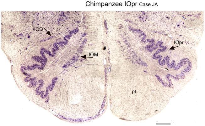
| Inferior olivary nucleus | Absent | Absent / forming | Prominent |
| CN nuclei | - | - | XII, X, IX nuclei present |
| Clinical syndrome | Hemiplegia cruciata | - | Dejerine, Wallenberg |
SUMMARY DIAGRAM OF KEY STRUCTURES AT PYRAMIDAL DECUSSATION LEVEL
POSTERIOR
  Gracilis nucleus (medial) | Cuneatus nucleus (lateral)
  Fasciculus gracilis        | Fasciculus cuneatus

  Central grey matter (around central canal)

  Spinal trigeminal nucleus + tract (lateral)
  Spinocerebellar tracts (DSCT medial, VSCT lateral - at periphery)
  Lateral spinothalamic tract (anterolateral)
  Descending sympathetics (lateral)

  [DECUSSATING PYRAMIDAL FIBERS] - crossing midline ventrally
  → obliterates anterior median fissure

ANTERIOR
APPLIED / EXAM POINTS TO REMEMBER
- Decussation of pyramids = motor decussation = great decussation = Mistichelli crossing - all the same thing
- 75-90% of fibers cross → lateral CST; 10-25% remain uncrossed → anterior CST
- Lesion at this level causes contralateral UMN hemiplegia below the neck (face spared - corticobulbar fibers already decussated above)
- Hemiplegia cruciata is the unique syndrome at exactly this level - diagonal paralysis
- Anterior median fissure is obliterated at this level - examiners love this fact
- Somatotopy: Lower limb lateral, upper limb medial within the pyramid
- Nucleus gracilis and cuneatus are forming at this level (fully developed one level up)
- Spinal nucleus of V visible here and continues down to C3
- The decussation occurs in an interdigitating pattern - bundles alternate
- Blood supply of medial medulla = vertebral artery / anterior spinal artery; lateral = PICA
Sources: Localization in Clinical Neurology, 8e (pp. 916-922) | Gray's Anatomy for Students (pp. 1317-1321) | Neuroanatomy through Clinical Cases, 3rd Ed. | Adams and Victor's Principles of Neurology, 12th Ed.
This covers every aspect examiners test at MBBS level: the structural anatomy of the section, the specific nuclei and tracts, vascular supply, and the three major clinical syndromes (hemiplegia cruciata, medial medullary syndrome, and Wallenberg syndrome) that correlate directly with structures at this level. Master the comparison table and the 10 applied points for full marks.