
Craniotomy and post surgery complications along with physiotherapy
craniotomy post surgery complications physiotherapy rehabilitation 2024
craniotomy complications postoperative management
craniotomy surgical procedure brain
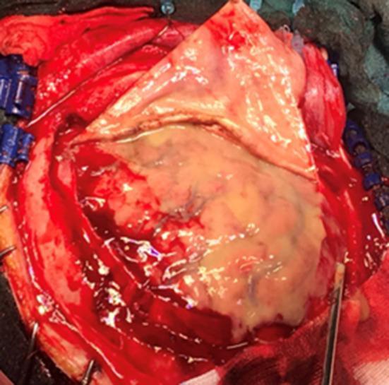
An intraoperative clinical photograph showing a neurosurgical procedure (craniotomy) for the management of an extensive subdural empyema. The image displays a wide surgical field with the dura mater retracted, revealing a thick, viscous, yellowish-white purulent collection that covers the majority of the visible cerebral hemisphere. The underlying and adjacent brain parenchyma exhibits significant congestion and hyperemia, characterized by a deep reddish hue and prominent vascularity, indicative of severe inflammatory response. Surgical instruments, including retractors and a suction tip, are visible at the margins of the craniotomy. This visual demonstrates the clinical manifestation of a large-scale intracranial infection and the necessity of surgical drainage to alleviate mass effect and control the infectious process within the subdural space.
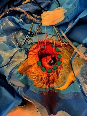
Clinical photograph of an intraoperative brain biopsy procedure. The image shows a prepared surgical field on the scalp, demarcated by sterile blue drapes and a clear adhesive incise drape. A circular craniotomy site is visible, with the scalp edges retracted using green Raney clips and sutures held by several hemostats/clamps. Within the surgical opening, the underlying cerebral tissue is exposed, revealing a prominent, deeply pigmented (blackish-brown) lesion localized to a gyrus, which is suspicious for metastatic melanoma. Moderate bleeding is observed around the incision margins, with blood tracking over the yellow-prepped scalp and onto the surrounding drapes. Surgical adjuncts visible include a clear suction tube, a saturated gauze sponge, and various cables for monitoring or cautery. This image demonstrates the visual presentation of a pigmented intracranial metastasis during neurosurgical intervention.
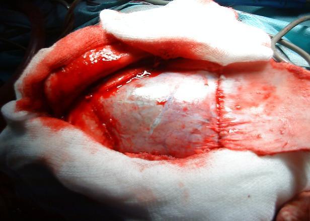
This clinical photograph captures a per-operative view of an open craniotomy during a neurosurgical procedure. The focal point of the image is the exposed brain surface, specifically the cerebral cortex, displaying characteristic gyri and sulci covered by the arachnoid mater. The surgical field is bounded by retracted scalp tissue and white surgical sponges or gauze used for hemostasis and to maintain field clarity. Significant sanguineous fluid (blood) is visible around the margins of the cranial opening. To the right of the primary cortical exposure, a linear suture line is visible on the reflected dural or scalp flap, indicating a phase of tissue approximation or closure. This image serves as an educational example of a large craniotomy, typical in procedures such as the Arana-Iniguez technique for the surgical management of intracranial lesions like hydatid cysts.
post craniotomy complications brain CT scan
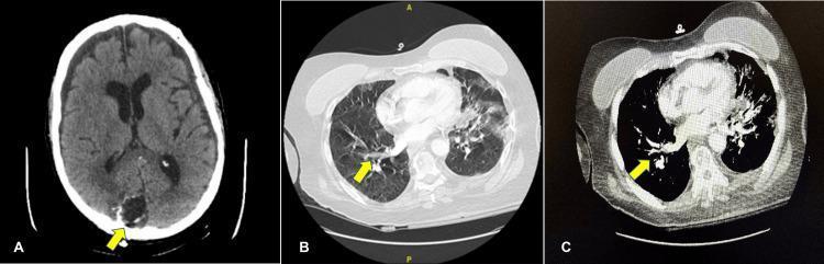
This composite of three axial computed tomography (CT) images illustrates post-treatment clinical findings in a patient with multi-organ pathology. Figure A is a non-contrast brain CT scan showing a right occipital craniotomy site. The surgical cavity is identifiable, surrounded by irregular linear hyperdensities (suggestive of acute post-operative hemorrhage) and areas of low-attenuation consistent with diffuse vasogenic edema over the parietal convexity. Figures B and C are CT angiography (CTA) images of the chest in lung and mediastinal windows, respectively. Yellow arrows indicate subsegmental filling defects within the right upper lobe pulmonary vasculature, diagnostic of small-volume pulmonary emboli (PE). The lung parenchyma adjacent to the emboli shows mild irregularities without dense consolidation. These images highlight the clinical challenge of managing systemic thromboembolic disease in the immediate post-operative period following neurosurgery, where the risk of intracranial hemorrhage complicates anticoagulation therapy. The educational focus is on identifying post-surgical intracranial complications and vascular filling defects on CTA.
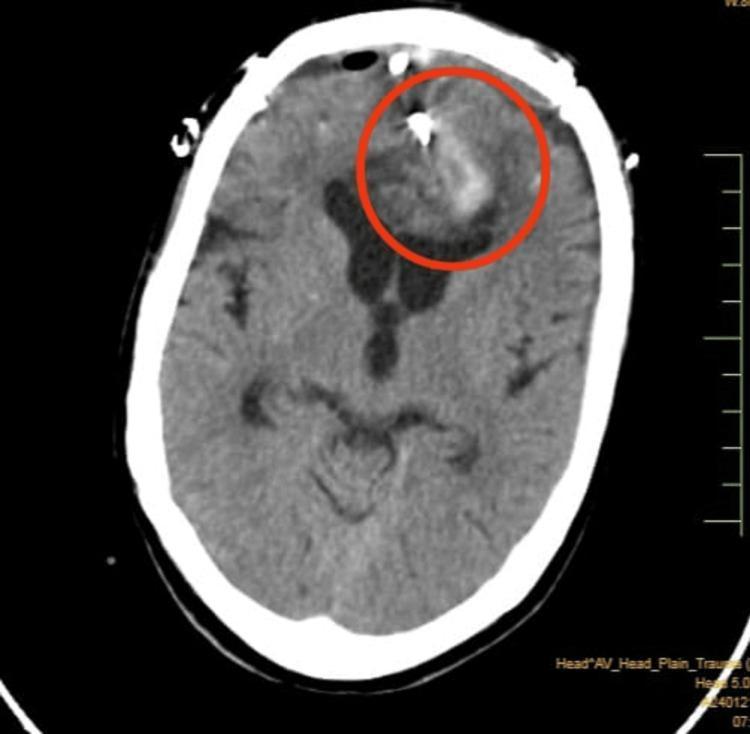
This axial non-contrast CT scan of the brain demonstrates a post-operative state following neurosurgical intervention in the bilateral frontal region. Key features include a bilateral frontal calvarial defect (craniotomy site) with overlying extracranial soft tissue swelling and a visible surgical drain tube. Intracranial findings are highlighted by a red circle, revealing a large, mixed-density intraparenchymal hematoma located in the interhemispheric and parafalcine regions, predominantly affecting the left frontal lobe. A hyperdense metallic focus, consistent with an aneurysm clip, is visible within the anterior interhemispheric region, associated with mild streak artifacts. There is evidence of mass effect, indicated by the effacement of the frontal horn of the left lateral ventricle. Small air-density foci (pneumocephalus) are scattered within the frontal region and adjacent to a thin, mixed-density subdural collection. The imaging illustrates typical post-surgical complications and the management of a ruptured intracranial aneurysm.
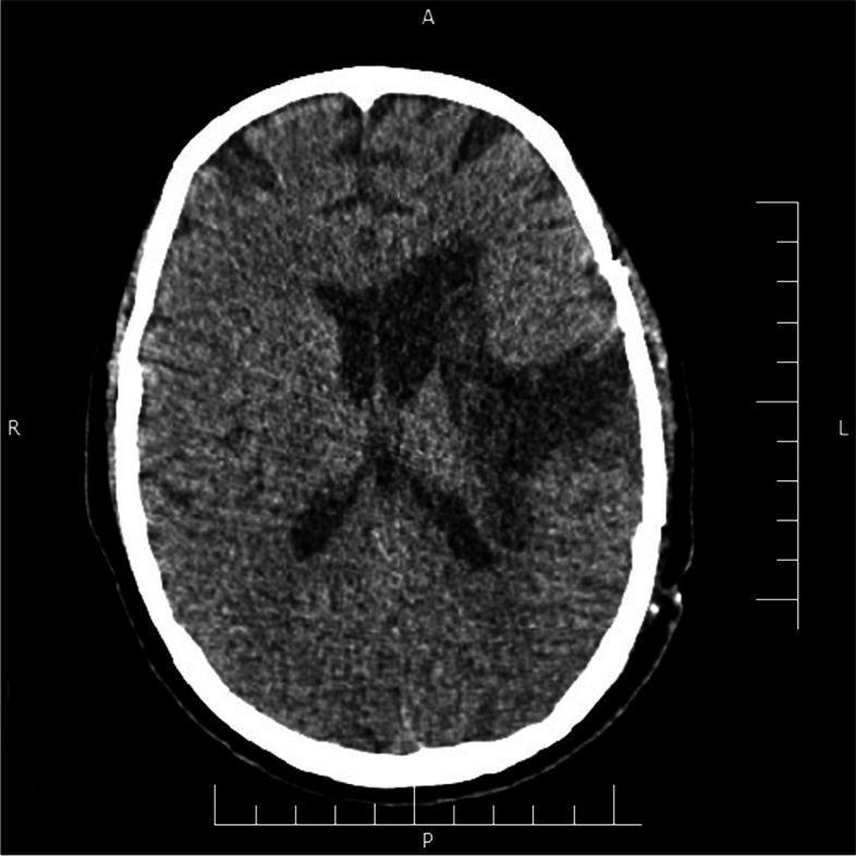
This diagnostic image is an axial non-contrast computed tomography (CT) scan of the brain, specifically a post-operative follow-up following evacuation of a left-sided intracranial hemorrhage. The image shows the brain at the level of the lateral ventricles. There is evidence of a left-sided craniotomy. In the left frontoparietal region and basal ganglia, there is a large area of hypoattenuation (low density), which represents encephalomalacia and evolving liquefactive necrosis in the site of the previous hematoma. The mass effect and midline shift observed in the acute phase have largely resolved, with the midline structures, including the septum pellucidum, returning to a near-normal position. The lateral ventricles appear symmetrical without evidence of acute intraventricular hemorrhage or hydrocephalus. No acute hyperdense areas suggestive of rebleeding are identified. This scan illustrates the expected radiological progression during the subacute phase of recovery following surgical hematoma evacuation in a patient with a history of COVID-19-associated neurological complications.
https://juniperpublishers.com/jojcs/JOJCS.MS.ID.555897.php
Craniotomy: Procedure, Post-Surgical Complications, and Physiotherapy
What is a Craniotomy?
- Intracranial hematomas (subdural, extradural, intracerebral)
- Brain tumors (primary or metastatic)
- Cerebral aneurysm clipping
- Arteriovenous malformations (AVMs)
- Brain abscess drainage
- Decompression for raised intracranial pressure (ICP)
- Epilepsy surgery (awake craniotomy)
- Traumatic brain injury (TBI)
Immediate Postoperative Care
| Parameter | Goal |
|---|---|
| Systolic BP | <160 mmHg (MAP <110 mmHg) to prevent rebleeding |
| Oxygenation | Maintain SpO₂; avoid hypoxia (raises ICP) |
| Head positioning | Elevated 30-45° to promote cerebral venous drainage |
| Neurologic status | Frequent GCS and pupillary checks |
| Pain | Multimodal analgesia (avoid excess narcotics); scalp block reduces need |
| Nausea/Vomiting | Serotonin antagonists (ondansetron, granisetron) + low-dose corticosteroids |
Post-Surgical Complications
Immediate (First 24-48 Hours)
- Incidence: ~0.5%, mainly within first 24 hours
- Triggered by hypertension (SBP >160 mmHg)
- Requires emergency re-exploration
- Results from surgical manipulation, venous occlusion, or ischemia
- Managed with head elevation, osmotherapy (mannitol/hypertonic saline), controlled ventilation
- Occur even in previously healthy patients without epilepsy history
- Brain manipulation and dural irritation are highly epileptogenic
- Treated acutely with IV benzodiazepines (lorazepam, midazolam); intubate if airway at risk
- Exclude metabolic causes (hypoglycemia, electrolyte imbalance)
- Air entry into the cranial cavity during surgery
- Usually self-limiting; tension pneumocephalus is a surgical emergency
Early Postoperative (Days 2-7)
- Through wound or into frontal sinuses (especially with bifrontal craniotomy or decompressive craniectomy)
- Can lead to meningitis if persistent
- Prevented by watertight dural closure using fascia lata or bovine pericardium
- Incidence: ~5%
- Risk factors: prolonged surgery, CSF leak, diabetes, obesity, re-operation
- Prophylactic antibiotics significantly reduce post-craniotomy meningitis risk
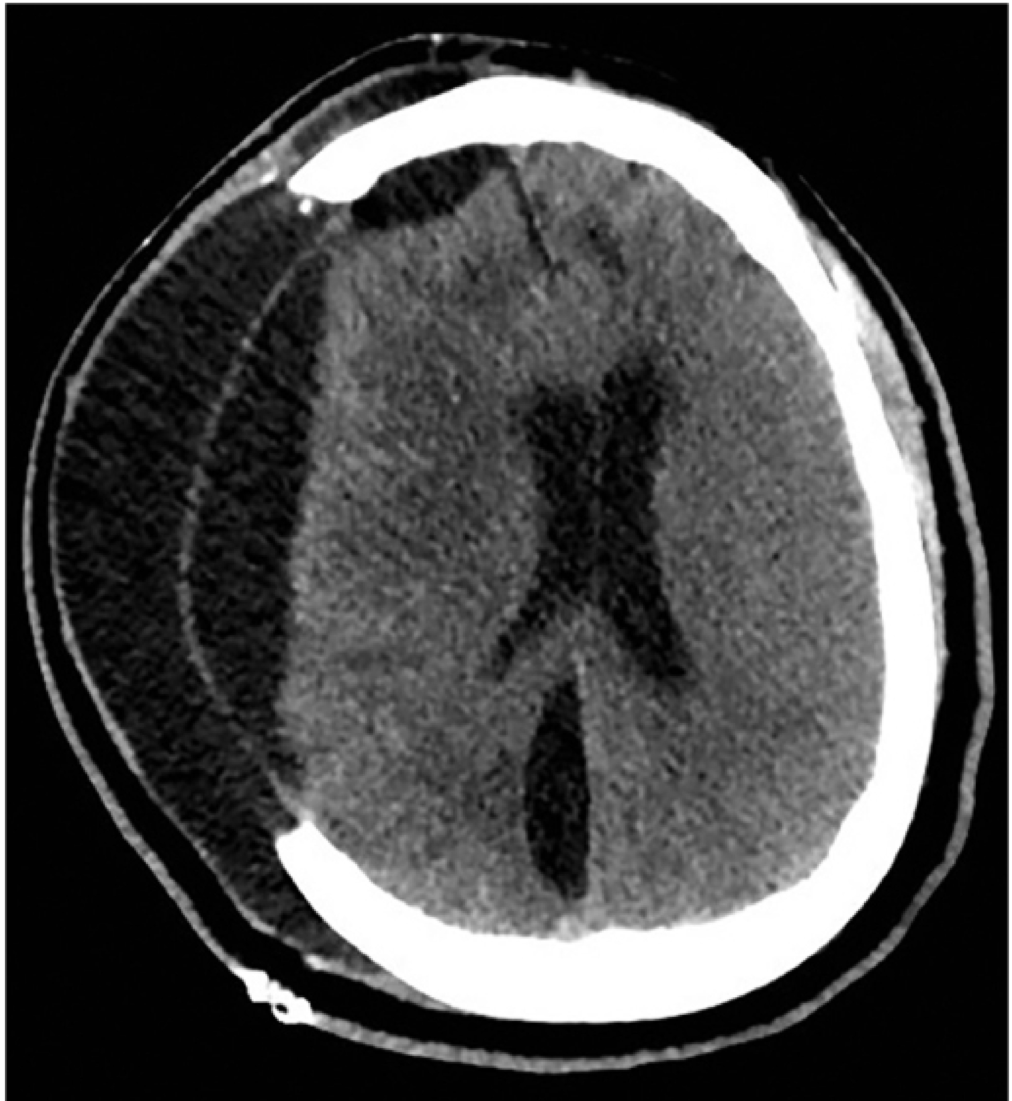
- Accumulation of CSF in the subdural space; common after decompressive craniectomy
- May communicate with frontal sinuses and leak
- Occur in ~3% within 30 days; peak at Day 3 post-op
- Prophylactic low-molecular-weight heparin can be started safely 24 hours after surgery in most cases
Subacute and Late Complications
- Motor weakness (hemiparesis/hemiplegia)
- Aphasia/dysphasia (if dominant hemisphere involved)
- Visual field defects
- Cranial nerve palsies (especially posterior fossa surgery)
- Cognitive impairment (memory, attention, executive function)
- Aseptic (chemical) or septic (bacterial)
- Pathogen often unidentified; metagenomic sequencing improving diagnosis
- Very common; usually not severe
- Managed with acetaminophen +/- NSAIDs; avoid excess opioids
- Communicating hydrocephalus can develop from blood/infection in the subarachnoid space
- May require VP shunting
- Late-onset seizures months after surgery; may require long-term anticonvulsants
- Occurs if superficial temporal artery is sacrificed in large frontoparietotemporal incisions
- Leads to wound breakdown and infection risk
- Depression, anxiety, PTSD
- Mood and behavioral changes
- Fatigue (very common)
Physiotherapy After Craniotomy
Goals of Physiotherapy
- Prevent secondary complications (DVT, respiratory complications, pressure sores, contractures)
- Restore motor function, balance, and gait
- Improve functional independence (FIM score)
- Enhance neuroplasticity and brain reorganization
Phase 1: ICU / Acute Phase (Days 1-7)
- Head elevated 30-45 degrees
- Frequent position changes to prevent pressure sores
- Correct limb positioning to prevent contractures
- Shoulder arm pouch for hemiplegic upper limb
- Chest percussion and vibration
- Airway clearance techniques
- Breathing exercises
- Suctioning if required (especially if prolonged ventilation)
- Particularly important as brain surgery can affect respiratory centers
- Passive ROM to all joints in multiple planes
- Prevents contractures and stimulates neural reorganization
- Begins even in unconscious or low-GCS patients
- Sensory stimulation (tactile, auditory, visual) - important for coma recovery
- Assessed using Coma Recovery Scale (CRS), RASS scale
- Gradual upright positioning to manage orthostatic hypotension
- Stimulates postural reflexes
Phase 2: Subacute / Ward Phase (Weeks 1-4)
- Progressive strengthening exercises
- Bobath / neurodevelopmental techniques for hemiplegia
- Hand grip exercises (key for FIM gain and reducing dependency)
- Weight-bearing and transfer training (bed to chair)
- Static and dynamic balance exercises
- Sitting and standing balance
- Brain surgery commonly disrupts static and dynamic balance
- Parallel bar walking progressing to supervised ambulation
- Assistive devices as needed
- Stair training when appropriate
- Activities of daily living (ADL) training
- Occupational therapy integration
- Communication exercises (alongside speech therapist if aphasia present)
Phase 3: Outpatient / Community Rehabilitation (Weeks 4+)
- Continued strengthening and aerobic conditioning
- Fine motor skills and coordination
- Cognitive rehabilitation (attention, memory, problem-solving)
- Return to work or school planning
- Driving assessment if applicable
- Home exercise programs
The Multidisciplinary Team
| Team Member | Role |
|---|---|
| Physiotherapist | Motor recovery, balance, gait, respiratory care |
| Occupational Therapist | ADLs, fine motor, home adaptations |
| Speech-Language Pathologist | Aphasia, dysarthria, dysphagia |
| Neuropsychologist | Cognitive and behavioral rehabilitation |
| Neurosurgeon / Neurologist | Monitoring, seizure management, imaging review |
| Neuro-ICU Nurses | Constant neurological monitoring |
| Dietitian | Nutritional support for healing and energy |
Outcome Measures Used in Craniotomy Rehabilitation
- GCS (Glasgow Coma Scale) - consciousness level
- FIM (Functional Independence Measure) - overall function
- ICU Mobility Scale - early mobility progress
- RASS (Richmond Agitation-Sedation Scale) - sedation/agitation
- Coma Recovery Scale (CRS) - response to stimulation
- RLAS (Rancho Los Amigos Scale) - cognitive-behavioral recovery
Key Clinical Pearls
- Early extubation and neurological assessment is superior to routine CT in detecting complications
- Blood pressure control is the single most important factor preventing post-craniotomy rebleeding
- DVT prophylaxis with LMWH should start at 24 hours post-op in most patients
- Seizure prophylaxis - evidence for routine prophylactic anticonvulsants is debated; check local guidelines
- Physiotherapy starting within 24 hours significantly reduces morbidity and hospital stay
- CSF leak prevention requires watertight dural closure; bifrontal craniotomies carry higher risk
- Superficial temporal artery preservation in large skin incisions prevents flap ischemia