
Draw a flowchart for immunity
A detailed educational flowchart of the immune system showing both innate and adaptive immunity. The flowchart starts with "Pathogen Enters Body" at the top, then branches into two main pathways: "Innate Immunity (Non-specific, Fast)" and "Adaptive Immunity (Specific, Slower)". Under Innate Immunity: Physical barriers (skin, mucus) → Inflammatory response → Phagocytes (Neutrophils, Macrophages) → Natural Killer Cells → Complement System → If pathogen persists, leads to Adaptive Immunity. Under Adaptive Immunity: Antigen Presenting Cells (Dendritic Cells, Macrophages) present antigens → MHC molecules → activates T Lymphocytes and B Lymphocytes in parallel. T Lymphocytes branch into: Helper T cells (CD4+) which activate B cells and Cytotoxic T cells (CD8+) which kill infected cells. B Lymphocytes → Plasma Cells → Antibody Production (IgM, IgG, IgA, IgE, IgD). Both pathways lead to Memory Cells for long-term protection. Final outcome boxes: "Pathogen Eliminated" and "Immunological Memory formed". Clean, professional medical diagram with color-coded boxes: blue for innate immunity, green for adaptive immunity, orange for cells, yellow for outcomes. Clear arrows showing flow and connections. White background, readable fonts.
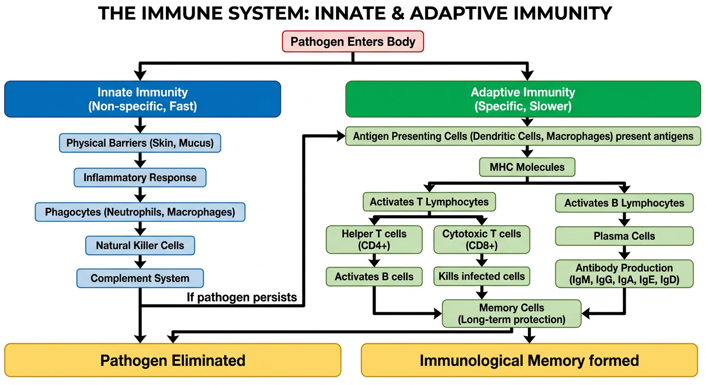
- First line: Physical/chemical barriers (skin, mucus, stomach acid)
- Second line: Inflammation, phagocytes (neutrophils, macrophages), natural killer cells, and the complement system
- If the pathogen persists, it triggers the adaptive response
- Antigen-presenting cells (dendritic cells, macrophages) present antigens via MHC molecules
- T lymphocytes: Helper T cells (CD4+) coordinate the response; Cytotoxic T cells (CD8+) directly kill infected cells
- B lymphocytes: Differentiate into plasma cells that secrete antibodies (IgM, IgG, IgA, IgE, IgD)
- Both arms generate memory cells for faster future responses
Create a detailed reference poster on adaptive vs innate immunity
A detailed, professional medical reference poster titled "INNATE vs ADAPTIVE IMMUNITY" for medical students. Portrait orientation, white background, clean academic layout. TOP SECTION: Large bold title "INNATE vs ADAPTIVE IMMUNITY" with a subtitle "A Comparative Reference Guide". A horizontal banner with a simple icon of a shield split in two colors — blue (left) and green (right). MAIN BODY: Two large side-by-side columns divided by a vertical line. LEFT COLUMN — INNATE IMMUNITY (blue theme): Header: "INNATE IMMUNITY" in bold blue Subtitle: "Non-specific | First Line of Defense" • Speed: Immediate (minutes to hours) • Specificity: Non-specific — same response to all pathogens • Memory: No immunological memory • Components: - Physical barriers: Skin, mucous membranes, cilia - Chemical barriers: Stomach acid, lysozyme, defensins - Cellular: Neutrophils, Macrophages, NK cells, Dendritic cells, Mast cells, Basophils, Eosinophils - Soluble factors: Complement proteins (C3, C5), cytokines (IL-1, IL-6, TNF-α), interferons • Recognition: Pattern Recognition Receptors (PRRs) — Toll-like receptors (TLRs) recognize PAMPs • Key processes: Phagocytosis, Inflammation, Fever, Opsonization, Complement activation • Limitations: Cannot distinguish between similar pathogens; no memory RIGHT COLUMN — ADAPTIVE IMMUNITY (green theme): Header: "ADAPTIVE IMMUNITY" in bold green Subtitle: "Specific | Second Line of Defense" • Speed: Days to weeks (first exposure); faster on re-exposure • Specificity: Highly specific — targets unique antigens • Memory: Long-lasting immunological memory (memory B and T cells) • Components: - T Lymphocytes: Helper T (CD4+), Cytotoxic T (CD8+), Regulatory T (Treg), Memory T cells - B Lymphocytes: Plasma cells (antibody-secreting), Memory B cells - Antibodies (Immunoglobulins): IgM, IgG, IgA, IgE, IgD — functions listed • Recognition: T-cell receptors (TCR) and B-cell receptors (BCR) recognize specific antigens via MHC • Key processes: Clonal selection, Clonal expansion, Antigen presentation via MHC I and MHC II, Somatic hypermutation, Class switching • Divisions: Humoral immunity (B cells, antibodies) vs Cell-mediated immunity (T cells) MIDDLE BRIDGE SECTION: A small central box labeled "BRIDGE BETWEEN BOTH" listing: - Dendritic cells present antigens to T cells - Cytokines from innate cells activate adaptive response - Complement links innate and adaptive - Innate response required to initiate adaptive BOTTOM SECTION: A comparison table with 6 rows and 3 columns: Feature | Innate Immunity | Adaptive Immunity Onset | Immediate | Days–Weeks Specificity | Non-specific | Antigen-specific Memory | None | Yes Cells | Neutrophils, Macrophages, NK cells | T cells, B cells Molecules | Complement, Cytokines, Interferons | Antibodies, TCR, BCR Role | First responder | Long-term protection FOOTER: Small text "Medical Immunology Reference | Innate & Adaptive Immunity Overview" Color scheme: Blue for innate (left), Green for adaptive (right), Gold/orange for bridge section. Professional typography, clear section headers, bullet points, well-spaced layout. High detail, no clutter.
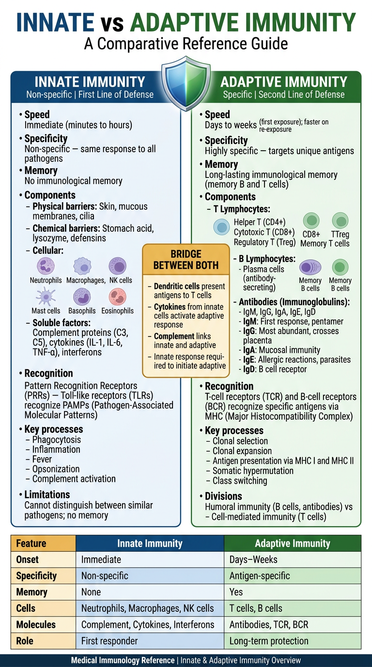
- Immediate response (minutes-hours), non-specific
- Cells: Neutrophils, Macrophages, NK cells, Dendritic cells, Mast cells
- Barriers: Physical (skin, cilia, mucus) and chemical (lysozyme, defensins, stomach acid)
- Recognition via Pattern Recognition Receptors (PRRs) / Toll-like receptors (TLRs) detecting PAMPs
- Soluble factors: Complement (C3, C5), Cytokines (IL-1, IL-6, TNF-α), Interferons
- No memory formed
- Delayed response (days-weeks), highly antigen-specific
- Cells: Helper T (CD4+), Cytotoxic T (CD8+), Regulatory T, Plasma B cells, Memory B & T cells
- Humoral arm: Antibodies (IgM, IgG, IgA, IgE, IgD)
- Cell-mediated arm: T cell-directed killing of infected cells
- Recognition via TCR/BCR + MHC class I and II presentation
- Forms long-lasting immunological memory (basis of vaccination)
- Dendritic cells physically link both systems by processing and presenting antigens to T cells
- Innate cytokines are required to initiate and shape the adaptive response
- Complement proteins serve roles in both arms
| Feature | Innate | Adaptive |
|---|---|---|
| Onset | Immediate | Days-Weeks |
| Specificity | Non-specific | Antigen-specific |
| Memory | None | Yes |
| Key cells | Neutrophils, Macrophages, NK | T cells, B cells |
| Key molecules | Complement, Cytokines | Antibodies, TCR, BCR |
According to guyton give me the entire short and simple summary of immunity
/textbooks/9780443111013/9780443111013_block10.md starting at line 1832. Let me get the full immunity section from Guyton, including its table of contents.Immunity - Summary According to Guyton & Hall (Medical Physiology)
1. Innate (Non-specific) Immunity
- Physical/mechanical barriers - skin, cilia, mucous membranes that trap pathogens before they enter
- Chemical barriers - stomach acid, lysozyme in secretions, defensins on mucosa
- Reticuloendothelial (Macrophage) System - tissue macrophages in lymph nodes, spleen, liver (Kupffer cells), lungs (alveolar macrophages), bone marrow, and subcutaneous tissue phagocytize invading organisms before they spread
- Vasodilation and increased local blood flow
- Increased capillary permeability - fluid leakage into tissues
- Migration of neutrophils and monocytes into tissue
- Tissue swelling
2. Acquired (Adaptive) Immunity
Why it's needed
Two Types
| Type | Also Called | Mediator | Mechanism |
|---|---|---|---|
| Humoral immunity | B-cell immunity | Circulating antibodies (globulins in plasma) | B lymphocytes produce antibodies that attack the invading agent |
| Cell-mediated immunity | T-cell immunity | Activated T lymphocytes | T cells are crafted in lymph nodes to directly destroy foreign agents |
3. Antigens - The Triggers of Acquired Immunity
- Each invading organism/toxin has unique chemical compounds called antigens (antibody generators)
- Must have a molecular weight of >8000 to be antigenic
- Usually proteins or large polysaccharides because they have recurring surface molecular groups called epitopes
- Antigens activate only the lymphocyte clones that carry matching surface receptors
4. Lymphocytes - The Core of Acquired Immunity
- Lymph nodes (primary location)
- Spleen, thymus, bone marrow, gastrointestinal submucosal tissue
- B lymphocytes - preprocessed in bone marrow; produce antibodies
- T lymphocytes - preprocessed in the thymus; mediate cell killing
Clonal Selection
5. Role of Macrophages in Activation
6. B-Lymphocyte System: Humoral Immunity
- Resting B cells carry ~100,000 antibody molecules on their surface
- On antigen exposure + T-helper cell signals, B cells transform into plasma cells
- Plasma cells secrete antibodies into blood and lymph
Antibodies (Immunoglobulins)
- Large protein molecules of the gamma globulin class
- Each antibody is specific - only reacts with the antigen that triggered it
- Classes: IgM, IgG, IgA, IgE, IgD
- IgM is produced first (primary response); IgG dominates on re-exposure
Mechanisms of Antibody Action
- Agglutination - cross-linking antigens on cells, causing clumping
- Precipitation - making soluble antigens insoluble so they can be phagocytized
- Neutralization - covering toxic sites on antigens, rendering them harmless
- Lysis - directly rupturing cell membranes of invaders (rare, needs complement)
- Opsonization - coating bacteria to make them more palatable to phagocytes
Memory
7. T-Lymphocyte System: Cell-Mediated Immunity
Helper T Cells (CD4+)
- Most numerous T cell type
- Secrete lymphokines (cytokines) that amplify the entire immune response
- Activate B cells to produce more antibodies
- Activate cytotoxic T cells
- Without helper T cells, the immune system is severely crippled (as in HIV/AIDS)
Cytotoxic T Cells (CD8+) - "Killer Cells"
- Directly attack and kill microorganisms and infected cells
- Bind tightly to target cells bearing the appropriate antigen
- Release perforins (hole-forming proteins) that punch pores in the target cell membrane
- Release cytotoxic substances directly into the attacked cell - cell swells and dissolves
- One cytotoxic T cell can kill multiple targets and move on
- Especially important for virus-infected cells, cancer cells, and transplanted cells
Regulatory T Cells (Tregs, CD4+)
- Suppress functions of cytotoxic and helper T cells
- Prevent excessive immune reactions that could damage the body's own tissues
- Important in immune tolerance and prevention of autoimmunity
- In cancer, Tregs can suppress anti-tumor immunity (current target of immunotherapy)
8. Complement System
- Classical pathway - triggered by antigen-antibody binding
- Alternative pathway - directly triggered by bacterial surfaces (innate immunity)
Effects of Complement Activation
- Opsonization - C3b coats bacteria, greatly enhancing phagocytosis (up to 100-fold)
- Lysis - Membrane Attack Complex (C5b6789) punches pores in bacterial membranes causing osmotic rupture
- Agglutination - clumps invading organisms together
- Viral neutralization - inactivates some viruses
- Chemotaxis - C5a attracts neutrophils and macrophages to the site
- Mast cell activation - C3a, C4a, C5a trigger release of histamine and heparin, promoting local inflammation
9. Immunological Tolerance (Self-Tolerance)
10. Passive Immunity
- Natural passive immunity - maternal IgG crosses the placenta; maternal IgA in breast milk protects the newborn
- Artificial passive immunity - injection of preformed antibodies (e.g., antitoxins, anti-venom, immunoglobulin preparations)
11. Sex Differences in Immunity
- Greater macrophage activation and phagocytic activity
- Higher interferon production after viral infections
- Stronger adaptive responses - more T cell expansion, stronger humoral responses, better vaccine responses
Quick Reference Table
| Feature | Innate | Acquired/Adaptive |
|---|---|---|
| Specificity | Non-specific | Highly specific |
| Speed | Immediate | Days-weeks (first exposure) |
| Memory | None | Yes (long-lasting) |
| Key cells | Neutrophils, Macrophages, NK cells | B cells, T cells |
| Key molecules | Complement, Cytokines, Interferons | Antibodies (IgG, IgM, IgA, IgE, IgD) |
| Trigger | PAMPs, tissue damage | Antigens (proteins, polysaccharides >8000 MW) |
Source: Guyton and Hall Textbook of Medical Physiology, Chapter 34-35 (Resistance of the Body to Infection; Immunity and Allergy)