
What causes pedal edema in pregnancy
Causes of Pedal Edema in Pregnancy
1. Massive Plasma Volume Expansion
- Creasy & Resnik's Maternal-Fetal Medicine, p. 208 & 213
- Brenner and Rector's The Kidney, p. 2140
2. RAAS Activation and Sodium/Water Retention
- Creasy & Resnik's Maternal-Fetal Medicine, p. 208
- Brenner and Rector's The Kidney, p. 2140
3. Reduced Colloid Osmotic Pressure (Hypoalbuminemia)
- Creasy & Resnik's Maternal-Fetal Medicine, p. 214
4. Mechanical Compression of the Inferior Vena Cava (IVC)
- Increases hydrostatic pressure in the lower limb veins
- Reduces venous return from the legs
- Promotes transudation of fluid into the interstitial space of the feet and ankles
- Robbins & Kumar Basic Pathology, p. 3343
- Campbell Walsh Wein Urology, p. 2583
5. Hormonal Effects (Progesterone and Estrogen)
- Progesterone promotes smooth muscle relaxation, reducing venous tone and increasing venous capacitance, which contributes to venous pooling in the lower limbs.
- Estrogen stimulates hepatic production of angiotensinogen and promotes aldosterone-mediated sodium retention, further expanding extracellular volume.
- Relaxin (a peptide hormone rising in pregnancy) also promotes systemic vasodilation and increases vascular permeability, contributing to the edematous tendency.
6. Increased Capillary Permeability
Summary: Starling Forces in Pregnancy
| Factor | Change in Pregnancy | Effect on Edema |
|---|---|---|
| Capillary hydrostatic pressure | Increased (IVC compression) | Promotes edema |
| Plasma colloid osmotic pressure | Decreased (dilutional hypoalbuminemia) | Promotes edema |
| Interstitial hydrostatic pressure | Relatively low | Promotes edema |
| Sodium/water retention (RAAS) | Increased | Promotes edema |
| Venous capacitance (progesterone) | Increased | Promotes edema |
Clinical Note
What is the mechanism of pain during wisdom tooth eruption
mechanism of pain wisdom tooth eruption pericoronitis inflammatory mediators
Mechanism of Pain During Wisdom Tooth Eruption
1. Mechanical Pressure on the Periodontal Ligament and Bone
2. Gingival Tissue Stretching and Rupture
- Bradykinin - a potent sensitizer of free nerve endings
- Prostaglandins (PGE2) - lower the pain threshold of nociceptors (peripheral sensitization)
- Histamine and serotonin - from mast cells and platelets in the traumatized tissue
- Substance P and CGRP - released from the nociceptor terminals themselves (neurogenic inflammation), causing local vasodilation and plasma extravasation (flare reaction)
3. Pericoronitis - The Major Source of Acute Pain
- The crown partially erupts but remains covered by a flap of gingival tissue called the operculum
- The operculum creates a deep, stagnant pocket between the tooth surface and the overlying gum
- Food debris, saliva, and oral bacteria (predominantly gram-negative anaerobes - Fusobacterium, spirochetes, Prevotella) accumulate in this pocket and cannot be cleaned
- Bacteria trigger a polymicrobial inflammatory cascade - the immune system releases IL-1β, TNF-α, IL-6, and prostaglandins into the tissue
- The resulting tissue edema and purulent exudate increase hydrostatic pressure within the closed space of the operculum, compressing local nerve fibers - a major source of the throbbing, pressure-type pain
- Trauma from mastication worsens this: the upper molar occludes directly against the swollen operculum during biting, mechanically re-injuring the inflamed tissue with every chew
- Rosen's Emergency Medicine, p. 860
- Tintinalli's Emergency Medicine, p. 1622-1623
- Scott-Brown's Otorhinolaryngology Head & Neck Surgery, p. 702
4. Trigeminal Nerve Sensitization and Referred Pain
- Trismus (jaw spasm/limitation of mouth opening) - reflex muscle guarding mediated by the masseteric and pterygoid branches
- Earache - referred pain via the auriculotemporal nerve
- Sore throat / odynophagia - if infection spreads to the pterygomandibular or parapharyngeal spaces
5. Spreading Infection (Severe Cases)
- Tissue destruction and rising inflammatory cytokines
- Nerve compression from expanding soft tissue swelling
- In extreme cases, Ludwig's angina (floor-of-mouth cellulitis) - a life-threatening emergency
Summary Table
| Mechanism | Pain Type | Key Mediators |
|---|---|---|
| Pressure on PDL/bone during eruption | Deep, dull ache | Mechanoreceptor activation |
| Gingival tearing/stretching | Sharp, acute | Bradykinin, PGE2, histamine, substance P |
| Pericoronitis (operculum infection) | Throbbing, severe | IL-1β, TNF-α, PGE2, bacterial toxins |
| Masticatory trauma to operculum | Pain on biting | Mechanical nociceptor activation |
| Central sensitization (V3) | Referred, diffuse | Trigeminal nucleus sensitization |
| Deep space infection | Constant, severe | Pus/edema pressure, cytokines |
Why NSAIDs Work So Well
- Rosen's Emergency Medicine, p. 861
- Tintinalli's Emergency Medicine, p. 1623
Can i take mefthal forte for the above?
Mefthal Forte mefenamic acid dicyclomine composition uses dental pain
Meftal Forte composition mefenamic acid 500mg dicyclomine 10mg uses side effects
https://pharmeasy.in/online-medicine-order/meftal-forte-tabl…
Can You Take Meftal Forte for Wisdom Tooth Pain?
What Is Meftal Forte?
| Drug | Composition |
|---|---|
| Meftal Forte | Mefenamic Acid 500 mg + Paracetamol 325 mg |
| Meftal Spas | Mefenamic Acid 250 mg + Dicyclomine 10 mg |
How Does Meftal Forte Work for Tooth Pain?
- Inhibits both COX-1 and COX-2 enzymes, blocking prostaglandin synthesis
- Prostaglandins (especially PGE2) are the major sensitizers of nociceptors in inflamed gingival tissue and the operculum in pericoronitis
- Directly counters the core mechanism of wisdom tooth pain
- Has both analgesic and anti-inflammatory effects - reduces both pain and swelling
- This is the same mechanism as ibuprofen (the textbook first-line NSAID for pericoronitis pain), just a different NSAID class
- Provides central analgesic effect (inhibits prostaglandin synthesis in the CNS, possibly via COX-3)
- Works synergistically with the NSAID - together they provide better pain relief than either alone (the combination covers both peripheral and central pain pathways)
Important Caveats
- Peptic ulcer or history of GI bleeding
- Asthma (NSAIDs can trigger bronchospasm in aspirin-sensitive asthmatics)
- Kidney or liver disease
- Currently pregnant (especially avoid in the third trimester - NSAIDs can cause premature closure of ductus arteriosus)
- Already taking blood thinners (warfarin, aspirin) or other NSAIDs
- Irrigation and cleaning under the operculum
- Antibiotics if there is spreading infection (penicillin or metronidazole are standard)
- Possible extraction of the impacted tooth
Bottom Line
Yes, Meftal Forte is a reasonable OTC choice for short-term relief of wisdom tooth pain. The mefenamic acid directly blocks the inflammatory prostaglandin cascade driving the pain. Take it after food, limit use to a few days, and see a dentist - pain relief is not a substitute for treating the underlying cause.
What all will be the symptoms of wisdom tooth eruption pain
Symptoms of Wisdom Tooth Eruption Pain
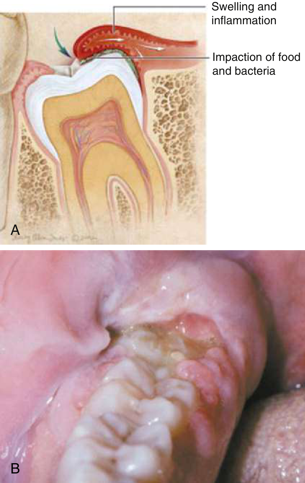
Local Symptoms (At the Tooth Site)
| Symptom | Description |
|---|---|
| Pain at the back of the mouth | Throbbing or dull ache at the lower/upper back jaw; typically the first and most prominent symptom |
| Swollen, red gum (operculum) | The gum flap overlying the erupting tooth becomes erythematous, edematous, and tender to touch |
| Pain worsened by biting/chewing | The upper tooth occludes directly against the inflamed operculum, re-injuring it with every bite |
| Bleeding gum | The inflamed operculum bleeds easily on contact or when probed |
| Pus or discharge | In pericoronitis, a purulent discharge may ooze from under the gum flap |
| Bad taste in the mouth | From purulent discharge and bacterial overgrowth in the stagnant pocket under the operculum |
- Rosen's Emergency Medicine, p. 860
Referred and Radiating Symptoms
| Symptom | Cause |
|---|---|
| Earache (otalgia) | Referred pain via the auriculotemporal nerve (branch of V3); the ear feels painful even though the problem is the tooth |
| Jaw or temple pain | Spread of pain along the inferior alveolar and masseteric branches of V3 |
| Sore throat / pain on swallowing | If infection tracks toward the pterygomandibular or parapharyngeal space |
Functional Symptoms
| Symptom | Cause |
|---|---|
| Trismus (difficulty opening the mouth) | Reflex muscle spasm of the masseter and medial pterygoid muscles; occurs when inflammation is near these muscles; a key sign of pericoronitis - Scott-Brown's Otorhinolaryngology, p. 702 |
| Difficulty chewing | Combination of trismus, pain on biting, and gum swelling |
| Difficulty swallowing (odynophagia) | If infection spreads to peritonsillar or parapharyngeal spaces |
Systemic Symptoms (in More Severe Cases)
| Symptom | Significance |
|---|---|
| Fever and malaise | Indicates spreading infection beyond the local gum tissue; warrants antibiotics |
| Facial swelling | Localized soft tissue edema over the jaw/cheek from the spreading inflammatory process |
| Regional lymphadenopathy | Swollen, tender lymph nodes under the jaw (submandibular) and neck - a clinical sign of infection, not simple eruption - Scott-Brown's, p. 702 |
| Headache | From systemic inflammation and central sensitization of the trigeminal nucleus |
Red Flag Symptoms (Seek Urgent Care)
- Progressive trismus - worsening inability to open the mouth
- High fever (>38.5°C) with chills
- Neck swelling or stiffness
- Difficulty swallowing or breathing
- Floor of mouth swelling - this suggests Ludwig's angina (bilateral cellulitis of the floor of the mouth), an airway emergency that can originate from third molar infections - Cummings Otolaryngology, p. 1953
If any of these are present, go to an emergency department immediately - not just a dentist.
How Symptoms Tend to Progress
Simple eruption         Pericoronitis              Spreading infection
─────────────────  →  ─────────────────────  →  ─────────────────────────
Mild ache               Throbbing pain             Severe pain + fever
Gum tenderness          Swollen operculum          Trismus + facial swelling
                        Bad taste + pus            Lymphadenopathy
                        Pain on biting             Dysphagia
                        Referred earache           [Ludwig's angina risk]
- Tintinalli's Emergency Medicine, p. 1622-1623
- Scott-Brown's Otorhinolaryngology Head & Neck Surgery, p. 702
- Rosen's Emergency Medicine, p. 860-861
How much iron is lost at the time of delivery
Iron Lost at the Time of Delivery
Blood loss at delivery: average 150 mg (range 90-310 mg)
- Creasy & Resnik's Maternal-Fetal Medicine, p. 1332 (Table 55.2)
Full Iron Balance of Pregnancy (The Complete Picture)
| Component | Average (mg) | Range (mg) |
|---|---|---|
| External iron loss (daily basal losses) | 170 | 150-200 |
| Expansion of red blood cell mass | 450 | 200-600 |
| Fetal iron | 270 | 200-370 |
| Iron in placenta and cord | 90 | 30-170 |
| Blood loss at delivery | 150 | 90-310 |
| Total requirement | 1130 | 580-1340 |
Key Points to Note
-
Iron lost at delivery (150 mg) represents the iron contained in the blood shed during childbirth. Each milliliter of blood contains approximately 0.5 mg of iron, so this corresponds to roughly 300 mL of blood loss at a normal vaginal delivery.
-
This figure is separate from the 270 mg transferred to the fetus and 90 mg in the placenta and cord - those are not "lost at delivery" in the hemorrhagic sense but are the physiological costs of supporting fetal growth throughout pregnancy.
-
A different framing from Creasy & Resnik's cardiovascular chapter sums it slightly differently:
- 500 mg for maternal RBC mass expansion
- 300 mg transferred to fetus
- 200 mg for daily losses during pregnancy
- Total ~1000 mg (a rounded figure used for clinical teaching)
- Creasy & Resnik's Maternal-Fetal Medicine, p. 208
-
The range matters clinically. A cesarean section or complicated vaginal delivery with postpartum hemorrhage can push blood loss iron well above 310 mg, which is why iron stores and hemoglobin are checked postnatally.
-
Most women enter pregnancy with marginal iron stores - studies show nearly 50% of primigravidas have minimal marrow iron stores in the first trimester. This means delivery blood loss on top of the cumulative demands of pregnancy leaves many women iron-depleted postpartum.
Clinical Implication

In this what can we answer for loss of iron
Filling the Blanks in Your Notes
150 mg (range: 90–310 mg)120–150 mgExplanation
| Item | Value |
|---|---|
| Total iron required during pregnancy | 1200 mg (your notes) |
| Fetus and placenta | 300 mg |
| Growing RBC of mother | 500 mg |
| Lost through sweat, urine, faeces | 200 mg |
| Lost at time of delivery | 150 mg |
| Saved due to amenorrhea | ~120–150 mg |
| Net requirement (1200 − 300 saved) | 900–1000 mg |
-
The 150 mg at delivery comes from blood lost during childbirth (~300 mL blood × 0.5 mg iron/mL = ~150 mg). This matches exactly with Creasy & Resnik's Maternal-Fetal Medicine, Table 55.2.
-
The amenorrhea saving is calculated from the fact that a woman normally loses 28–36 mL of blood per menstrual cycle = ~14–18 mg of iron per month. Over 9 months of pregnancy amenorrhea: 9 × ~14–16 mg ≈ 120–150 mg saved.
-
Your notes then do the math: 1200 mg total cost − ~300 mg saved (amenorrhea + other offsets) = 900–1000 mg net requirement during pregnancy.
Lost at delivery = 150 mg Saved by amenorrhea = 120–150 mg
What is bite in wisdom tooth cherya
What did you mean by 'cherya'? Which of these are you asking about?
bite force wisdom tooth third molar chewing pain occlusal trauma operculum
Bite (Chewing/Occlusal Force) and Wisdom Tooth Pain
The Core Problem - Occlusal Trauma on the Operculum
The upper molar bites directly down onto the swollen operculum, re-injuring the inflamed tissue with each chew.
- Rosen's Emergency Medicine, p. 860 - "This condition [pericoronitis] is exacerbated by trauma from mastication."
Why Bite/Chewing Hurts More Than Rest Pain
| Situation | What Happens | Pain Level |
|---|---|---|
| At rest | Inflammatory mediators (PGE2, bradykinin) sensitize nerve endings | Dull, background ache |
| On biting/chewing | Upper molar mechanically crushes the inflamed operculum | Sharp, severe, sudden spike in pain |
| On jaw opening (trismus) | Pterygoid and masseter muscles, already in reflex spasm, are stretched | Pain + restricted opening |
The Neuroscience of Bite Force and Dental Pain
-
Normally help fine-tune bite force and give you the sensation of what you are chewing
-
When the surrounding tissue is inflamed (pericoronitis), these receptors become peripherally sensitized - meaning even a light bite that normally wouldn't hurt now triggers intense pain
-
Dental pain is carried by A-delta fibers (sharp, bright pain on biting) and C fibers (dull, burning background pain)
-
Cummings Otolaryngology, p. 3981-3983
The Vicious Cycle of Chewing and Pericoronitis
Eat/bite
    ↓
Upper molar strikes inflamed operculum
    ↓
Mechanical injury → more tissue damage
    ↓
More inflammation, more swelling
    ↓
More sensitization of nerve endings
    ↓
Even lighter biting causes more pain
    ↓
(Repeat with every meal)
Practical Implications
- Eat soft foods during a flare - hard, crunchy foods that require strong biting directly worsen pericoronitis
- Chew on the opposite side to avoid trauma to the affected operculum
- NSAIDs (like Meftal Forte) work here because they reduce prostaglandin-mediated sensitization, so biting becomes less painful
- The definitive solution is removal of the upper wisdom tooth (if it is the one biting the operculum) or extraction of the lower wisdom tooth itself - this eliminates the occlusal trauma entirely
Can you give an image for the above
A detailed dental diagram showing occlusal trauma during wisdom tooth eruption. The lower third molar (wisdom tooth) is partially erupted with an inflamed red operculum (gum flap) covering part of the crown. The upper molar is shown biting down onto the inflamed operculum during chewing, causing mechanical trauma. Labels pointing to: operculum (inflamed gum flap), partially erupted wisdom tooth, food debris trapped under operculum, upper molar biting on operculum, swelling and inflammation, inferior alveolar nerve (V3). A second inset diagram shows the vicious cycle: bite force → operculum injury → more inflammation → more pain sensitization. Clean medical illustration style with anatomical cross-section view of the jaw.
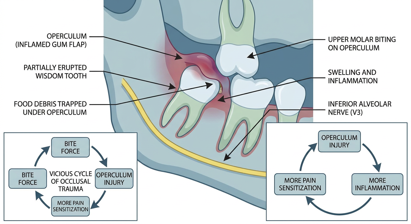
- The partially erupted lower wisdom tooth with the inflamed operculum (red gum flap) covering its crown
- The upper molar biting down onto the swollen operculum during chewing - this is the direct mechanical trauma
- Food and bacteria trapped under the gum flap
- The inferior alveolar nerve (V3) that carries the pain signals
- The vicious cycle - each bite injures the operculum further, driving more inflammation and pain sensitization
What home remedies help wisdom tooth pain (salt rinses, ice)?
salt water rinse ice pack clove oil home remedies wisdom tooth pericoronitis evidence
https://www.medicalnewstoday.com/articles/319461
Home Remedies for Wisdom Tooth Pain
Important upfront: Home remedies provide temporary symptom relief only. They do not cure pericoronitis or impaction. If pain is severe, spreading, or accompanied by fever/trismus, see a dentist.
✅ 1. Salt Water (Saline) Rinse - Most Recommended
- Flushes food debris and bacteria out from under the operculum - directly addressing the source of pericoronitis
- Creates a hyperosmotic environment that reduces bacterial growth
- Mild anti-inflammatory effect on the gum tissue
- Promotes healing of inflamed mucosa
- Dissolve ½ teaspoon of salt in 1 cup (240 mL) of warm water
- Swish gently (not forcefully) around the back of the mouth for 30-60 seconds
- Spit out - do not swallow
- Repeat 2-3 times a day, especially after meals
- Tintinalli's Emergency Medicine, p. 1623
- Rosen's Emergency Medicine, p. 861
✅ 2. Ice Pack (Cold Compress) - Good for Swelling
- Causes vasoconstriction - reduces blood flow to the area, limiting swelling and edema
- Numbs the area by slowing nerve conduction velocity (cold-induced analgesia)
- Reduces inflammatory exudate accumulation
- Wrap an ice pack or bag of frozen peas in a cloth or towel (never apply ice directly to skin)
- Apply to the outside of the jaw/cheek over the affected side
- 15 minutes on, 15 minutes off - repeat as needed
- Most useful in the first 24-48 hours of a flare
✅ 3. Clove Oil (Eugenol) - Effective Topical Numbing
- Contains eugenol - a natural COX inhibitor and sodium channel blocker that produces local anesthesia
- Directly numbs the nerve endings in the inflamed gum tissue
- Has some antibacterial activity
- Dip a cotton ball or cotton swab in a small amount of clove oil
- Hold it gently against the painful gum for a few minutes
- Do not swallow - eugenol can irritate the throat and stomach in large amounts
- Alternatively, place a whole clove between the back teeth near the painful area and let it sit
✅ 4. Chlorhexidine Mouthwash - Stronger Than Salt Water
- Broad-spectrum antimicrobial - kills the gram-negative anaerobes driving pericoronitis
- More effective than saline for bacterial reduction
- Available OTC as 0.12%-0.2% chlorhexidine gluconate mouthwash (e.g., Hexidine, Clohex)
- Swish 10-15 mL for 30-60 seconds, twice daily
- Do not rinse with water immediately after - this reduces effectiveness
- Rosen's Emergency Medicine, p. 861
✅ 5. OTC Pain Medications
| Drug | Dose | Why it helps |
|---|---|---|
| Ibuprofen (Brufen, Advil) | 400-600 mg every 6-8 hrs with food | COX inhibitor - reduces prostaglandin-driven inflammation AND pain |
| Paracetamol (Calpol, Crocin) | 500-650 mg every 6 hrs | Central analgesic |
| Meftal Forte | 1 tablet every 8 hrs with food | Mefenamic acid (NSAID) + Paracetamol combination |
| Topical benzocaine gel (e.g., Dentogel) | Apply small amount to gum | Numbs surface nerve endings directly |
✅ 6. Soft Diet + Eating on the Opposite Side
- Avoid hard, crunchy foods (chips, toast, nuts) - they directly traumatize the operculum
- Eat soft foods: rice, yogurt, soups, mashed foods
- Chew only on the side opposite the painful wisdom tooth
- This breaks the vicious cycle of mechanical re-injury with every meal
❌ What Does NOT Work
| Remedy | Why it doesn't help |
|---|---|
| Warm salt water alone overnight | Temporary only - bacteria repopulate quickly |
| Antibiotics without dentist review | Self-medicating antibiotics is inappropriate and builds resistance |
| Ignoring the pain | Pericoronitis can spread to deep fascial spaces if untreated |
| Alcohol (whiskey on gums) | Minimal numbing; irritates already-inflamed tissue |
Quick Summary
Best home trio for wisdom tooth pain:
1. Salt water rinse after every meal (clears debris)
2. Ice pack on cheek (reduces swelling)
3. Ibuprofen / Meftal Forte (blocks inflammation)
+ Clove oil / chlorhexidine for extra relief