
Feline esophagus
feline esophagus histology anatomy
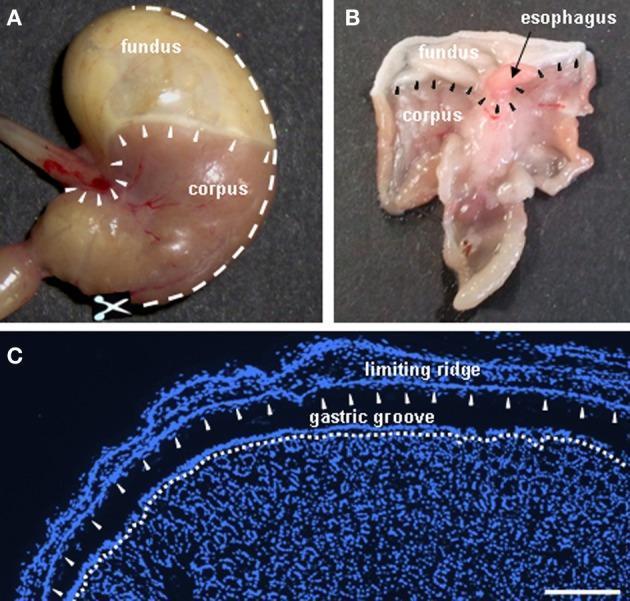
This educational image panel illustrates the gross anatomy and histology of the murine stomach, specifically highlighting the boundary between the non-glandular and glandular mucosa. Panel A shows an intact murine stomach where the fundus (lighter, smoother appearance) is distinguished from the corpus (darker, vascularized appearance). White arrowheads delineate the 'limiting ridge,' a structural fold encircling the esophageal orifice and separating the two regions. Panel B displays the stomach dissected along the greater curvature, exposing the internal mucosal surface and the relationship between the esophagus, fundus, and corpus. Panel C is a DAPI-stained fluorescent cryosection (nuclei in blue) showing the microanatomy of the fundus/corpus boundary. It clearly demonstrates the 'limiting ridge' as a tissue strip overarching the 'gastric groove.' The most apical cell layer of the corpus mucosa, which forms the distal wall of this groove, is indicated by a white broken line and arrowheads. This region is a known site for specialized chemosensory brush cells. Scale bar for panel C represents 200 μm.
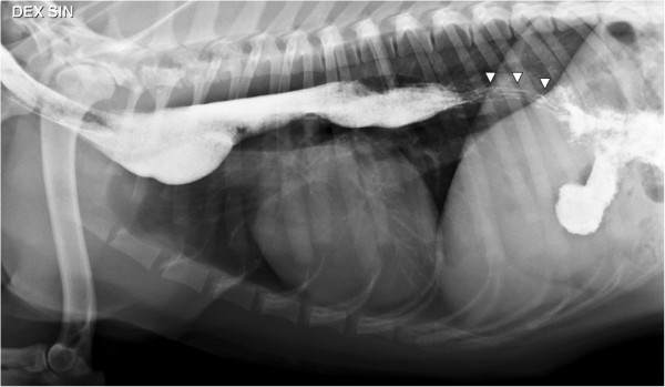
**Imaging Modality:** Lateral thoracic radiograph; positive-contrast esophagram (barium study). **Anatomical Region:** Thorax, specifically the esophagus and cranial abdomen. **Observed Pathology:** * **Megaesophagus:** Generalized, moderate-to-severe esophageal dilation extending from the cervical region to the diaphragm. * **Esophageal Diverticulum:** Focal ventral saccular distension of the esophagus located in the cranial mediastinum, just cranial to the cardiac silhouette. * **Herringbone Pattern:** Characteristic "striped" or "herringbone" mucosal pattern visible in the distal (caudal) thoracic esophagus (indicated by arrowheads), representing normal feline esophageal anatomy where the distal third is composed of smooth muscle with circumferential mucosal folds. **Characteristic Visual Features:** * Radio-opaque barium contrast outlines the dilated esophageal lumen. * Significant contrast retention within the thoracic esophagus. * Contrast passage into the stomach (gastrogram) is present. * No evidence of aspiration pneumonia is visible in the visualized lung fields. **Diagnostic Features:** The combination of generalized dilation with a focal ventral pouching (diverticulum) and a normal distal feline herringbone pattern. These findings are consistent with chronic esophageal dysmotility and secondary structural changes.
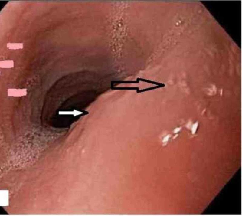
An endoscopic view of the human esophagus demonstrating hallmark features of eosinophilic esophagitis (EoE). The diagnostic image reveals a significantly narrowed esophageal lumen due to a benign-appearing stenosis, estimated at approximately 1 cm in diameter. Key diagnostic findings include the presence of multiple circumferential esophageal rings, also known as trachealization or feline esophagus, which appear as raised, concentric mucosal bands. The mucosa exhibits typical inflammatory changes, including subtle longitudinal furrowing and irregularities around the stenotic region. Black and white arrows indicate the localized area of narrowing and the distal rings. This visual evidence supports a clinical diagnosis of EoE, often associated with dysphagia and food bolus impaction. The image is categorized under Gastroenterology and serves as an educational example of structural esophageal changes related to chronic allergic inflammation.
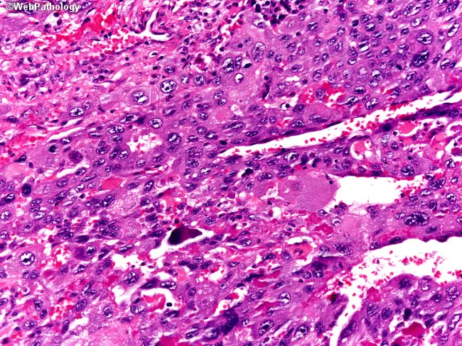
Histology: Formalin-fixed Barrett esophagus biopsy examined by light microscopy. The section is stained with Hematoxylin and Eosin (H&E). It shows a biphasic trophoblastic tumor consisting of cytotrophoblasts admixed with prominent syncytiotrophoblastic giant cells in a hemorrhagic background. The cytotrophoblasts are round to polygonal, with vesicular nuclei, indistinct cytoplasmic borders, and mild to moderate pleomorphism; mitotic activity may be evident. The syncytiotrophoblastic giant cells display abundant cytoplasm, multinucleation, and marked nuclear variability, creating a characteristic multinucleated syncytial pattern. There is extensive hemorrhage and occasional necrotic foci within the lesion, with nests and sheets of tumor cells infiltrating the esophageal mucosa and extending into the submucosa in an irregular, infiltrative fashion. In the Barrett context, this represents choriocarcinomatous differentiation in an esophageal lesion, an unusual but documented association. The differential diagnosis includes squamous cell carcinoma, adenosquamous carcinoma, neuroendocrine carcinoma, and sarcomatoid carcinoma, among others. Clinically, such histology carries implications for prognosis and therapy, often prompting systemic treatment regimens aligned with gestational trophoblastic disease or mixed esophageal carcinomas, in conjunction with endoscopic and imaging staging. Immunohistochemistry, when performed, commonly demonstrates trophoblastic markers (e.g., hCG) supporting the diagnosis. This image highlights biphasic trophoblastic differentiation and hemorrhagic context critical for accurate, timely diagnosis.
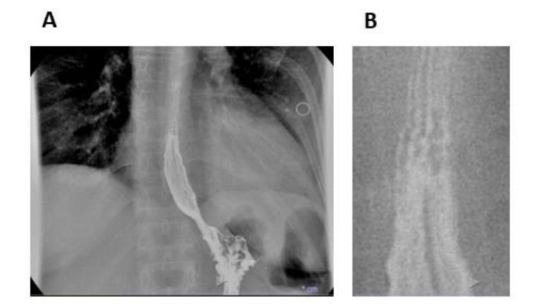
This diagnostic image set features a videofluoroscopic swallowing study and esophageal transit evaluation using barium contrast. Image A presents a posterior-anterior fluoroscopic view of the thoracic esophagus, revealing an altered esophageal profile characterized by a mildly spiculed or serrated outline along its margins, rather than a smooth mucosal surface. Image B provides a magnified detail of the proximal barium column, where subtle horizontal lines are visible across the esophageal lumen. These radiographic findings, known as 'feline esophagus' or transient esophageal rings, are classic markers of eosinophilic esophagitis (EoE). The images demonstrate the use of contrast radiography in detecting structural and mucosal changes associated with chronic esophageal inflammation, assisting in the differentiation of motility disorders and inflammatory conditions. This content is suitable for medical students and gastroenterology specialists studying diagnostic imaging of dysphagia and esophageal pathologies.
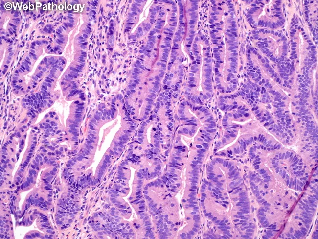
Histology image of Barrett esophagus with low-grade dysplasia illustrating intestinal metaplasia of the distal esophageal mucosa. The material is a formalin-fixed, paraffin-embedded biopsy section stained with hematoxylin and eosin and viewed at high magnification. The mucosa shows tubular glands with goblet cells characteristic of Barrett mucosa, embedded in lamina propria and terminating at the muscularis mucosae. In LGD, cytologic atypia is evident: mild to moderate nuclear pleomorphism, increased nuclear-to-cytoplasmic ratio, hyperchromasia, and pseudostratification of nuclei within dysplastic gland bases. Architectural crowding with elongated, irregular glands and occasional outpouchings is compatible with dysplastic change. Normal-appearing Barrett epithelium with intestinal metaplasia is present but lacks high-grade features or invasion. There is no invasion beyond the mucosa; no desmoplasia or submucosal invasion is seen. Clinically, Barrett esophagus with LGD carries a higher risk of progression to high-grade dysplasia or adenocarcinoma and supports intensified endoscopic surveillance or therapeutic eradication strategies. This image is relevant for educational comparison of LGD versus non-dysplastic BE and for radiologic-pathologic correlation, pathology slide review, and multidisciplinary case discussion in gastroenterology and pathology. No invasion beyond the muscularis mucosa is evident, underscoring mucosal-confined dysplasia. The image aids teaching on LGD criteria, biopsy interpretation, and risk stratification guiding endoscopic management today.
"Feline Esophagus"
What It Is
Radiologic Appearance
Primary Association: Eosinophilic Esophagitis (EoE)
| Feature | Detail |
|---|---|
| Pathogenesis | Immune/allergic disorder; often associated with atopy (eczema, asthma, allergic rhinitis) |
| Population | More common in children and young adult males |
| Symptoms | Dysphagia, food impaction, vomiting, chest/epigastric pain; infants show feeding intolerance |
| Endoscopic findings | Mucosal rings (feline/ringed esophagus), longitudinal furrowing, white papules (eosinophilic abscesses), strictures |
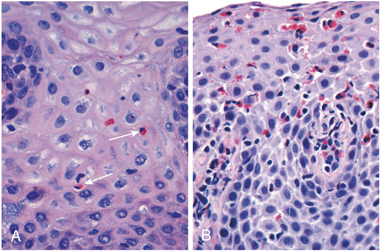
| Histology | >15 eosinophils per high-power field (x40); basal zone hyperplasia; papillary lengthening |
| Diagnosis | Suggestive history + biopsy (≥5 specimens for ~100% sensitivity) + normal pH study or PPI-refractoriness |
Distinction from GERD and Other Rings
- The feline esophagus pattern is transient - rings may disappear on repeat endoscopy or when the esophagus is not fully distended, unlike the fixed Schatzki ring of GERD
- Clinical Gastrointestinal Endoscopy (3e) notes: "Often confused with feline esophagus (transient plications). Best appreciated when esophagus is fully insufflated"
- There can be substantial overlap between GERD and EoE endoscopic appearances
- In GERD, a "ringed" or feline pattern can also appear, making biopsy essential for definitive differentiation
Key Histologic Image (EoE)
Note on Actual Feline (Cat) Esophageal Anatomy
Eosinophilic esphgitis
"eosinophilic esophagitis"[MeSH Terms] AND treatment
eosinophilic esophagitis endoscopy rings furrowing histology eosinophils
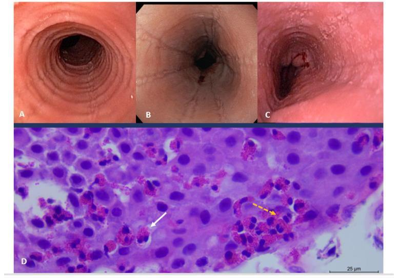
This composite educational image illustrates the diagnostic features of eosinophilic esophagitis (EoE) through endoscopy and histopathology. Upper panels (A, B, and C) display clinical endoscopic views of the esophagus. Panel A reveals trachealization, characterized by prominent circular rings. Panel B shows longitudinal linear furrows and narrowings. Panel C demonstrates mucosal fragility (crepe-paper esophagus) with visible erosions and minor surface bleeding. The lower panel (D) is a high-power light microscopy image of an H&E stained esophageal biopsy. It shows dense intraepithelial inflammation with numerous eosinophils (indicated by the dashed orange arrow), exceeding the diagnostic threshold of 15 eosinophils per high-power field. A solid white arrow points to an eosinophilic microabscess, a collection of eosinophils within the squamous epithelium. The image serves as a clinical reference for gastroenterology and pathology, highlighting the correlation between macroscopic structural changes and microscopic inflammatory infiltrates in chronic allergic esophagitis.
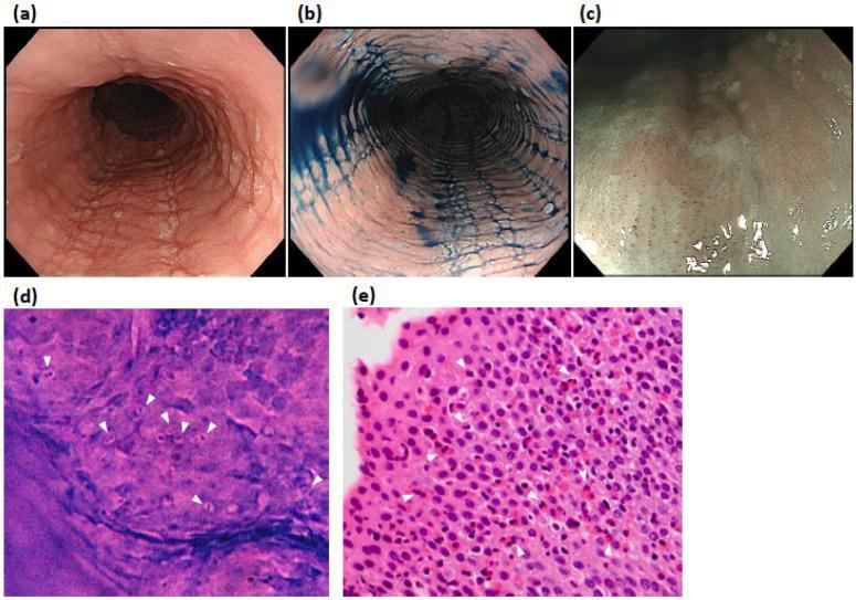
This composite educational image illustrates diagnostic modalities for Eosinophilic Esophagitis (EoE) in a 52-year-old patient. Panels (a) and (b) present White Light Endoscopy (WLE) and indigocarmine chromoendoscopy, respectively, showing classic endoscopic features: linear furrows, white exudates, and concentric rings (trachealization) within the esophageal lumen. Panel (c) utilizes Narrow-Band Imaging Magnifying Endoscopy (NBI-ME), demonstrating beige-colored mucosa and characteristic dot-shaped intrapapillary capillary loops (IPCLs) without visible cyan vessels. Panel (d) displays high-magnification endocytoscopy (x520), an in vivo imaging technique showing multiple cells with purple bilobed nuclei and nonstained peripheries (white arrowheads), characteristic of eosinophilic infiltration. Panel (e) provides histological correlation (H&E stain) from a biopsy of the same site, confirming dense eosinophilic infiltration (>50 eosinophils per high-power field) with prominent bilobed nuclei (white arrowheads). The image provides a comprehensive comparison between gross endoscopic findings, advanced in vivo microscopy, and gold-standard histopathology for gastroenterology education.
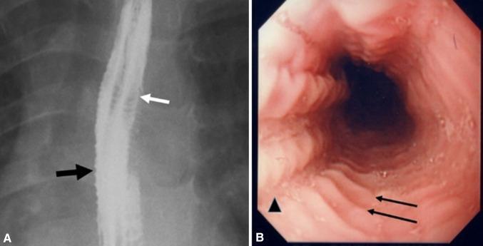
This dual-panel image displays diagnostic findings characteristic of Eosinophilic Esophagitis (EoE). Panel A is a lateral view of a barium esophagram showing a 'ringed' or 'corrugated' esophagus. A white arrow highlights multiple concentric, closely spaced esophageal rings, while a black arrow indicates mucosal irregularity and a narrow-caliber lumen. Panel B is a corresponding endoscopic image of the same patient, providing a luminal view. The endoscopy reveals prominent transverse concentric rings (feline esophagus appearance), indicated by black arrows, and longitudinal mucosal furrowing, indicated by an arrowhead. These features represent the classic remodeling and inflammatory changes seen in pediatric and adult patients presenting with dysphagia and food impaction. The side-by-side comparison demonstrates the radiological and endoscopic correlation of fixed esophageal rings and architectural distortion of the esophageal wall.
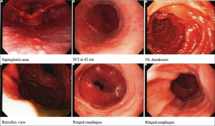
This composite of six clinical photographs demonstrates various endoscopic views from an upper gastrointestinal endoscopy (Esophagogastroduodenoscopy). The primary educational focus is on panels 5 and 6, which exhibit a 'ringed esophagus' (trachealization), a classic endoscopic hallmark of eosinophilic esophagitis (EoE). These panels reveal concentric, circular mucosal ridges or rings that cause a corrugated appearance and luminal narrowing in the distal esophagus. The esophageal mucosa appears erythematous with subtle longitudinal furrowing and small white exudates, representing eosinophilic microabscesses. Other panels include the supraglottic area, the squamocolumnar junction (SCJ) at 42 cm, a retroflexed view of the cardia, and the normal appearing (NL) duodenum. This visual material is critical for training in gastroenterology, specifically for identifying inflammatory conditions of the esophagus and differentiating them from mechanical strictures or gastroesophageal reflux disease (GERD).
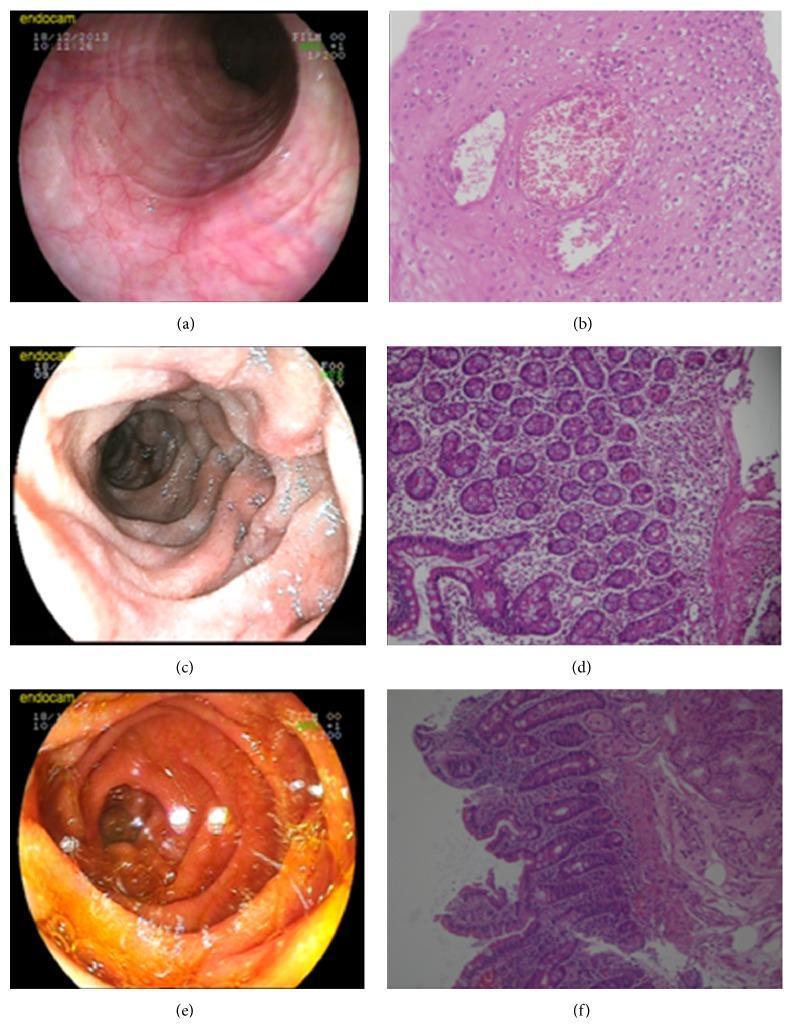
This composite figure illustrates the endoscopic and histological findings of eosinophilic gastroenteritis across three stages (initial presentation and follow-up). (a) Endoscopy of the esophagus shows classic 'felinization' characterized by concentric mucosal rings. (b) Corresponding H&E-stained esophageal biopsy (200x) displays a dense eosinophilic microabscess within the squamous epithelium. (c) Duodenal endoscopy reveals significant mucosal edema, congestion, and luminal narrowing. (d) H&E-stained duodenal biopsy (200x) demonstrates marked eosinophilic infiltration and inflammatory cells within the lamina propria and submucosa, disrupting normal architecture. (e) Follow-up endoscopy after three months of steroid therapy shows marked resolution of the duodenal edema and congestion. (f) Follow-up duodenal histology (200x) reveals restoration of normal mucosal architecture with resolved eosinophilic infiltration. The figure effectively contrasts diagnostic markers of eosinophilic esophagitis and duodenitis with post-treatment outcomes, emphasizing both gross endoscopic signs and microscopic cellular infiltration patterns.
PMID: 37491157
PMID: 39998943
Eosinophilic Esophagitis (EoE)
Definition
- Symptoms of esophageal dysfunction (dysphagia, food impaction, etc.)
- ≥15 eosinophils per high-power field (HPF, x40) in one or more esophageal biopsy specimens
- Absence of other causes of esophageal eosinophilia (GERD, Crohn disease, infections, autoimmune disorders)
Epidemiology
| Feature | Detail |
|---|---|
| Incidence | 0.1 - 1.2 per 10,000 worldwide |
| Gender | Strongly male predominant (up to 76% in adults) |
| Race | More common in Caucasian / non-Hispanic whites |
| Peak age | 20s-30s; mean diagnosis age ~34 years |
| Trend | Increasing incidence over past 2 decades, paralleling rise in atopic disorders |
Pathophysiology
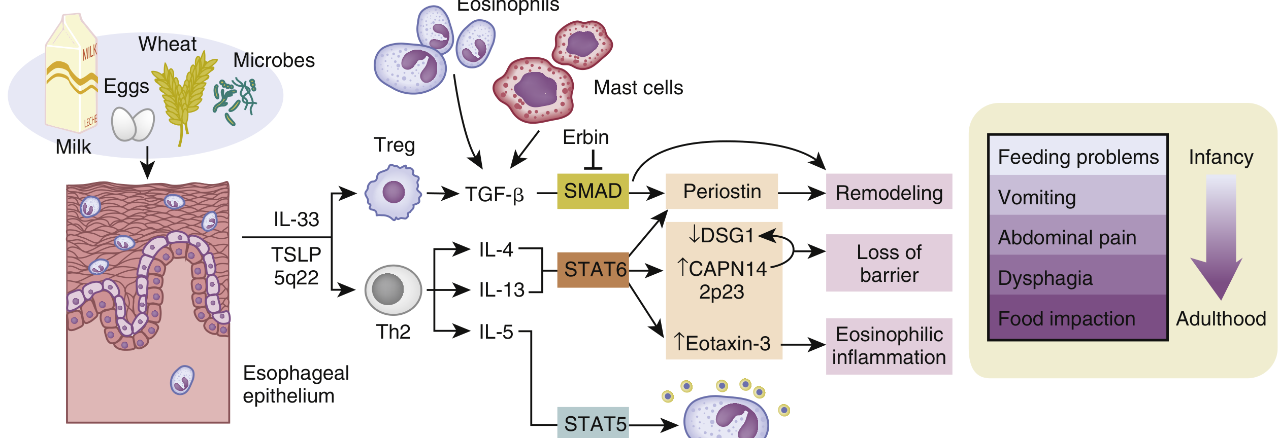
- Genetic factors: Variants in TSLP (thymic stromal lymphopoietin) and CAPN14 (calpain 14) confer susceptibility
- Trigger: Dietary antigens (food) and environmental allergens breach an impaired esophageal epithelial barrier
- Immune cascade: Innate activation (TSLP, IL-33) → Th2 skewing → IL-4, IL-5, IL-13 overproduction → eosinophil recruitment and degranulation
- Tissue damage: Eosinophil degranulation releases toxic proteins; chronic inflammation leads to fibrosis and stricture formation
- Atopic comorbidities: ~70% rhinitis, ~40% asthma, ~30% eczema, ~50% IgE-mediated food allergy
Disease Subtypes (Endotypes)
| Endotype | Features |
|---|---|
| EoE1 | Mild; normal-appearing esophagus; mild histologic, endoscopic, and molecular changes |
| EoEe2 | Inflammatory; highest expression of inflammatory cytokines and steroid-responsive genes; may have a refractory steroid phenotype |
| EoEe3a | Fibrostenotic; narrow-caliber esophagus; highest degree of endoscopic and histologic severity; lowest epithelial differentiation gene expression |
Clinical Presentation
- Reflux/vomiting, irritability, food refusal
- Early satiety, failure to thrive, feeding intolerance
- Dysphagia (most common presenting symptom)
- Food bolus impaction - most common cause in young adults
- Chest pain (central, not responding to antacids)
- Heartburn/GERD-like symptoms that are PPI-refractory
- Upper abdominal pain
Endoscopic Findings
| Finding | Significance |
|---|---|
| Mucosal rings / trachealization (feline esophagus) | Most characteristic sign |
| Longitudinal furrowing | Common |
| White papules/plaques | Represent eosinophilic microabscesses |
| Mucosal fragility (crepe-paper esophagus) | Tears easily on scope passage |
| Strictures | Sign of chronic fibrostenotic remodeling |
| Narrow-caliber esophagus | Advanced disease |
Histology
- Diagnostic threshold: >15 eos/hpf (some studies use ≤6/hpf for stricter remission criteria)
- Basal zone hyperplasia and papillary lengthening also seen
- Changes are patchy - at least 5 biopsies needed (single biopsy sensitivity only 55%; 5 biopsies raises sensitivity to ~100%)
Diagnosis
- Suggestive clinical history
- Endoscopy with ≥5 biopsy specimens (proximal + distal esophagus)
- Histology: ≥15 eos/hpf
- Exclude GERD (normal pH study OR failure to respond to high-dose PPIs)
- Allergy workup: elevated total IgE (80%), peripheral eosinophilia (50%), SPT / specific IgE testing
Treatment
1. Dietary Therapy
- Elemental diet (amino acid-based formula) - most effective but impractical long-term
- Empiric elimination diet - typically the "6-food elimination diet" (milk, wheat, egg, soy, nuts, seafood); milk is the most common trigger (~2/3 of patients), followed by egg and wheat
- Allergy-testing guided elimination
2. Pharmacological Therapy
| Drug | Notes |
|---|---|
| Proton pump inhibitors (PPIs) | First-line; effective in ~50% for induction and maintenance; a 2025 systematic review (Lucendo et al., Clin Gastroenterol Hepatol 2025) confirms their role |
| Topical corticosteroids - swallowed fluticasone or viscous budesonide | Induce remission in 50-80%; risk of esophageal candidiasis in up to 20% |
| Budesonide orally disintegrating tablet (BOT) | Best-ranked agent for symptom + endoscopic improvement in a 2023 network meta-analysis in Gut (17 RCTs, 1813 subjects) |
| Dupilumab (IL-4/IL-13 receptor antagonist) | FDA-approved - the only approved biologic for EoE; real-world meta-analysis (2025, PMID 39998943) showed 89.2% symptom improvement and dramatic reduction in eosinophil counts |
| Systemic corticosteroids | Reserved for refractory or severe cases |
3. Endoscopic Dilation
- For established strictures causing dysphagia
- Must be performed in small increments over multiple sessions - risk of mucosal tears and esophageal perforation
- Relieves symptoms but does not reduce eosinophilic inflammation
Key Points to Remember
- No association with Barrett esophagus or esophageal adenocarcinoma
- Normal endoscopy does not exclude EoE - always biopsy if clinically suspected
- EoE is the leading cause of spontaneous esophageal perforation (Boerhaave-like) and food bolus impaction in young adults
- Dupilumab (anti-IL-4Ra) is the only FDA-approved drug; biologics are especially valuable in refractory EoE