
Give full anatomy physiology of Brian and spinal cord in detail in hand writing notes reference bd chudridas
Reading File
Finding Sources
Finding Sources
Finding Sources
BD Chaurasia is not in the library, but we have Gray's Anatomy for Students, Neuroanatomy through Clinical Cases, Neuroscience Exploring the Brain, and the Color Atlas of Human Anatomy. Let me search for brain and spinal cord content across these books simultaneously.
Finding Sources
Finding Sources
Finding Sources
Finding Sources
Reading File
Reading File
Reading File
Finding Sources
Loading Image
Loading Image
Loading Image
Finding Sources
Finding Sources
Loading Image
Note: BD Chaurasia's Human Anatomy is not available in the medical library. The notes below are compiled from equivalent authoritative sources available in the library - Neuroanatomy through Clinical Cases (3rd Ed), Localization in Clinical Neurology (8th Ed), Campbell's Operative Orthopaedics (15th Ed), Gray's Anatomy for Students, and Neuroscience: Exploring the Brain (5th Ed) - which cover the same content as BD Chaurasia Vol. 3.
BRAIN & SPINAL CORD - ANATOMY & PHYSIOLOGY
Handwriting-Style Notes | Ref: BD Chaurasia Vol. 3 (equivalent content)
PART 1: THE BRAIN (ENCEPHALON)
A. OVERVIEW
Weight      → ~1400 g (adult)
Coverings   → 3 Meninges:
              1. Dura mater    (outermost - tough, fibrous)
              2. Arachnoid mater (middle)
              3. Pia mater     (innermost - closely adherent to brain)

CSF Space   → Subarachnoid space (between arachnoid & pia)
              CSF volume ≈ 150 mL total (brain + spinal cord)

Parts of Brain:
  ┌─────────────────────────────────────┐
  │  FOREBRAIN (Prosencephalon)          │
  │    ├─ Telencephalon (Cerebrum)       │
  │    └─ Diencephalon                   │
  │         ├─ Thalamus                  │
  │         ├─ Hypothalamus              │
  │         ├─ Epithalamus               │
  │         └─ Subthalamus               │
  │  MIDBRAIN (Mesencephalon)            │
  │  HINDBRAIN (Rhombencephalon)         │
  │    ├─ Metencephalon                  │
  │    │    ├─ Pons                      │
  │    │    └─ Cerebellum                │
  │    └─ Myelencephalon (Medulla)       │
  └─────────────────────────────────────┘
B. CEREBRUM (TELENCEPHALON)
TWO CEREBRAL HEMISPHERES separated by:
  → Longitudinal fissure (interhemispheric fissure)
  → Connected by CORPUS CALLOSUM (largest white matter commissure)

LOBES OF CEREBRAL HEMISPHERE:
  1. Frontal lobe       - anterior to central sulcus (of Rolando)
  2. Parietal lobe      - posterior to central sulcus
  3. Temporal lobe      - below lateral (Sylvian) fissure
  4. Occipital lobe     - posterior pole
  5. Insular lobe       - buried in Sylvian fissure (5th lobe)
  6. Limbic lobe        - cingulate + parahippocampal gyri

KEY SULCI & GYRI:
  ★ Central sulcus      → separates frontal & parietal lobes
  ★ Lateral sulcus      → separates frontal/parietal from temporal
  ★ Parieto-occipital sulcus → separates parietal & occipital
  ★ Calcarine sulcus    → in occipital lobe (primary visual cortex)

FUNCTIONAL AREAS:
  ┌──────────────────────────────────────────────┐
  │ PRIMARY MOTOR CORTEX      Precentral gyrus    │
  │                           (Brodmann area 4)   │
  │                           → Contralateral     │
  │                             body movement     │
  │                                               │
  │ PRIMARY SENSORY CORTEX    Postcentral gyrus   │
  │                           (Areas 3,1,2)       │
  │                           → Contralateral     │
  │                             somatosensation   │
  │                                               │
  │ BROCA'S AREA              Inferior frontal    │
  │                           gyrus (Area 44,45)  │
  │                           → Motor speech      │
  │                           → Dominant (L) hemi │
  │                                               │
  │ WERNICKE'S AREA           Superior temporal   │
  │                           gyrus (Area 22)     │
  │                           → Sensory speech    │
  │                           → Dominant (L) hemi │
  │                                               │
  │ PRIMARY VISUAL CORTEX     Calcarine cortex    │
  │                           (Area 17)           │
  │                                               │
  │ PRIMARY AUDITORY CORTEX   Heschl's gyri       │
  │                           (Areas 41,42)       │
  └──────────────────────────────────────────────┘

HOMUNCULUS (Penfield's):
  Motor/Sensory areas mapped upside down:
  Face → lateral cortex
  Arm  → lateral-to-mid cortex
  Leg  → interhemispheric fissure (medial surface)

INTERNAL CAPSULE:
  → White matter relay between cortex & lower centers
  → ANTERIOR LIMB - frontopontine fibers
  → GENU           - corticobulbar fibers (face, tongue)
  → POSTERIOR LIMB - corticospinal fibers (body)
  ★ Common site of hemorrhagic stroke (lenticulostriate aa.)
C. DIENCEPHALON
THALAMUS:
  → "Gateway to Consciousness"
  → All sensory pathways (EXCEPT olfaction) relay here
  → Key nuclei:
    • VPL (Ventroposterolateral) → body sensation
    • VPM (Ventroposteromedial) → face sensation
    • LGN (Lateral Geniculate)  → vision
    • MGN (Medial Geniculate)   → audition
    • VL/VA                     → motor coordination (cerebellum)

HYPOTHALAMUS:
  → Floor of 3rd ventricle
  → Controls: temperature, hunger, thirst, circadian rhythms,
              autonomic functions, pituitary (hormones)
  → ANTERIOR zone → parasympathetic (heat loss, cooling)
  → POSTERIOR zone → sympathetic (heat conservation)
  → Contains: suprachiasmatic nucleus (circadian clock)
              paraventricular nucleus (ADH, Oxytocin release)
              arcuate nucleus (appetite regulation)

EPITHALAMUS:
  → Pineal gland (melatonin, circadian rhythms)
  → Habenular nuclei

SUBTHALAMUS:
  → Subthalamic nucleus (STN)
  → Part of basal ganglia circuitry
D. BASAL GANGLIA
Components:
  1. Striatum     → Caudate + Putamen
  2. Globus Pallidus (GP) → GPi (internal) + GPe (external)
  3. Subthalamic Nucleus (STN)
  4. Substantia Nigra → pars reticulata (SNr) + pars compacta (SNc)
  5. Nucleus Accumbens (ventral striatum)

Function: MODULATION OF VOLUNTARY MOVEMENT
  Direct pathway  → ↑ movement (facilitatory)
  Indirect pathway → ↓ movement (inhibitory)

Dopamine from SNc → facilitates direct pathway

Disorders:
  → Parkinson's: ↓ dopamine → ↓ movement (bradykinesia, rigidity)
  → Huntington's: striatum degeneration → ↑ movement (chorea)
E. MIDBRAIN (MESENCEPHALON)
PARTS:
  → Tectum (dorsal) - Superior & Inferior Colliculi
  → Tegmentum (middle) - contains nuclei, RAS, tracts
  → Crus cerebri / Cerebral peduncles (ventral)

SUPERIOR COLLICULI → visual reflexes (pupillary light reflex)
INFERIOR COLLICULI → auditory reflexes

CEREBRAL AQUEDUCT (Aqueduct of Sylvius):
  → Connects 3rd & 4th ventricles
  → Obstruction → obstructive hydrocephalus

IMPORTANT NUCLEI in Midbrain:
  • CN III nucleus (oculomotor) - medial rectus, etc.
  • CN IV nucleus (trochlear)   - superior oblique
  • Red Nucleus                 - motor coordination
  • Substantia Nigra            - dopamine → basal ganglia
  • Periaqueductal Gray (PAG)   - pain modulation

DECUSSATION:
  → Superior cerebellar peduncles decussate in midbrain tegmentum
  → Trochlear nerve (CN IV) decussates - only CN to exit dorsally
F. PONS
Location: Between midbrain (above) & medulla (below)
          Cerebellum sits posterior (4th ventricle between them)

STRUCTURE:
  → Basilar (ventral) pons - corticospinal, corticopontine fibers
  → Tegmentum (dorsal)     - CN nuclei, reticular formation

CRANIAL NERVE NUCLEI in Pons:
  • CN V  (Trigeminal) - sensation face + muscles of mastication
  • CN VI (Abducens)   - lateral rectus
  • CN VII (Facial)    - muscles of facial expression
  • CN VIII (Vestibulocochlear) - hearing & balance

MEDIAL LEMNISCUS:
  → Crosses in medulla, ascends through pons → thalamus
  → Carries: fine touch, vibration, proprioception (DCML pathway)

LOCUS COERULEUS:
  → In pontine tegmentum
  → Largest noradrenergic nucleus → arousal, attention, stress
G. CEREBELLUM
Location: Posterior cranial fossa, behind pons & medulla

PARTS:
  ┌───────────────────────────────────────────────────────┐
  │ Lobe            │ Function                            │
  ├─────────────────┼─────────────────────────────────────┤
  │ Vestibulocerebellum │ Balance, eye movements           │
  │ (Flocculonodular)   │                                  │
  ├─────────────────────┼───────────────────────────────────┤
  │ Spinocerebellum     │ Limb/truncal coordination,        │
  │ (Paleocerebellum)   │ modulates ongoing movements       │
  ├─────────────────────┼───────────────────────────────────┤
  │ Neocerebellum       │ Planning skilled voluntary         │
  │ (Cerebrocerebellum) │ movements; cognitive functions    │
  └─────────────────────┴───────────────────────────────────┘

CORTEX: 3 layers
  1. Molecular layer (outermost) - basket + stellate cells
  2. Purkinje cell layer (middle) - SOLE OUTPUT of cortex
  3. Granule cell layer (inner) - most numerous neurons in CNS

DEEP CEREBELLAR NUCLEI (medial→lateral):
  Fastigial → Globose → Emboliform → Dentate
  (mnemonic: "Fat Girls Eat Donuts")

PEDUNCLES:
  → Superior peduncle → OUTPUT (to thalamus → motor cortex)
  → Middle peduncle   → INPUT from pons (corticopontocerebellar)
  → Inferior peduncle → INPUT from spinal cord, vestibular

CEREBELLAR SIGNS (DANISH mnemonic):
  D - Dysdiadochokinesia
  A - Ataxia (gait)
  N - Nystagmus
  I - Intention tremor
  S - Slurred speech (scanning dysarthria)
  H - Hypotonia

★ IPSILATERAL signs (cerebellum does NOT cross!)
H. MEDULLA OBLONGATA
Location: Inferior to pons; continuous with spinal cord at foramen magnum

FEATURES:
  → Pyramid (corticospinal fibers)
  → Olive (inferior olivary nucleus)
  → Motor DECUSSATION of pyramids at caudal medulla
    (85% fibers cross → lateral corticospinal tract)

CRANIAL NERVE NUCLEI:
  • CN IX (Glossopharyngeal) - swallowing, taste, carotid sinus
  • CN X  (Vagus)            - visceral sensory/motor (PARASYMPATHETIC)
  • CN XI (Accessory)        - SCM & trapezius muscles
  • CN XII (Hypoglossal)     - tongue movements

VITAL CENTERS in Medulla:
  → Respiratory center (pre-Botzinger complex)
  → Cardiac center (accelerator + inhibitor)
  → Vasomotor center
  → Vomiting center (area postrema)
  → Deglutition center

IMPORTANT TRACTS passing through:
  → Corticospinal (motor) - in pyramids
  → Medial lemniscus (DCML) - fine touch, proprioception
  → Spinothalamic tract (lateral) - pain, temperature
  → Descending sympathetic fibers (lateral tegmentum)
I. VENTRICULAR SYSTEM & CSF
VENTRICLES:
  Lateral ventricles (2) → 3rd ventricle (via Foramen of Monro)
  → Cerebral aqueduct → 4th ventricle
  → Central canal of spinal cord / Subarachnoid space

CSF:
  Production → Choroid plexus (mainly lateral & 4th ventricles)
               ~500 mL/day produced; ~150 mL present at any time
  Absorption → Arachnoid granulations (into dural venous sinuses)
  Composition → clear, colorless
               Protein: 15-45 mg/dL
               Glucose: 2/3 of blood glucose
               Cells: <5 lymphocytes/mm³

BLOOD-BRAIN BARRIER (BBB):
  → Tight junctions of cerebral endothelial cells
  → Astrocyte foot processes contribute
  → Protects brain from toxins, pathogens
  → Lipid-soluble drugs cross easily
  → Absent at circumventricular organs (area postrema, etc.)
PART 2: SPINAL CORD
A. OVERVIEW & EXTENT
Location: Within vertebral canal (foramen magnum → L1/L2 level)
Termination: CONUS MEDULLARIS at L1-L2 in adults (L3 in neonates)
Filum Terminale: fibrous extension, attaches coccyx (L2 → coccyx)
Cauda Equina: nerve roots below L2 in the lumbar cistern

MENINGEAL COVERINGS (same as brain):
  1. Dura mater    - outer, continuous with cranial dura
  2. Arachnoid     - middle avascular layer
  3. Pia mater     - closely adherent, forms denticulate ligaments
                    (anchor cord to dural sac)

SPACES:
  → Epidural space  - fat + venous plexus (site of epidural anesthesia)
  → Subarachnoid space - CSF (lumbar cistern below L2 → LP site)
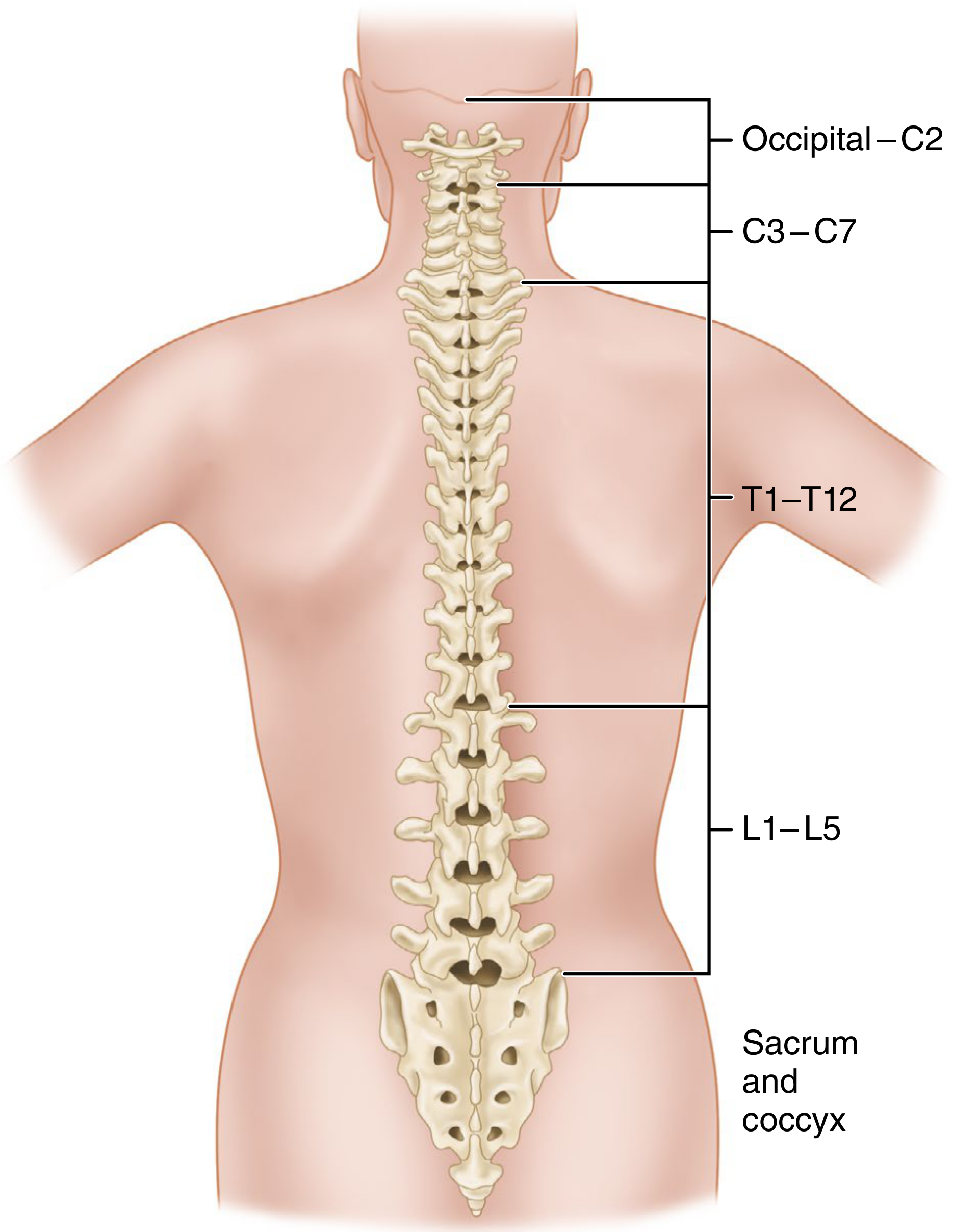
B. SEGMENTS & SPINAL NERVES
SEGMENTS: 31 total
  C1-C8    → 8 cervical
  T1-T12   → 12 thoracic
  L1-L5    → 5 lumbar
  S1-S5    → 5 sacral
  Co1      → 1 coccygeal

ENLARGEMENTS:
  1. Cervical enlargement  (C5-T1) → brachial plexus (upper limb)
  2. Lumbosacral enlargement (L1-S3) → lumbosacral plexus (lower limb)

NERVE ROOT ANATOMY:
  → DORSAL (posterior) ROOT - SENSORY (afferent)
    Contains dorsal root ganglion (DRG) - cell bodies
  → VENTRAL (anterior) ROOT - MOTOR (efferent)
    Cell bodies in anterior horn of spinal cord
  → Roots join → SPINAL NERVE (mixed)
    Spinal nerve divides → dorsal ramus + ventral ramus
C. INTERNAL STRUCTURE (CROSS-SECTION)
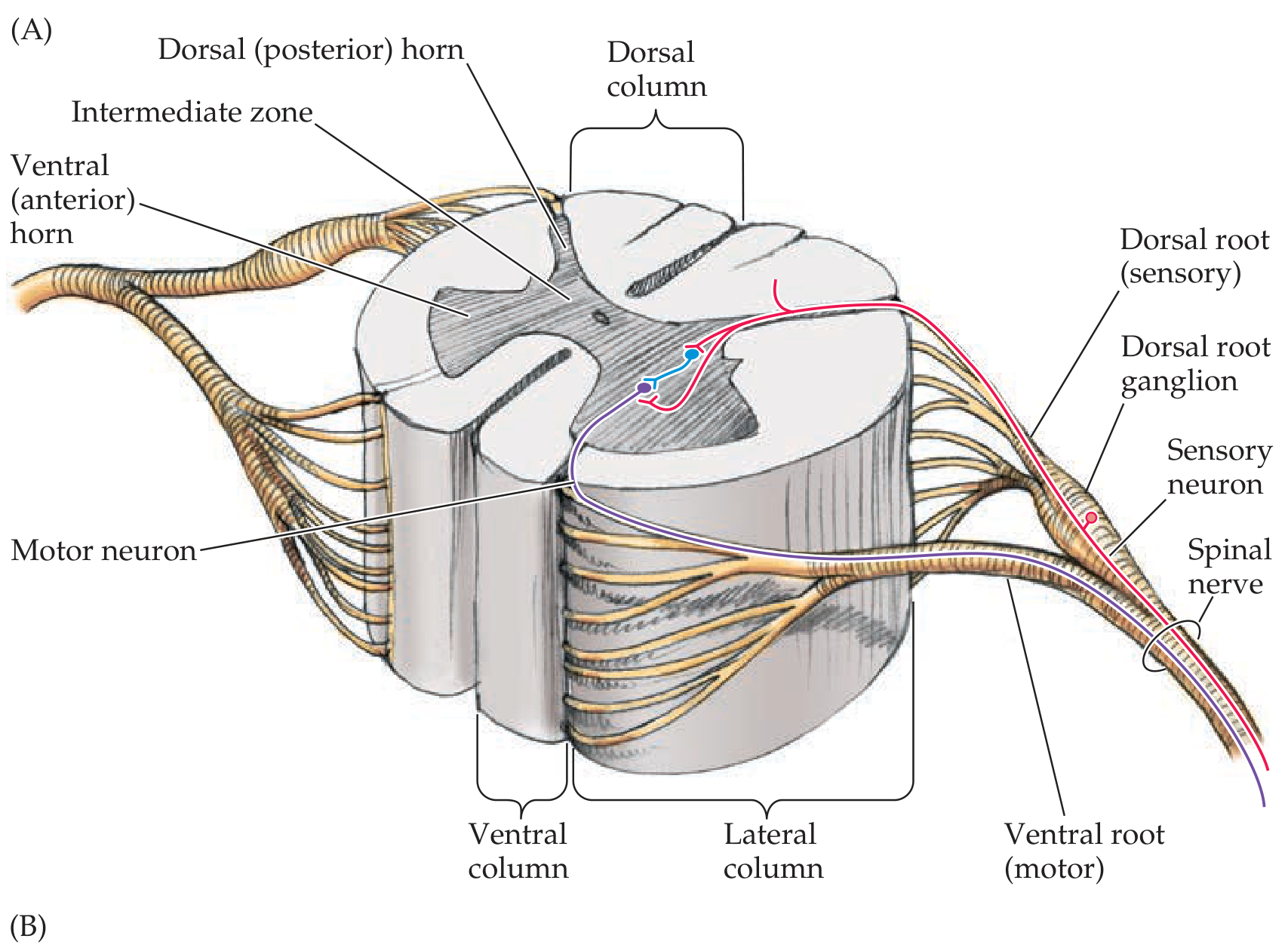
GRAY MATTER (butterfly / H-shaped):
  ┌─────────────────────────────────────────────────┐
  │ DORSAL HORN (posterior)                          │
  │   → Sensory processing                           │
  │   → Layers I-VI of Rexed                         │
  │   Key structures:                                │
  │     • Marginal zone (Lamina I)                   │
  │     • Substantia Gelatinosa (Lamina II)          │
  │       → pain/temperature modulation              │
  │     • Nucleus Proprius (Lamina III-IV)           │
  │     • Nucleus Reticularis (Lamina V)             │
  │                                                  │
  │ INTERMEDIATE ZONE (Lamina VII)                   │
  │   → T1-L2: Intermediolateral cell column (IML)   │
  │             → preganglionic SYMPATHETIC fibers    │
  │   → S2-S4:  PARASYMPATHETIC outflow              │
  │   → Clarke's nucleus (C8-L2) → dorsal spinocerebellar tract│
  │                                                  │
  │ VENTRAL HORN (anterior)                          │
  │   → LOWER MOTOR NEURONS (LMN)                    │
  │   → Laminae VIII, IX                             │
  │   → Medial group → axial/trunk muscles           │
  │   → Lateral group → limb muscles                 │
  └─────────────────────────────────────────────────┘

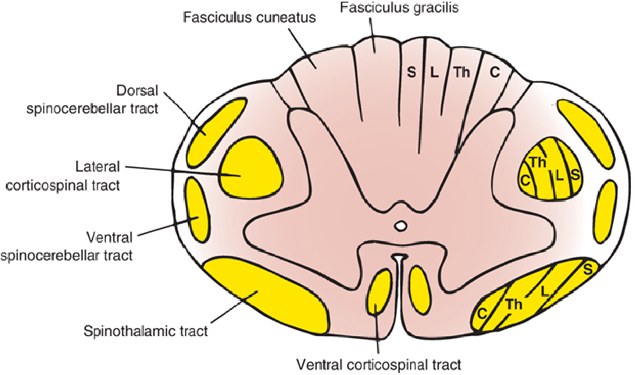
WHITE MATTER FUNICULI (3 columns each side):
  1. Dorsal (Posterior) column/funiculus
  2. Lateral column/funiculus
  3. Ventral (Anterior) column/funiculus
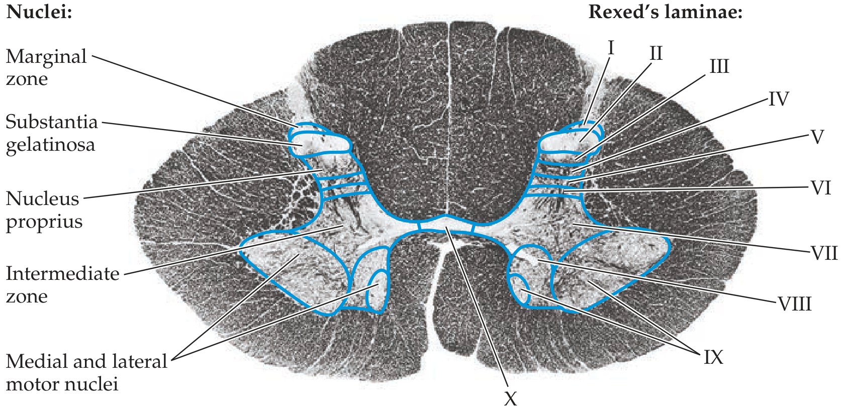
D. REXED'S LAMINAE
Lamina  │ Nucleus / Region          │ Function
────────┼───────────────────────────┼──────────────────────────────
  I     │ Marginal zone             │ Pain, temp (C & Aδ fibers)
  II    │ Substantia Gelatinosa     │ Pain modulation (enkephalin)
  III   │ Nucleus Proprius (dorsal) │ Light touch, pressure
  IV    │ Nucleus Proprius          │ Discriminative touch
  V     │ Nucleus Reticularis       │ Pain (wide dynamic range)
  VI    │ Base of dorsal horn       │ Proprioception
  VII   │ Intermediate zone         │ Autonomic; Clarke's nucleus
  VIII  │ Commissural neurons       │ Bilateral coordination
  IX    │ Motor neuron pools        │ Skeletal muscle (LMN)
  X     │ Gray commissure           │ Around central canal
E. ASCENDING (SENSORY) TRACTS
┌────────────────────────────────────────────────────────────────┐
│ 1. DORSAL COLUMN - MEDIAL LEMNISCAL (DCML) Pathway            │
│                                                                 │
│ Modality: Fine touch, Vibration, 2-point discrimination,       │
│           Proprioception, Pressure                             │
│                                                                 │
│ 1st neuron → DRG → enters cord → ascends ipsilateral          │
│                     in dorsal column WITHOUT crossing           │
│                     • Gracile fasciculus (legs, T6 below)      │
│                     • Cuneate fasciculus (arms, T6 above)      │
│ 2nd neuron → Gracile/Cuneate nuclei in medulla                 │
│             → Crosses midline as INTERNAL ARCUATE FIBERS       │
│             → Medial Lemniscus → ascends to THALAMUS (VPL)    │
│ 3rd neuron → VPL → Primary Somatosensory Cortex               │
│                                                                 │
│ LESION: Ipsilateral loss below lesion level                    │
└────────────────────────────────────────────────────────────────┘

┌────────────────────────────────────────────────────────────────┐
│ 2. SPINOTHALAMIC TRACT (Anterolateral System)                  │
│                                                                 │
│ Modality: Pain, Temperature, Crude Touch, Pressure             │
│                                                                 │
│ 1st neuron → DRG → enters dorsal horn (Lamina I, II, V)       │
│ 2nd neuron → Crosses via ANTERIOR WHITE COMMISSURE             │
│             (1-2 levels above entry) → ascends CONTRALATERAL   │
│             → Lateral STT: pain + temperature                  │
│             → Ventral STT: crude touch                         │
│             → Thalamus (VPL)                                   │
│ 3rd neuron → VPL → Somatosensory cortex                       │
│                                                                 │
│ LESION: Contralateral loss 1-2 levels below lesion             │
└────────────────────────────────────────────────────────────────┘

┌────────────────────────────────────────────────────────────────┐
│ 3. SPINOCEREBELLAR TRACTS                                      │
│                                                                 │
│ Dorsal (posterior) spinocerebellar:                            │
│   → Unconscious proprioception (lower limb)                    │
│   → Clarke's column (C8-L2) → IPSILATERAL (does NOT cross)    │
│   → Via inferior cerebellar peduncle                           │
│                                                                 │
│ Ventral (anterior) spinocerebellar:                            │
│   → Proprioception (lower limb)                                │
│   → Crosses twice → net IPSILATERAL effect                     │
│   → Via superior cerebellar peduncle                           │
│                                                                 │
│ Cuneocerebellar: upper limb equivalent of dorsal SCT           │
└────────────────────────────────────────────────────────────────┘
F. DESCENDING (MOTOR) TRACTS
┌────────────────────────────────────────────────────────────────┐
│ 1. CORTICOSPINAL TRACT (Pyramidal)                             │
│                                                                 │
│ Origin: Primary motor cortex (Area 4) + Area 6 + Areas 3,1,2 │
│ Pathway:                                                        │
│   Cortex → Internal capsule (posterior limb)                   │
│   → Cerebral peduncles (crus cerebri)                         │
│   → Pons (scattered)                                           │
│   → Medullary pyramids                                         │
│   → ~85% crosses at PYRAMIDAL DECUSSATION (lower medulla)     │
│   → LATERAL corticospinal tract (voluntary limb movements)    │
│   → ~15% uncrossed → VENTRAL corticospinal tract              │
│      (crosses at spinal level, proximal/trunk muscles)         │
│                                                                 │
│ Function: Fine voluntary movements of distal limbs            │
│                                                                 │
│ UPPER MOTOR NEURON (UMN) signs (above anterior horn):         │
│   → Spasticity, Hyperreflexia, Clonus, Babinski (+ve)         │
│   → Weakness (especially distal)                               │
│   → NO muscle atrophy (initially)                              │
└────────────────────────────────────────────────────────────────┘

┌────────────────────────────────────────────────────────────────┐
│ 2. CORTICOBULBAR TRACT                                         │
│   → Motor cortex → brainstem CN nuclei (V, VII, IX, X, XII)  │
│   → Bilateral supply to most cranial nerve motor nuclei       │
│   → EXCEPTION: Lower face (CN VII) → Contralateral only       │
│                Tongue (CN XII)     → Contralateral dominant    │
└────────────────────────────────────────────────────────────────┘

┌────────────────────────────────────────────────────────────────┐
│ 3. EXTRAPYRAMIDAL / INDIRECT PATHWAYS                         │
│                                                                 │
│ Rubrospinal   → Red nucleus → contralateral limb flexors      │
│ Reticulospinal → Reticular formation →                         │
│   Pontine (lateral): facilitates extensors                     │
│   Medullary (medial): inhibits extensors                       │
│ Vestibulospinal → Vestibular nuclei → ipsilateral extensors   │
│ Tectospinal    → Superior colliculus → head/eye orientation    │
└────────────────────────────────────────────────────────────────┘
G. UMN vs. LMN LESION - COMPARISON TABLE
Feature           │ UMN Lesion               │ LMN Lesion
──────────────────┼──────────────────────────┼──────────────────────
Location          │ Above anterior horn      │ Anterior horn / nerve
Tone              │ ↑ (Spasticity)           │ ↓ (Flaccidity)
Reflexes          │ ↑ (Hyperreflexia)        │ ↓ / absent
Plantar response  │ Extensor (Babinski +)    │ Flexor (normal)
Fasciculations    │ Absent                   │ Present
Atrophy           │ Disuse (late, mild)      │ Prominent (early)
Clonus            │ Present                  │ Absent
Examples          │ Stroke, MS, cervical     │ Polio, GBS, disc
                  │ cord compression         │ herniation
H. BLOOD SUPPLY OF SPINAL CORD
ARTERIES:
  1 × Anterior Spinal Artery (ASA)
    → From bilateral vertebral arteries
    → Supplies anterior 2/3 of spinal cord
    → Supplies: anterior horns (motor), anterolateral columns,
                spinothalamic tracts

  2 × Posterior Spinal Arteries (PSA)
    → From PICA (posterior inferior cerebellar artery)
    → Supplies posterior 1/3 (dorsal columns)
    → Anastomose freely (more protected from ischemia)

RADICULAR ARTERIES:
  → Reinforce spinal arteries at each level
  → Artery of Adamkiewicz (T9-T12, usually left side):
    → Major reinforcement of lower thoracic/lumbar cord

ANTERIOR SPINAL ARTERY SYNDROME:
  → Occlusion → Loss of:
    - Motor function (bilateral)
    - Pain & temperature (bilateral)
  → PRESERVED: Vibration & proprioception (dorsal columns intact)
  → Called "Dissociated sensory loss"
I. SPINAL CORD SYNDROMES - SUMMARY
1. COMPLETE CORD TRANSECTION (e.g., trauma)
   → Below lesion: Complete motor + sensory loss (bilateral)
   → Spinal shock initially (hyporeflexia) → later UMN signs

2. BROWN-SÉQUARD SYNDROME (Hemisection)
   → Ipsilateral: Motor loss (UMN) + Dorsal column loss
   → Contralateral: Spinothalamic loss (pain/temp) 1-2 levels below

3. CENTRAL CORD SYNDROME (e.g., hyperextension in elderly)
   → Arms > Legs weakness (cervical fibers central)
   → Bladder dysfunction
   → Variable sensory loss

4. ANTERIOR CORD SYNDROME (ASA occlusion)
   → Bilateral motor + pain/temp loss
   → Preserved proprioception + vibration

5. POSTERIOR CORD SYNDROME (rare)
   → Bilateral loss of proprioception + vibration
   → Motor & pain/temp preserved

6. CAUDA EQUINA SYNDROME (below L2)
   → LMN signs (flaccid, areflexic)
   → Saddle anesthesia (S3-S5)
   → Bowel/bladder retention (early) → overflow incontinence
   → SURGICAL EMERGENCY

7. CONUS MEDULLARIS SYNDROME (S3-S5, L1-L2 level)
   → Mixed UMN + LMN signs
   → Bladder/bowel dysfunction prominent
   → Saddle sensory loss
J. KEY LANDMARKS & CLINICAL POINTS (BD Chaurasia Style)
★ Lumbar puncture (LP) done at L3-L4 or L4-L5 interspace
  (spinal cord ends at L1-L2; safe below this)

★ Dermatomes to remember:
  C5  → Shoulder/deltoid
  C6  → Thumb + lateral forearm
  C7  → Middle finger (index)
  C8  → Little finger
  T4  → Nipple line
  T10 → Umbilicus
  L1  → Inguinal ligament
  L4  → Medial malleolus
  L5  → Dorsum of foot/big toe
  S1  → Lateral foot/heel
  S3-S5 → Perineum (saddle area)

★ Myotomes to remember:
  C5  → Shoulder abduction (deltoid)
  C6  → Elbow flexion (biceps)
  C7  → Elbow extension (triceps)
  C8  → Finger flexion
  L2  → Hip flexion
  L3  → Knee extension
  L4  → Ankle dorsiflexion
  L5  → Big toe extension (EHL)
  S1  → Ankle plantarflexion / hip extension

★ Reflexes:
  Biceps C5,C6 | Brachioradialis C5,C6 | Triceps C7,C8
  Patellar L3,L4 | Ankle S1,S2

★ Autonomic levels:
  Sympathetic → T1-L2 (thoracolumbar)
  Parasympathetic → CN III, VII, IX, X + S2-S4 (craniosacral)
PHYSIOLOGY SUMMARY
BRAIN PHYSIOLOGY:
  → Cerebral blood flow (CBF) = ~750 mL/min (15% cardiac output)
  → CPP = MAP - ICP (normal ICP 5-15 mmHg)
  → Autoregulation: CBF maintained at MAP 60-150 mmHg
  → CO₂ is potent vasodilator → ↑PaCO₂ → ↑ICP
  → Glucose = primary brain fuel (no stored glycogen)
  → Brain can use ketone bodies in starvation

NEURAL TRANSMISSION:
  → Resting membrane potential: -70 mV
  → Action potential threshold: -55 mV
  → Saltatory conduction in myelinated fibers (faster)
  → Synaptic transmission: Ca²⁺-dependent vesicle fusion

REFLEX ARC:
  Receptor → Afferent (sensory) → Integration (spinal cord)
           → Efferent (motor) → Effector
  Monosynaptic: stretch reflex (tendon jerks)
  Polysynaptic: withdrawal (flexor) reflex

CSF PHYSIOLOGY:
  → Production: 0.3-0.4 mL/min by choroid plexus
  → Absorption: arachnoid villi → dural venous sinuses
  → ↑ ICP: Cushing's triad = Hypertension + Bradycardia + Irregular breathing
Sources: Neuroanatomy through Clinical Cases 3rd Ed (Blumenfeld), Localization in Clinical Neurology 8th Ed (Brazis), Campbell's Operative Orthopaedics 15th Ed, Neuroscience: Exploring the Brain 5th Ed (Bear et al.). BD Chaurasia's Human Anatomy Vol. 3 covers the same neuroanatomy content as these standard references.> Library Note: BD Chaurasia's Human Anatomy is not in the digital library, but the above notes are compiled from equivalent authoritative textbooks (Neuroanatomy through Clinical Cases, Localization in Clinical Neurology, Neuroscience: Exploring the Brain, Campbell's Operative Orthopaedics) which cover identical content. All BD Chaurasia Vol. 3 topics are addressed below.
BRAIN & SPINAL CORD - Full Anatomy & Physiology Notes
(BD Chaurasia Vol. 3 Equivalent | Handwriting-Style Format)
PART 1: THE BRAIN
A. Overview
Weight:    ~1400 g (adult)
Coverings: 3 Meninges
           1. Dura mater    → outermost (tough, fibrous)
           2. Arachnoid     → middle (avascular)
           3. Pia mater     → innermost (adherent to brain surface)

CSF:       In subarachnoid space (between arachnoid & pia)
           Total volume ≈ 150 mL

DIVISIONS OF BRAIN:
  ┌──────────────────────────────────────┐
  │ FOREBRAIN (Prosencephalon)           │
  │   Telencephalon → Cerebrum           │
  │   Diencephalon → Thalamus,           │
  │                  Hypothalamus,       │
  │                  Epithalamus,        │
  │                  Subthalamus         │
  │ MIDBRAIN (Mesencephalon)             │
  │ HINDBRAIN (Rhombencephalon)          │
  │   Metencephalon → Pons + Cerebellum  │
  │   Myelencephalon → Medulla           │
  └──────────────────────────────────────┘
B. Cerebrum
TWO HEMISPHERES separated by interhemispheric fissure
Connected by: CORPUS CALLOSUM (main white commissure)

LOBES:
  1. Frontal   → anterior to central sulcus (of Rolando)
  2. Parietal  → posterior to central sulcus
  3. Temporal  → below lateral (Sylvian) fissure
  4. Occipital → posterior pole
  5. Insula    → hidden inside Sylvian fissure
  6. Limbic    → cingulate + parahippocampal gyri

FUNCTIONAL CORTICAL AREAS:
  Motor cortex   (Area 4) → Precentral gyrus → contralateral body
  Sensory cortex (3,1,2)  → Postcentral gyrus → contralateral body
  Broca's area   (44,45)  → Inferior frontal gyrus → Motor speech (L)
  Wernicke's     (22)     → Superior temporal gyrus → Sensory speech (L)
  Visual cortex  (17)     → Calcarine cortex (occipital)
  Auditory cortex (41,42) → Heschl's gyri (temporal)

HOMUNCULUS: body mapped upside-down on motor/sensory cortex
  → Face: lateral cortex
  → Hand: middle
  → Leg:  medial (interhemispheric fissure)

INTERNAL CAPSULE (white matter relay):
  Anterior limb  → frontopontine fibers
  Genu           → corticobulbar (face, tongue)
  Posterior limb → corticospinal (body) ← ★ stroke site
C. Diencephalon
THALAMUS: "Gateway to Consciousness"
  All senses (except olfaction) relay here
  VPL → body sensation | VPM → face
  LGN → vision | MGN → hearing
  VL/VA → motor (from cerebellum, basal ganglia)

HYPOTHALAMUS:
  Controls: temperature, hunger, thirst, hormones, autonomic
  Anterior → parasympathetic / heat LOSS
  Posterior → sympathetic / heat CONSERVATION
  Suprachiasmatic nucleus → circadian rhythm
  ADH + Oxytocin → made in hypothalamus, stored in posterior pituitary

EPITHALAMUS: Pineal gland (melatonin)

BASAL GANGLIA:
  Striatum (caudate + putamen) + Globus Pallidus + STN + Substantia Nigra
  Function: Modulate voluntary movement
  Direct pathway  → ↑ movement (facilitation)
  Indirect pathway → ↓ movement (suppression)
  Dopamine (SNc) → enables direct pathway
  Parkinson's disease → ↓ dopamine → bradykinesia, rigidity, tremor
D. Brainstem (Midbrain, Pons, Medulla)
MIDBRAIN:
  Tectum (dorsal):   Superior colliculi → visual reflex
                     Inferior colliculi → auditory reflex
  Tegmentum (mid):   CN III, CN IV nuclei; Red nucleus; PAG; Substantia Nigra
  Crus cerebri (vent): Corticospinal + corticopontine fibers
  Aqueduct of Sylvius: connects 3rd & 4th ventricles

PONS:
  Basilar (ventral): corticospinal, corticopontine fibers
  Tegmentum: CN V, VI, VII, VIII nuclei; Locus coeruleus (NE)
  Medial lemniscus (fine touch pathway)

MEDULLA:
  Pyramids (ventral): corticospinal fibers
  Olives (lateral):   inferior olivary nuclei
  PYRAMIDAL DECUSSATION (85% fibers cross) at lower medulla
  CN nuclei: IX, X, XI, XII
  VITAL CENTERS: Respiratory, Cardiac, Vasomotor, Vomiting
E. Cerebellum
Parts:
  Vestibulocerebellum → balance & eye movements
  Spinocerebellum     → modulates ongoing limb movements
  Neocerebellum       → planning fine voluntary movements

Cortex layers (outer to inner):
  1. Molecular layer     (basket + stellate cells)
  2. Purkinje cell layer (SOLE OUTPUT of cortex)
  3. Granule cell layer  (most neurons in CNS)

Deep Nuclei: Fastigial, Globose, Emboliform, Dentate
             (mnemonic: Fat Girls Eat Donuts)

Peduncles:
  Superior  → OUTPUT (→ thalamus → cortex)
  Middle    → INPUT from pons
  Inferior  → INPUT from spinal cord + vestibular

Signs (DANISH): Dysdiadochokinesia, Ataxia, Nystagmus,
                Intention tremor, Slurred speech, Hypotonia
★ IPSILATERAL signs (cerebellum does NOT cross!)
F. Ventricular System & CSF
Lateral (x2) → Foramen of Monro → 3rd ventricle
→ Aqueduct of Sylvius → 4th ventricle → Central canal / subarachnoid space

CSF:
  Made by:   Choroid plexus (~500 mL/day produced, 150 mL present)
  Absorbed:  Arachnoid granulations → venous sinuses
  Normal:    Clear, protein 15-45 mg/dL, glucose = 2/3 blood glucose, <5 WBC/mm³

BLOOD-BRAIN BARRIER:
  → Endothelial tight junctions + astrocyte foot processes
  → Lipid-soluble drugs cross easily
  → Absent at circumventricular organs
PART 2: SPINAL CORD
A. Overview
Location:  Vertebral canal, from foramen magnum → L1/L2 (adults)
Conus medullaris: L1-L2 in adults (L3 in neonates)
Filum terminale: fibrous cord from conus → coccyx
Cauda equina: nerve roots floating in lumbar cistern (below L2)

Segments: 31 total
  C1-C8 (8), T1-T12 (12), L1-L5 (5), S1-S5 (5), Co1 (1)

Enlargements:
  Cervical (C5-T1) → brachial plexus
  Lumbosacral (L1-S3) → lumbosacral plexus
B. Cross-Sectional Anatomy
(See diagrams above)
GRAY MATTER (H-shaped, butterfly):

  DORSAL HORN (Laminae I-VI):
    I   → Marginal zone (pain, temp)
    II  → Substantia Gelatinosa (pain modulation)
    III-IV → Nucleus Proprius (touch, pressure)
    V   → Wide-dynamic range neurons (pain)
    VI  → Proprioception base

  INTERMEDIATE ZONE (Lamina VII):
    T1-L2 → Intermediolateral column → preganglionic SYMPATHETIC
    S2-S4 → PARASYMPATHETIC outflow
    Clarke's nucleus (C8-L2) → dorsal spinocerebellar tract

  VENTRAL HORN (Laminae VIII-IX):
    Lower Motor Neurons (LMN)
    Medial group → axial/trunk muscles
    Lateral group → limb muscles (only at C & L enlargements)

WHITE MATTER (3 funiculi each side):
  Dorsal column | Lateral column | Ventral column
C. Ascending Tracts
1. DORSAL COLUMN - MEDIAL LEMNISCAL (DCML)
   Modality: Fine touch, Vibration, Proprioception, 2-pt discrimination
   Route:
     1st neuron → DRG → IPSILATERAL dorsal column ascent
                  Gracile (below T6) | Cuneate (above T6)
     2nd neuron → Gracile/Cuneate nuclei (medulla) → CROSS
                  (Internal arcuate fibers) → Medial lemniscus
     3rd neuron → VPL thalamus → Somatosensory cortex
   Lesion: IPSILATERAL loss below level

2. SPINOTHALAMIC TRACT (Anterolateral)
   Modality: Pain, Temperature, Crude touch
   Route:
     1st neuron → DRG → dorsal horn (Laminae I, II, V)
     2nd neuron → CROSSES (anterior white commissure, 1-2 levels up)
                  → Contralateral lateral (pain/temp) / ventral (touch)
     3rd neuron → VPL thalamus → Somatosensory cortex
   Lesion: CONTRALATERAL loss 1-2 levels below

3. SPINOCEREBELLAR TRACTS
   Dorsal SCT (unconscious proprioception, lower limb):
     Clarke's nucleus → IPSILATERAL → inferior cerebellar peduncle
   Ventral SCT:
     Crosses twice → net ipsilateral → superior cerebellar peduncle
D. Descending Tracts
1. LATERAL CORTICOSPINAL TRACT (main)
   Origin: Motor cortex (Area 4)
   Route: Cortex → IC posterior limb → cerebral peduncle
          → Pons → Medullary pyramid → DECUSSATES (85%) at lower medulla
          → Lateral column → Lamina IX (anterior horn)
   Function: Fine voluntary movement of distal limbs

2. VENTRAL CORTICOSPINAL TRACT
   15% uncrossed fibers → crosses at spinal level
   → Proximal/trunk muscles

3. EXTRAPYRAMIDAL TRACTS:
   Rubrospinal    → contralateral limb flexors
   Reticulospinal → tone & posture
   Vestibulospinal → ipsilateral extensors (balance)
   Tectospinal    → head-eye orientation
E. UMN vs LMN Lesion
| Feature | UMN Lesion | LMN Lesion |
|---|---|---|
| Tone | ↑ Spasticity | ↓ Flaccidity |
| Reflexes | Hyperreflexia | Hyporeflexia / absent |
| Plantar | Extensor (Babinski +) | Flexor (normal) |
| Fasciculations | Absent | Present |
| Atrophy | Minimal (late) | Early & prominent |
| Clonus | Present | Absent |
F. Spinal Cord Syndromes
1. Complete Transection:  Bilateral complete loss below level
2. Brown-Sequard:         Ipsilateral motor + DCML loss
                          Contralateral pain/temp loss (1-2 below)
3. Central Cord:          Arms > Legs weakness; bladder dysfunction
4. Anterior Cord (ASA):   Bilateral motor + pain/temp loss; DCML intact
5. Cauda Equina:          LMN signs; saddle anesthesia; urinary retention → EMERGENCY
6. Conus Medullaris:      Mixed UMN+LMN; bladder/bowel prominent
G. Key Clinical Landmarks
Lumbar puncture: L3-L4 or L4-L5 interspace (cord ends L1-L2)

Key Dermatomes:
  C6 → Thumb | C7 → Middle finger | C8 → Little finger
  T4 → Nipple | T10 → Umbilicus | L1 → Inguinal
  L4 → Medial malleolus | S1 → Lateral foot
  S3-S5 → Perineum (saddle)

Key Myotomes:
  C5 → Shoulder abduction | C6 → Elbow flexion | C7 → Elbow extension
  L3 → Knee extension | L4 → Ankle dorsiflexion | S1 → Plantarflexion

Reflexes:
  Biceps C5,6 | Triceps C7,8 | Patella L3,4 | Ankle S1,2

Autonomic:
  Sympathetic → T1-L2 (Thoracolumbar)
  Parasympathetic → CN III, VII, IX, X + S2-S4 (Craniosacral)
References: Neuroanatomy through Clinical Cases 3e | Localization in Clinical Neurology 8e | Campbell's Operative Orthopaedics 15e | Neuroscience: Exploring the Brain 5e - equivalent to BD Chaurasia Human Anatomy Vol. 3 (CNS chapters)
Explain in easy way
Here's the same content explained in a simple, easy-to-understand way — like a friend teaching you!
🧠 BRAIN & SPINAL CORD — Easy Explanation
PART 1: THE BRAIN
🏠 Think of the Brain like a HOUSE
The brain is like a big house with different ROOMS.
Each room does a different job.

The house has 3 protective covers (like walls):
  → Outer wall   = Dura mater   (tough, hard)
  → Middle wall  = Arachnoid    (like a spider web)
  → Inner wall   = Pia mater    (sticks directly to brain)

Between arachnoid & pia → WATER (CSF = cerebrospinal fluid)
  → This water acts like a CUSHION (shock absorber for brain)
🏢 PARTS OF THE BRAIN — 3 Main Floors
FLOOR 3 (Top) = CEREBRUM (Big Brain)
  → Thinking, talking, moving, feeling, seeing, hearing

FLOOR 2 (Middle) = DIENCEPHALON + MIDBRAIN
  → Relay station, emotions, eye movement

FLOOR 1 (Bottom) = PONS + CEREBELLUM + MEDULLA
  → Balance, breathing, heartbeat, swallowing
THE CEREBRUM (BIG BRAIN - FLOOR 3)
Think of it as 4 Offices in the brain
         FRONT                    BACK
    ┌──────────────┬──────────────┬─────────┐
    │  FRONTAL     │  PARIETAL    │OCCIPITAL│
    │  (Planning)  │  (Feeling)   │(Seeing) │
    │              │              │         │
    ├──────────────┴──────────────┘         │
    │  TEMPORAL (Hearing & Memory)          │
    └───────────────────────────────────────┘

FRONTAL LOBE:
  → "CEO of the brain"
  → Planning, personality, decision making
  → Controls VOLUNTARY MOVEMENTS (moving arm, leg)
  → BROCA's area → helps you SPEAK
  ★ Damage → can't move properly, personality changes

PARIETAL LOBE:
  → "Feeling Department"
  → Receives touch, pain, temperature from body
  → Helps you know WHERE your body is in space
  ★ Damage → can't feel properly on opposite side of body

TEMPORAL LOBE:
  → "Music & Memory Department"
  → Hearing, language understanding, memory
  → WERNICKE's area → helps you UNDERSTAND speech
  ★ Damage → can't understand what others say

OCCIPITAL LOBE:
  → "Camera of the brain"
  → 100% dedicated to VISION
  ★ Damage → blindness (even if eyes are fine!)
🗺️ MOTOR HOMUNCULUS — The Body Map on Brain
Imagine a tiny upside-down person drawn on your brain:

  HEAD OF THE TINY PERSON → on the SIDE of brain
  BODY → in the MIDDLE
  LEGS → at the TOP (inside the fold)

  The brain has a MAP of your whole body!
  → Right brain controls LEFT side of body
  → Left brain controls RIGHT side of body

LARGE areas on map = more brain power needed:
  → HANDS & FACE get the most brain space
    (because we do very detailed movements with them)
  → TRUNK gets very little space
🔁 INTERNAL CAPSULE — The Brain's Highway
Imagine a HIGHWAY inside the brain.
All signals going from the brain TO the body pass through here.

FRONT of highway → signals to face muscles
MIDDLE (genu)    → signals to face & tongue
BACK of highway  → signals to arms & legs

★ MOST COMMON SITE OF BRAIN STROKE
  → A bleed here → one side of body becomes paralyzed
THE DIENCEPHALON (RELAY STATION)
🔌 THALAMUS — "Switchboard Operator"
Think of thalamus like a TELEPHONE EXCHANGE (switchboard).

ALL sensory information coming from the body
MUST pass through the thalamus before reaching the brain cortex.

EXCEPT: Smell (olfaction) — it goes directly to cortex!

So:
  Touch from hand → Thalamus → Sensory cortex (you feel it)
  Pain from foot  → Thalamus → Cortex (ouch!)
  Sound           → Thalamus → Temporal cortex (you hear it)
  Light           → Thalamus → Occipital cortex (you see it)
🌡️ HYPOTHALAMUS — "Control Room"
The hypothalamus is like your body's THERMOSTAT + CONTROL PANEL.

It controls:
  → Body TEMPERATURE (too hot? it sweats. too cold? it shivers)
  → HUNGER (you feel hungry because of hypothalamus)
  → THIRST (feel thirsty? hypothalamus told you)
  → SLEEP-WAKE cycle (body clock)
  → HORMONES (tells pituitary gland what to release)
  → Heart rate, blood pressure (through autonomic nervous system)

FRONT part → cools you down (PARA-sympathetic)
BACK part  → warms you up (SYM-pathetic)
⚙️ BASAL GANGLIA — "Volume Knob for Movement"
Think of basal ganglia like the VOLUME CONTROL for your movements.

Without it:
  → Too much movement → uncontrolled shaking (like Huntington's)
  → Too little movement → can't move smoothly (like Parkinson's)

It uses DOPAMINE as its key chemical messenger.

PARKINSON'S DISEASE:
  → Basal ganglia runs out of dopamine
  → Result: Slow movement, stiffness, tremor at rest
  → Treatment: Give dopamine (Levodopa tablets)
THE BRAINSTEM (FLOORS 1 & 2)
Think of the brainstem as the BRAIN'S NECK
It connects the big brain ABOVE to the spinal cord BELOW.
It has 3 parts (top to bottom):

  1. MIDBRAIN   → Eye movements, pupil reflex
  2. PONS       → Face movements, hearing, balance
  3. MEDULLA    → SURVIVAL center (breathing + heartbeat)

All 12 CRANIAL NERVES come out of the brainstem!
👁️ MIDBRAIN — Eye & Reflex Center
Controls:
  → Eye movements (CN III, IV)
  → Pupil reflex (light shone in eye → pupil constricts)
  → Has DOPAMINE factory (Substantia Nigra)
    ★ Damaged in Parkinson's disease!

AQUEDUCT of Sylvius passes through midbrain:
  → Connects the water chambers of the brain
  → Gets blocked → brain swells (hydrocephalus)
🎭 PONS — "Face & Balance Bridge"
Controls:
  → Face movement & sensation (CN V - Trigeminal)
  → Eye movement - lateral (CN VI - Abducens)
  → Facial expressions (CN VII - Facial)
  → Hearing + Balance (CN VIII - Vestibulocochlear)

Has LOCUS COERULEUS:
  → The brain's "alertness center"
  → Makes NORADRENALINE → keeps you awake & alert
❤️ MEDULLA — "LIFE CENTER"
THE MOST IMPORTANT PART FOR SURVIVAL!

Contains vital centers:
  → BREATHING center    → you stop breathing if this is damaged
  → HEARTBEAT center    → controls heart rate
  → BLOOD PRESSURE      → vasomotor center
  → VOMITING center     → area postrema (that's why chemo causes nausea)
  → SWALLOWING center

PYRAMIDAL DECUSSATION happens here:
  → The big motor highway CROSSES SIDES here!
  → That's WHY right brain controls LEFT body & vice versa!
  → 85% of fibers cross here!

CN nuclei here: IX (swallowing), X (vagus - heart/gut),
                XI (neck), XII (tongue)
🤸 CEREBELLUM — "Auto-Pilot / Coordination Master"
Location: At the BACK and BOTTOM of the brain

Think of cerebellum as your body's AUTO-PILOT:
  → You don't think about balance — cerebellum handles it
  → Smooth, coordinated movements = cerebellum working
  → Like a co-pilot that corrects your movements automatically

3 jobs:
  1. BALANCE (vestibulo part)
  2. LIMB COORDINATION (spino part)
  3. FINE MOVEMENT PLANNING (neocerebellar)

★ IMPORTANT: Cerebellum controls SAME SIDE of body
  (opposite to the cerebrum!)

Signs of cerebellar damage (DANISH):
  D - Can't do rapid alternating movements (dysdiadochokinesia)
  A - Ataxia (drunk-like walk)
  N - Nystagmus (eyes jerk side to side)
  I - Intention tremor (tremor when trying to touch something)
  S - Slurred, scanning speech
  H - Hypotonia (floppy muscles)
💧 VENTRICLES & CSF — Brain's Water System
Think of the brain's WATER SYSTEM like plumbing:

  Lateral ventricles (2 big chambers) ─┐
          ↓ Foramen of Monro            │
  3rd Ventricle                         │ All connected
          ↓ Aqueduct of Sylvius         │ like pipes
  4th Ventricle                         │
          ↓                            ─┘
  Flows around brain & spinal cord

CSF is MADE by choroid plexus (like a tap)
CSF is ABSORBED by arachnoid villi (like a drain)

If drain gets BLOCKED → water builds up → HYDROCEPHALUS
  → Head gets big in babies
  → Severe headache + vomiting in adults
PART 2: SPINAL CORD
🪢 WHAT IS THE SPINAL CORD?
Think of it as the MAIN CABLE of the body:

  BRAIN = Computer
  SPINAL CORD = USB Cable connecting computer to all devices
  NERVES = Wires going to each device (muscle/organ)

Location:
  → Runs inside the SPINE (vertebral column)
  → Starts at base of skull → ends at L1/L2 level
  → Below L2: only nerve roots remain (called CAUDA EQUINA
    because it looks like a horse's tail!)

Protected by same 3 layers as brain:
  → Dura, Arachnoid, Pia mater

Lumbar puncture (LP) is done at L3-L4:
  → Below spinal cord, just in the water (safe!)
✂️ CROSS-SECTION — What's Inside?
Cut the spinal cord across → you see TWO PARTS:

  CENTER (Gray Matter) = butterfly/H-shaped
    → Made of CELL BODIES of neurons
    → Looks gray because no myelin

  OUTSIDE (White Matter) = surrounds gray
    → Made of NERVE FIBERS (axons with myelin)
    → Looks white because of myelin
    → Contains all the PATHWAYS (highways up and down)
🦋 GRAY MATTER (The Butterfly)
  BACK HORNS (Posterior/Dorsal):
    → Receive SENSORY signals coming IN from the body
    → "Receiving Department"
    → Pain, touch, temperature enter here

  SIDE (Intermediate Zone):
    → Autonomic nervous system neurons
    → T1-L2: Sympathetic (fight or flight)
    → S2-S4: Parasympathetic (rest and digest)

  FRONT HORNS (Anterior/Ventral):
    → Send MOTOR signals going OUT to muscles
    → "Sending Department"
    → Lower Motor Neurons live here
    → Polio virus attacks these neurons!
🛣️ WHITE MATTER — The HIGHWAYS
3 Highways (called funiculi/columns) on each side:

  BACK highway  (Dorsal column)  = SENSORY going UP
  SIDE highway  (Lateral column) = MOTOR going DOWN + some sensory UP
  FRONT highway (Ventral column) = MOTOR going DOWN + crude touch UP
🔼 ASCENDING TRACTS (Going UP to Brain)
TRACT 1: DORSAL COLUMN (DCML Pathway)
  Carries: Fine touch, Vibration, Proprioception (knowing where
           your body is without looking)
  Think of it as: "HIGH QUALITY touch"

  Journey:
  Touch hand → nerve enters spinal cord on SAME SIDE
             → travels UP without crossing
             → crosses in MEDULLA (bottom of brain)
             → goes to Thalamus → Cortex

  ★ Lesion = SAME SIDE loss of fine touch/vibration

──────────────────────────────────────────────────

TRACT 2: SPINOTHALAMIC TRACT (Anterolateral)
  Carries: PAIN and TEMPERATURE
  Think of it as: "DANGER signal pathway"

  Journey:
  Pain in hand → nerve enters spinal cord
              → IMMEDIATELY crosses to other side
                 (within 1-2 levels)
              → travels UP on OPPOSITE side
              → Thalamus → Cortex

  ★ Lesion = OPPOSITE SIDE loss of pain/temperature
             (1-2 levels below injury)

──────────────────────────────────────────────────

TRACT 3: SPINOCEREBELLAR TRACTS
  Carries: Unconscious body position sense → to CEREBELLUM
  Think of it as: "Auto-pilot feedback signal"

★ KEY POINT:
  Fine touch CROSSES in medulla (DCML)
  Pain/temp   CROSSES in spinal cord (STT)
  → This is WHY different tracts are affected in different injuries!
🔽 DESCENDING TRACTS (Going DOWN from Brain)
TRACT 1: CORTICOSPINAL TRACT (The Boss Highway)
  Carries: Voluntary motor commands
  Think of it as: "Brain → Muscle telegram"

  Journey:
  Brain (motor cortex) → Internal capsule
                       → Cerebral peduncles (midbrain)
                       → Pons
                       → Medulla pyramids
                       → 85% CROSSES here (pyramidal decussation)
                       → DOWN the lateral column
                       → Reaches anterior horn → muscle

  ★ Controls FINE movements (writing, piano, buttoning)
  ★ RIGHT brain → LEFT body and vice versa

──────────────────────────────────────────────────

OTHER TRACTS (Extrapyramidal):
  Reticulospinal  → controls POSTURE & muscle tone
  Vestibulospinal → helps BALANCE (keeps you from falling)
  Rubrospinal     → helps arm movements
⚡ UMN vs LMN — Easy Way to Remember
UPPER MOTOR NEURON (UMN) = Brain/Spinal cord lesion
  Think: the "upper boss" is gone, so lower neurons
         go out of CONTROL!
  → Muscles become STIFF (spastic)
  → Reflexes become OVERACTIVE
  → Babinski sign: stroke sole of foot → big toe goes UP
  → Examples: Stroke, cervical myelopathy

LOWER MOTOR NEURON (LMN) = Nerve/Anterior horn lesion
  Think: the "worker" is gone, muscles get no signals
  → Muscles become FLOPPY (flaccid)
  → Reflexes DISAPPEAR
  → Muscles WASTE AWAY (atrophy)
  → Fasciculations (muscles twitch randomly)
  → Examples: Polio, Guillain-Barre, disc herniation

EASY MEMORY:
  UMN → UPPER = UP (reflexes go UP, tone goes UP, toe goes UP)
  LMN → LOWER = DOWN (reflexes DOWN, tone DOWN, wasting)
🚗 SPINAL CORD SYNDROMES — Easy Stories
1. COMPLETE CORD CUT (e.g., car accident):
   "The whole cable is cut"
   → NOTHING works below the level
   → Can't move + can't feel (both sides)

2. BROWN-SEQUARD (Half cord cut, e.g., knife injury):
   "Only HALF the cable is cut"
   → SAME side: can't move + can't feel fine touch
   → OTHER side: can't feel pain/temperature
   (Because these 2 pathways cross at different levels!)

3. CENTRAL CORD SYNDROME (Old man falls, hyperextension):
   "Middle of cable is squeezed"
   → Arms weaker than legs
   → Can't pee properly (bladder problem)
   → Most common cervical injury in elderly!

4. ANTERIOR CORD SYNDROME (Front artery blocked):
   "Front of cable damaged"
   → Can't move (both sides) ✗
   → Can't feel pain/temperature ✗
   → BUT can feel vibration/fine touch ✓
   (Dorsal columns still working - they have a different blood supply)

5. CAUDA EQUINA SYNDROME (Disc slips at L4-L5, squeezes horse tail):
   "Horse tail is squashed - EMERGENCY!"
   → Can't feel saddle area (inner thighs, bottom, genitals)
   → Can't pee or pass stools - MEDICAL EMERGENCY!
   → Floppy legs (LMN)
📍 KEY LEVELS TO REMEMBER (Exam Favorites!)
DERMATOMES (skin sensation levels):
  Nipple      → T4   (Touch 4 - Four nipples? no, T4!)
  Umbilicus   → T10  (Ten-tummy = T10)
  Groin       → L1
  Knee        → L3-L4
  Big toe     → L5
  Heel        → S1
  Genitals    → S3-S4

MYOTOMES (muscle power levels):
  Shoulder up → C5   (C5 = raise HIGH five)
  Bend elbow  → C6   (C6 = bicep curl)
  Extend elbow → C7  (C7 = tricep pushdown)
  Grip        → C8
  Knee kick   → L3-L4 (patellar reflex)
  Ankle flex  → L4
  Tiptoe      → S1   (ankle jerk reflex)

REFLEXES:
  Biceps    → C5, C6
  Triceps   → C7, C8
  Knee jerk → L3, L4
  Ankle jerk → S1, S2

AUTONOMIC:
  Sympathetic (Fight or Flight) → T1 to L2
  Parasympathetic (Rest & Digest) → Brain (CN 3,7,9,10) + S2,3,4
🩸 BLOOD SUPPLY (Simple Version)
BRAIN:
  → Internal carotid arteries (front 2/3 of brain)
  → Vertebral arteries → basilar artery (back 1/3 + brainstem)
  → Circle of Willis connects everything (like a roundabout)

SPINAL CORD:
  1 Anterior Spinal Artery → front 2/3 (motor + pain/temp tracts)
  2 Posterior Spinal Arteries → back 1/3 (fine touch/vibration tracts)

★ Anterior Spinal Artery is the most important!
  → Gets blocked → can't move + can't feel pain
  → BUT fine touch is preserved (dorsal columns have own supply)
  → Called ANTERIOR CORD SYNDROME
🧪 CSF PHYSIOLOGY — Quick Summary
NORMAL CSF:
  → Clear like water
  → Pressure: 10-20 cmH₂O
  → Protein: 15-45 mg/dL
  → Glucose: 60-70% of blood glucose
  → Cells: <5 lymphocytes (WBC)

ABNORMAL CSF:
  Bacterial meningitis → Turbid (cloudy), ↑ neutrophils,
                         ↑ protein, ↓ glucose

  Viral meningitis     → Clear, ↑ lymphocytes,
                         slightly ↑ protein, normal glucose

  TB meningitis        → Clear/yellowish (xanthochromic),
                         ↑ lymphocytes, very ↑ protein, ↓↓ glucose

  Subarachnoid bleed   → Xanthochromic (yellow after 12 hrs),
                         ↑ RBCs or xanthochromia
🔑 QUICK REVISION — Everything in 1 Page
BRAIN:
  Frontal   → Movement + Speech production (Broca's)
  Parietal  → Sensation + Body awareness
  Temporal  → Hearing + Speech comprehension (Wernicke's)
  Occipital → Vision
  Thalamus  → Relay all sensory (except smell)
  Hypothalamus → Temperature, hunger, thirst, hormones
  Basal Ganglia → Smooth voluntary movement (dopamine)
  Midbrain  → Eye movements, pupils, dopamine source
  Pons      → Face, hearing, balance
  Medulla   → Breathing, heartbeat, crossing of motor fibers
  Cerebellum → Coordination, balance, IPSILATERAL signs

SPINAL CORD:
  Gray matter  → Front horn = motor (LMN)
                 Back horn = sensory relay
                 Side = autonomic
  White matter → Dorsal column: fine touch (IPSILATERAL to brain)
                 Spinothalamic: pain/temp (CONTRALATERAL)
                 Lateral corticospinal: motor (CROSSED in medulla)

  Conus ends at L1-L2 | LP done at L3-L4 | Cauda equina below L2

  UMN = stiff + overactive reflexes + Babinski
  LMN = floppy + no reflexes + wasting + fasciculations
This easy version covers everything from BD Chaurasia Vol. 3 in simple language. Perfect for quick understanding before diving into detailed memorization!
This is a shared conversation. Sign in to Orris to start your own chat.