
Mri findings in hsv encephalitis
Reading File
HSV encephalitis (HSE) shows characteristic MRI findings predominantly involving the temporal lobes and limbic structures. Key findings include T2/FLAIR hyperintensity in medial temporal lobes, insular cortex, and cingulate gyrus, typically asymmetric and bilateral. DWI restriction and cortical/subcortical edema are also seen.
Searching sources...
Finding Sources
Finding Sources
Searching PubMed
"herpes simplex encephalitis"[MeSH Terms] AND "MRI" AND neuroimaging
Good results. Let me read the full textbook section and fetch the MRI image.
Reading File
Loading Image
Now let me get more detail from the diagnosis and additional imaging sections:
Reading File
Finding Sources
Now let me read the detailed radiology section from Bradley & Daroff and the radiology textbook:
Reading File
Reading File
Let me also get the Harrison's section and the Plum & Posner section for DWI/additional imaging details:
Reading File
Reading File
I also have the Harrison's FLAIR + DWI MRI image URL. Let me fetch that too:
Loading Image
I now have comprehensive information from multiple authoritative textbooks and two actual MRI images. Here is the complete answer:
MRI Findings in HSV Encephalitis
MRI is the most sensitive imaging modality for detecting HSV encephalitis (HSE), identifying lesions far earlier than CT.
Characteristic Regions Involved
HSV-1 has a striking predilection for limbic structures due to its route of entry via the olfactory pathway and reactivation from the trigeminal ganglion:
| Region | Structures |
|---|---|
| Mesiotemporal lobe | Amygdala, hippocampus, uncus, parahippocampal gyrus |
| Inferofrontal lobe | Cingulate gyrus, orbital frontal cortex |
| Insular cortex | Claustrum region, lateral temporal border |
- Distribution is typically unilateral or asymmetrically bilateral
- Mesiotemporal > lateral temporal involvement - this is a key differentiator from other encephalitides
- Gray matter predominance over white matter - another distinguishing feature from autoimmune/paraneoplastic limbic encephalitis
MRI Signal Characteristics by Sequence
T2-weighted / FLAIR (most important)
- Increased (hyperintense) signal in the affected regions
- FLAIR is superior to standard T2 for detecting subtle early involvement
- Cortical swelling and gyral expansion often present
T1-weighted
- Mildly hypointense signal in affected areas
- Can show cortical/gyral pattern of involvement
Diffusion-weighted imaging (DWI)
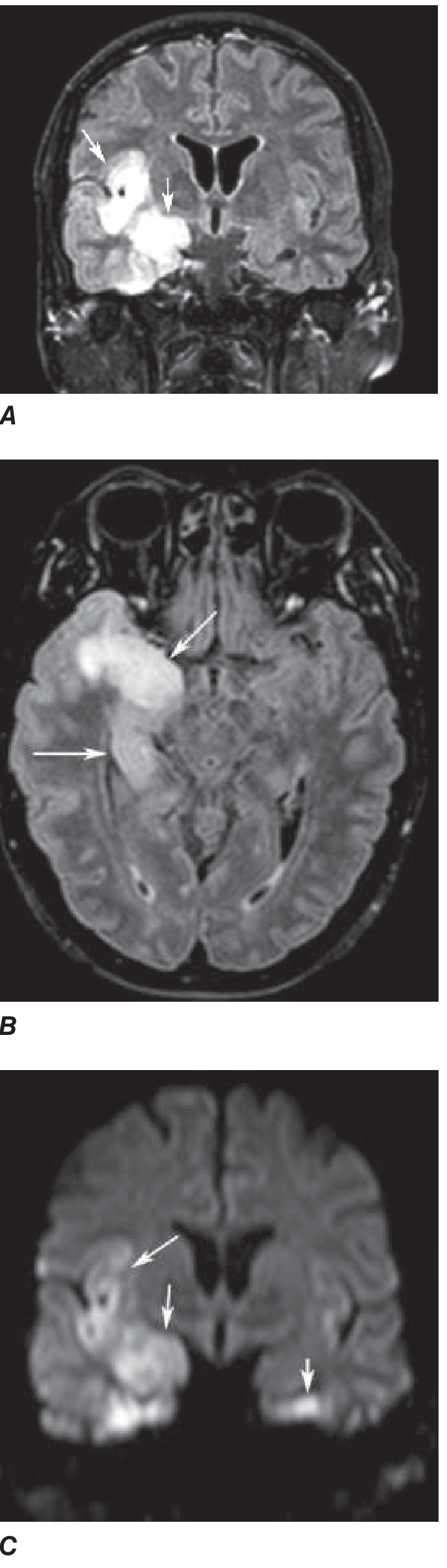
- Restricted diffusion (bright on DWI, dark on ADC) in the medial temporal lobe and hippocampus
- Represents cytotoxic edema and early neuronal death
- DWI may be positive even before FLAIR becomes abnormal in very early cases
- As seen in Harrison's image (Panel C): restricted diffusion in the right medial temporal lobe/hippocampus with subtle involvement on the left side
Gadolinium contrast (T1+C)
- Early gadolinium enhancement may occur but is not universal
- Enhancement reflects blood-brain barrier breakdown
- Leptomeningeal or cortical pattern of enhancement
CT comparison
- CT is less sensitive than MRI for early/mild disease
- However, because HSE can be hemorrhagic, CT may detect hemorrhage more reliably than MRI in some cases
Actual MRI Images
Figure 1 - FLAIR MRI (Goldman-Cecil): Right mesiotemporal hyperintensity (Panel A) and bilateral interfrontal/cingulate/right insular cortex signal change (Panel B):
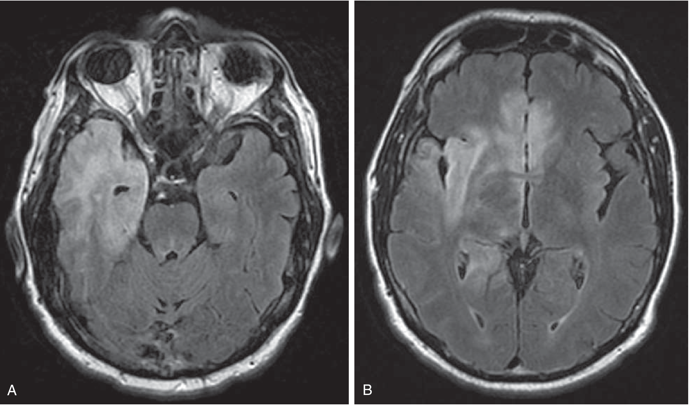
Figure 2 - Harrison's (FLAIR + DWI): Coronal FLAIR (A), axial FLAIR (B), and coronal DWI (C) showing right medial temporal/insular high signal with restricted diffusion, and subtle left temporal involvement on DWI:
Additional Features
- EEG correlation: Periodic high-voltage 1-Hz sharp waves from one or both temporal lobes are highly characteristic and correlate with the temporal lobe MRI changes
- Functional imaging: Hyperperfusion in the temporal lobe on SPECT/PET
- Normal MRI does not exclude HSE - frank encephalitis can occur with normal imaging findings, especially very early in disease
- Hemorrhagic transformation: Small areas of cortical hemorrhage can be seen, as the pathology is a necrotizing hemorrhagic encephalitis
Key Differentiating Points (from other limbic encephalitides)
| Feature | HSV Encephalitis | Autoimmune Limbic Encephalitis |
|---|---|---|
| Onset | Acute (hours-days) | Subacute (weeks) |
| Location | Mesiotemporal, insular, cingulate | Similar but often bilateral |
| Laterality | Unilateral or asymmetric | Often bilateral, more symmetric |
| Gray/white matter | Gray matter predominant | Can be similar |
| Hemorrhage | May be present | Rare |
| ADC restriction | Common | Less common |
Post-HSE Complication
Late autoimmune encephalitis (anti-NMDA receptor antibody mediated) can occur 1-7 weeks after HSE, showing worsening or new MRI changes with a clinical relapse - Goldman-Cecil Medicine.
Sources: Goldman-Cecil Medicine (International Ed.) | Harrison's Principles of Internal Medicine, 22E | Plum & Posner's Diagnosis and Treatment of Stupor and Coma | Bradley & Daroff's Neurology in Clinical Practice
This is a shared conversation. Sign in to Orris to start your own chat.