
Can you give me diagrams of scapula,clavicle,humerus,ulna,radius and carpal bones from moore’s anatomy
Reading File
Now let me search for Moore's anatomy and the relevant bone diagrams in parallel.
Finding Sources
Finding Sources
Moore's Clinically Oriented Anatomy is not in the library, but Gray's Anatomy for Students is available. Let me search for all the relevant bone sections and their images in Gray's.
Finding Sources
Finding Sources
Moore's is not in the library, but Gray's Anatomy for Students is available with all the bones. Let me now read the relevant sections to find the image URLs. I'll read multiple blocks in parallel.
Reading File
Reading File
I found images. Let me now read more sections to get all the remaining bone diagrams (scapula, humerus, carpal bones).
Reading File
Reading File
Reading File
I have humerus and radius/ulna images found. I need the scapula diagram (Fig. 7.21) and carpal bones diagram (Fig. 7.94). The scapula figure was referenced in block3 but that file returned empty on the extended offset. Let me search block3 for the scapula image and also get the carpal bones image from block4.
Finding Sources
Reading File
Reading File
I have all the images I need except the scapula (block3 was empty when read beyond offset 7438 - but the scapula figure ref was there). The scapula image was referenced in block3 but appeared to be in block4 as well (winged scapula ref in block4). Let me search block4 around line 1 for the scapula diagram (Fig. 7.21 was referenced in block3 text).
Reading File
Reading File
I now have all the images needed. Let me also get the radius shaft/distal image and ulna shaft image. I can see ulna already (Fig. 7.81 at line 2043). Let me get the radius shaft image.
Reading File
Reading File
I now have all the diagrams I need. Let me find the radius shaft/distal image (Fig. 7.80) which should be just before line 2053.
Reading File
I now have all the diagrams. Let me now fetch all the images in parallel to display them.
Loading Image
Loading Image
Loading Image
Loading Image
Loading Image
Loading Image
Loading Image
Loading Image
All diagrams retrieved. Here they all are, sourced from Gray's Anatomy for Students (Moore's Clinically Oriented Anatomy is not in our textbook library, but Gray's contains identical high-quality labeled diagrams for all the bones you need):
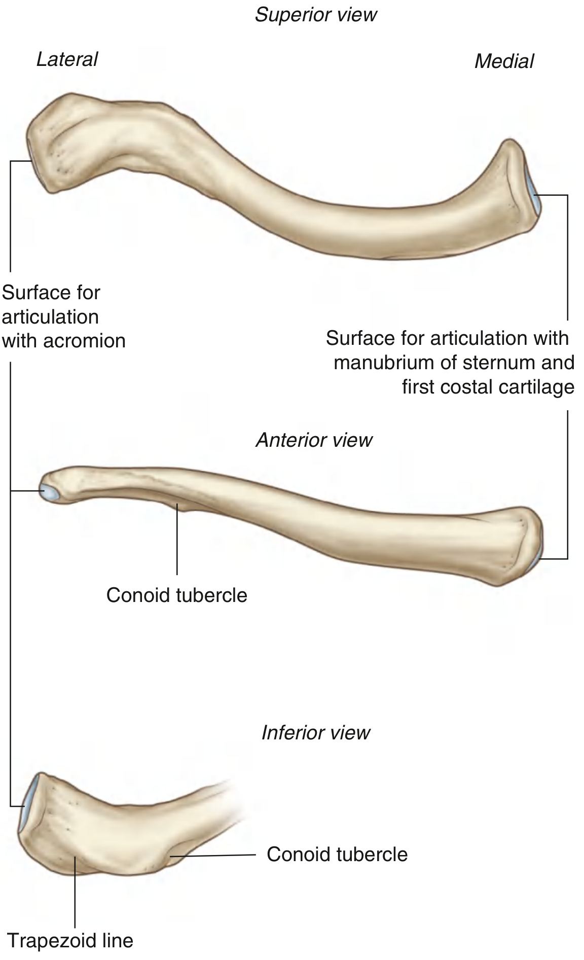
1. Clavicle
Fig. 7.20 - Right Clavicle (superior, anterior, and inferior views)
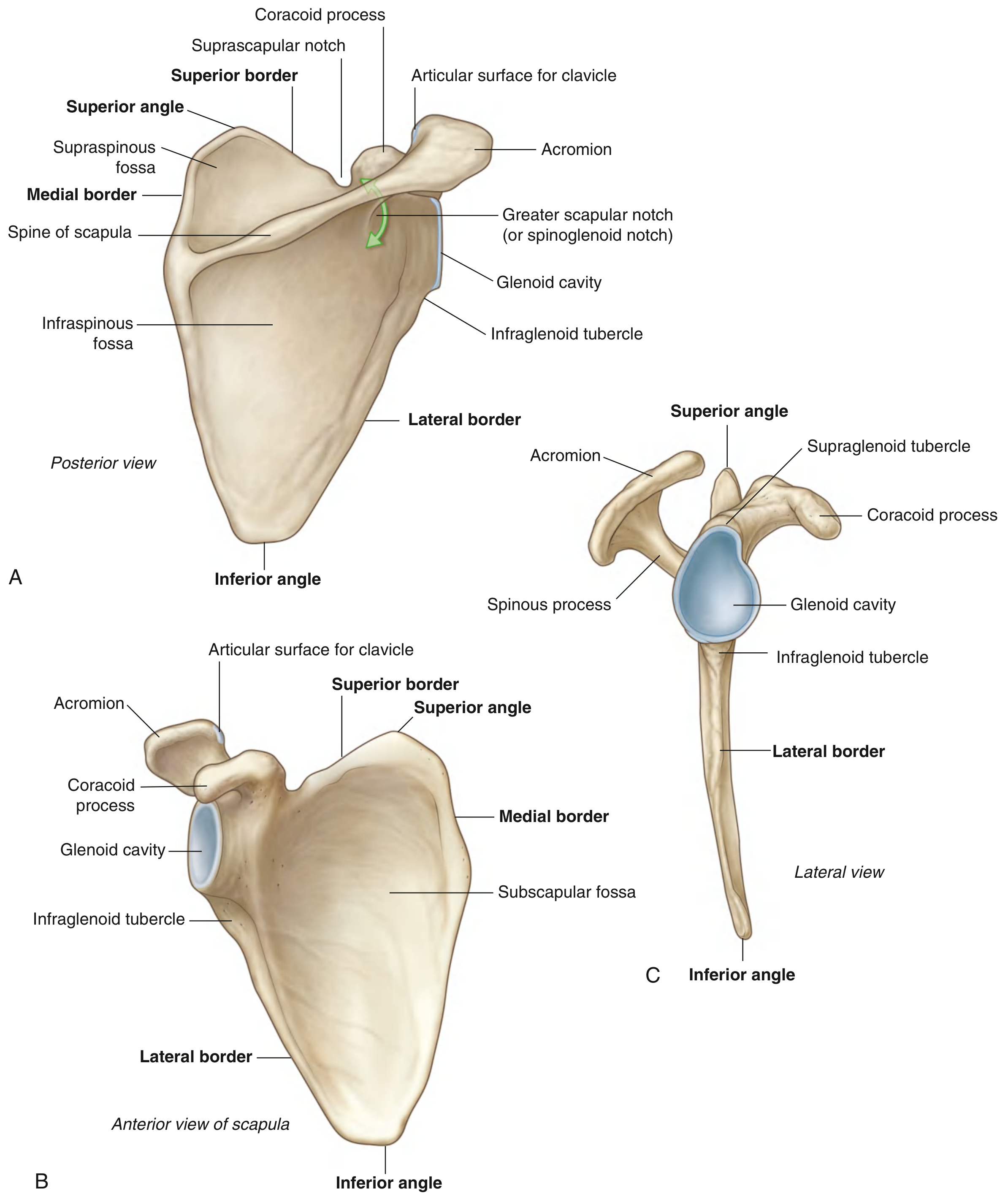
2. Scapula
Fig. 7.21 - (A) Posterior view, (B) Anterior/costal surface, (C) Lateral view
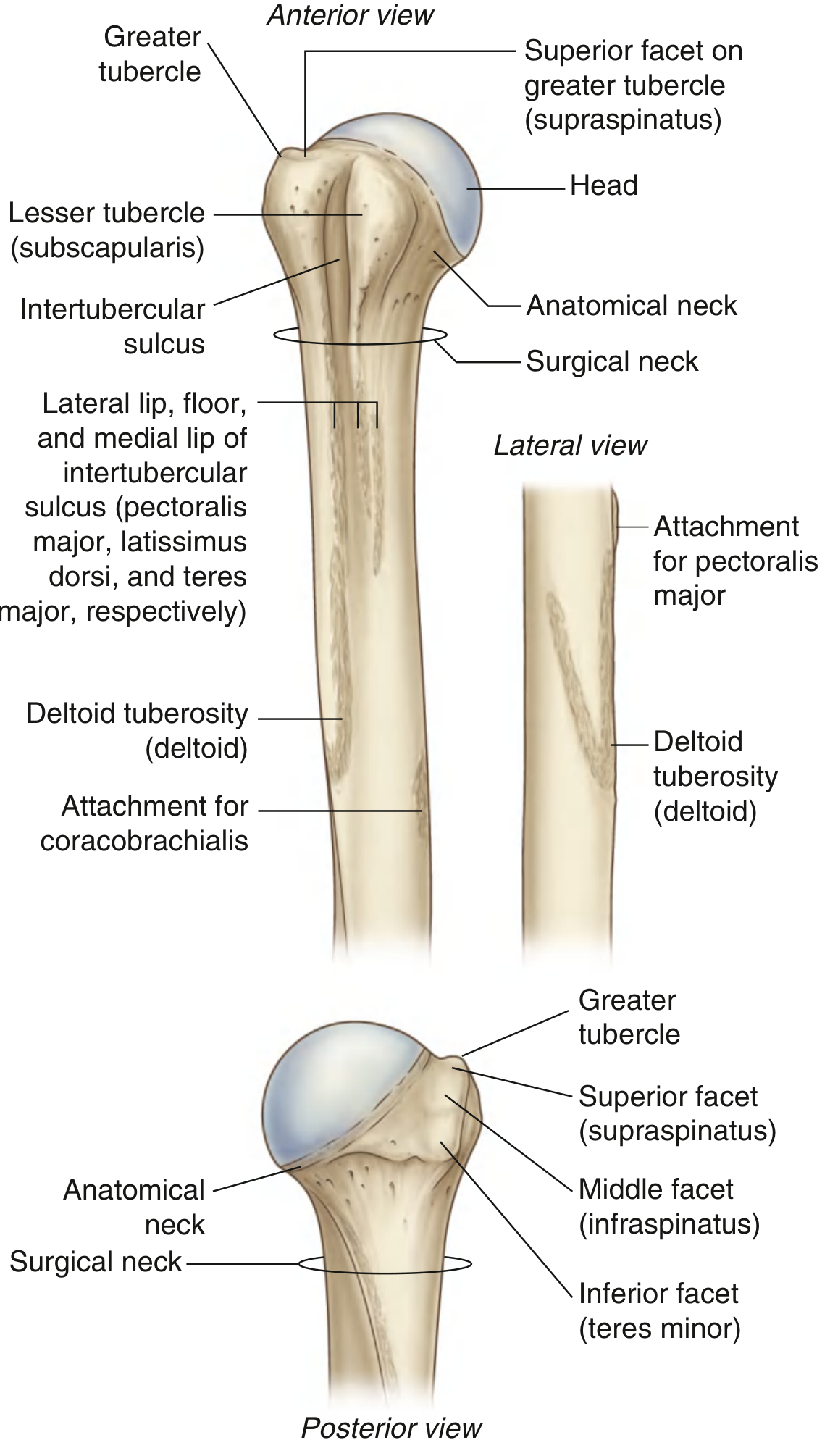
3. Humerus - Proximal End
Fig. 7.22 - Proximal End of Right Humerus (anterior and posterior views)
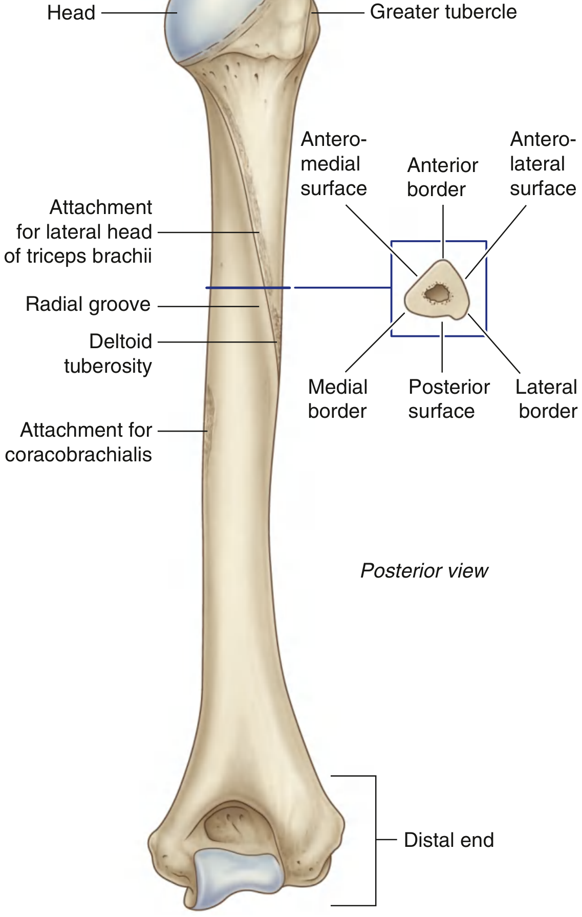
4. Humerus - Shaft and Distal End
Fig. 7.60 - Shaft and Distal End of Right Humerus (posterior view with cross-section)
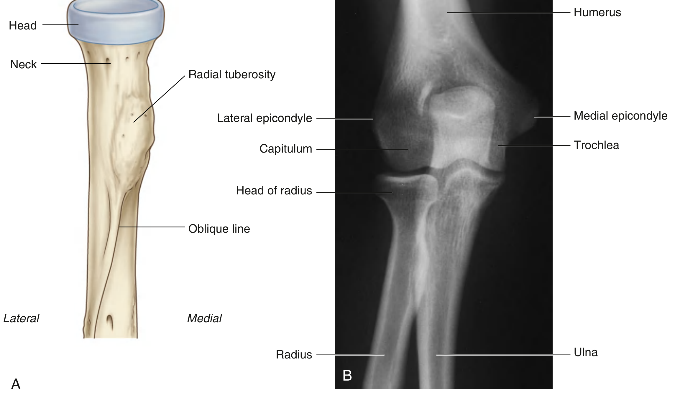
5. Radius - Proximal End
Fig. 7.62A - Proximal end of the radius with elbow radiograph
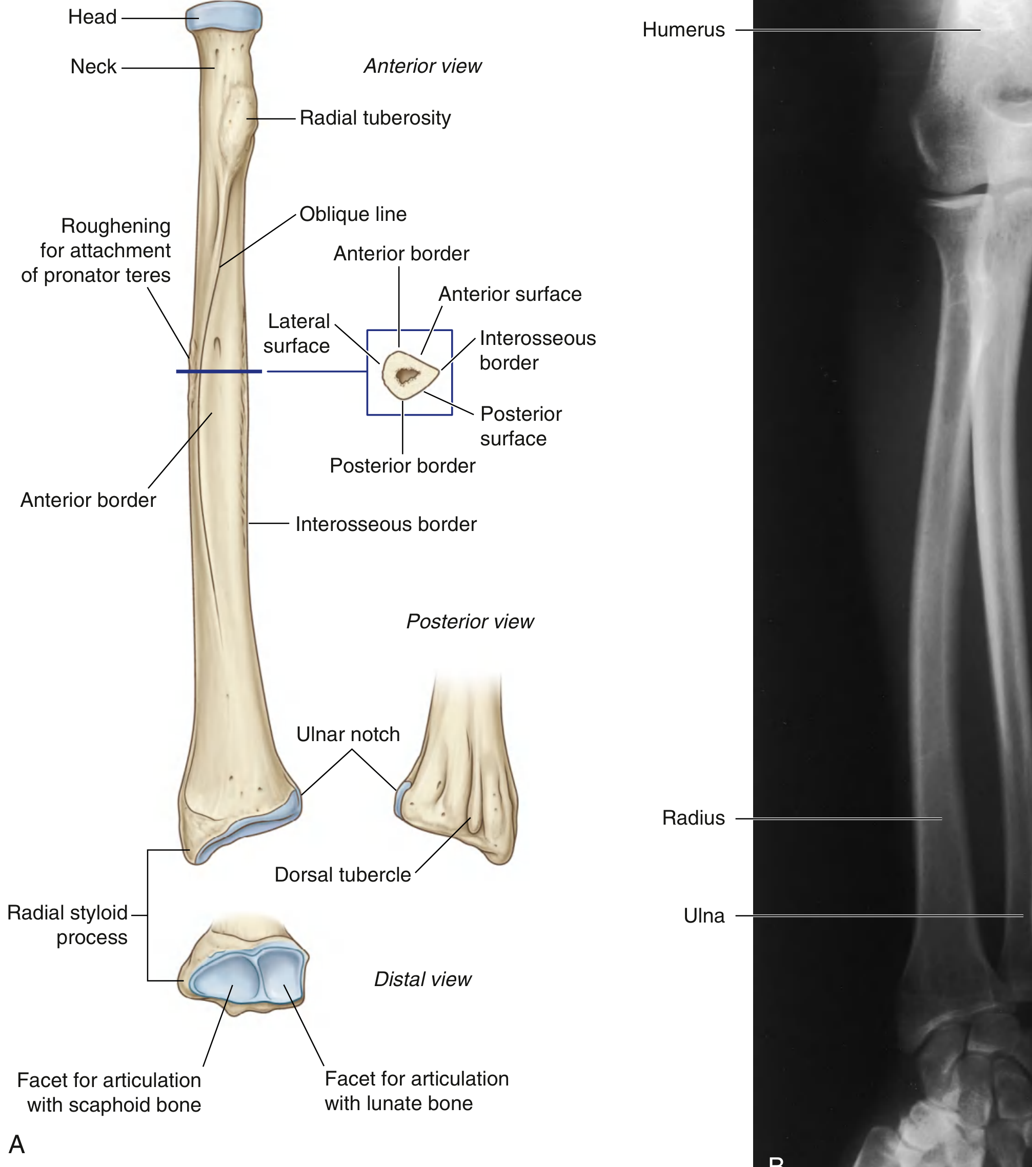
6. Radius - Shaft and Distal End
Fig. 7.80 - Radius full length (anterior view, cross-section, distal surface)
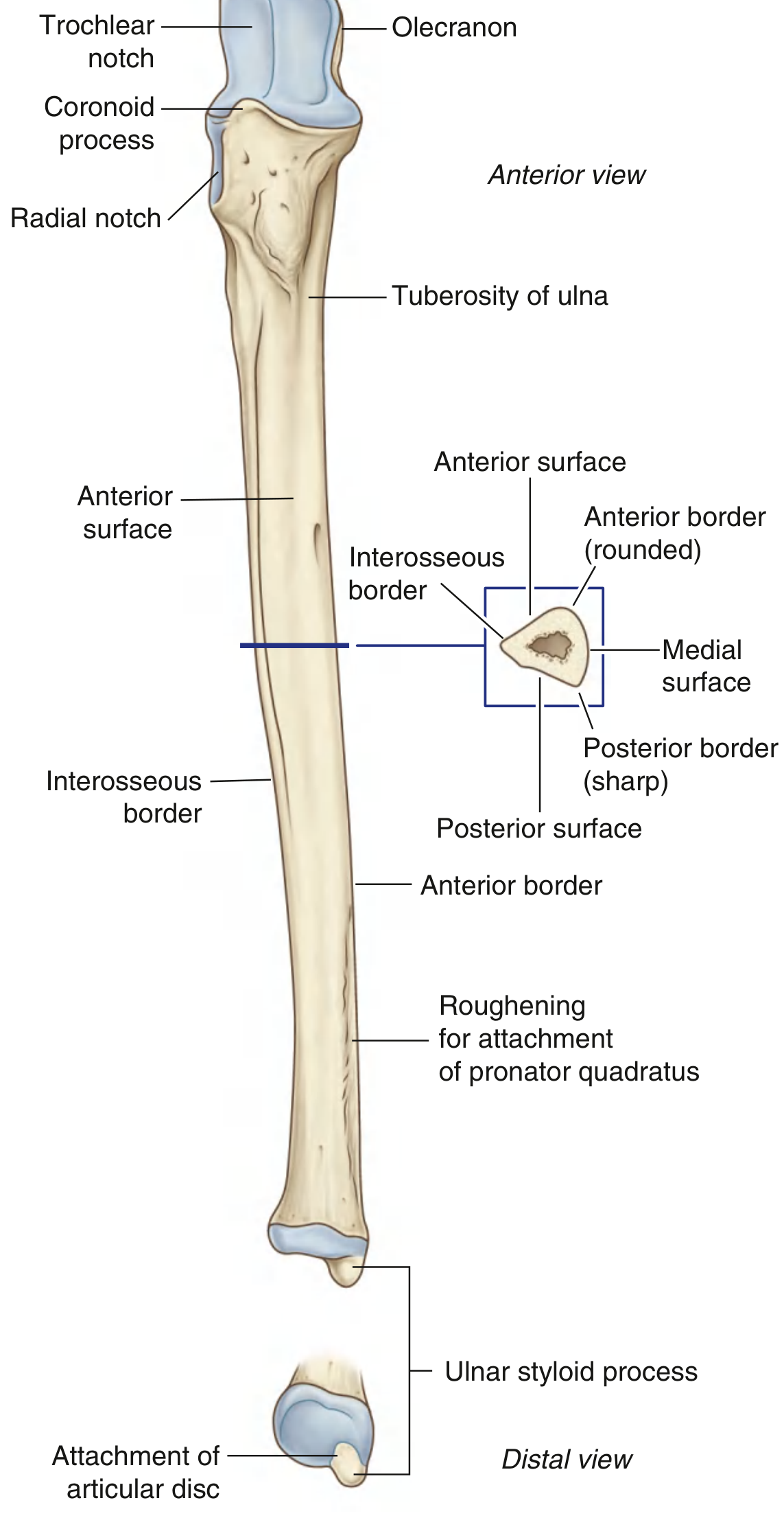
7. Ulna - Full Length
Fig. 7.81 - Right Ulna (anterior view, cross-section, distal end)
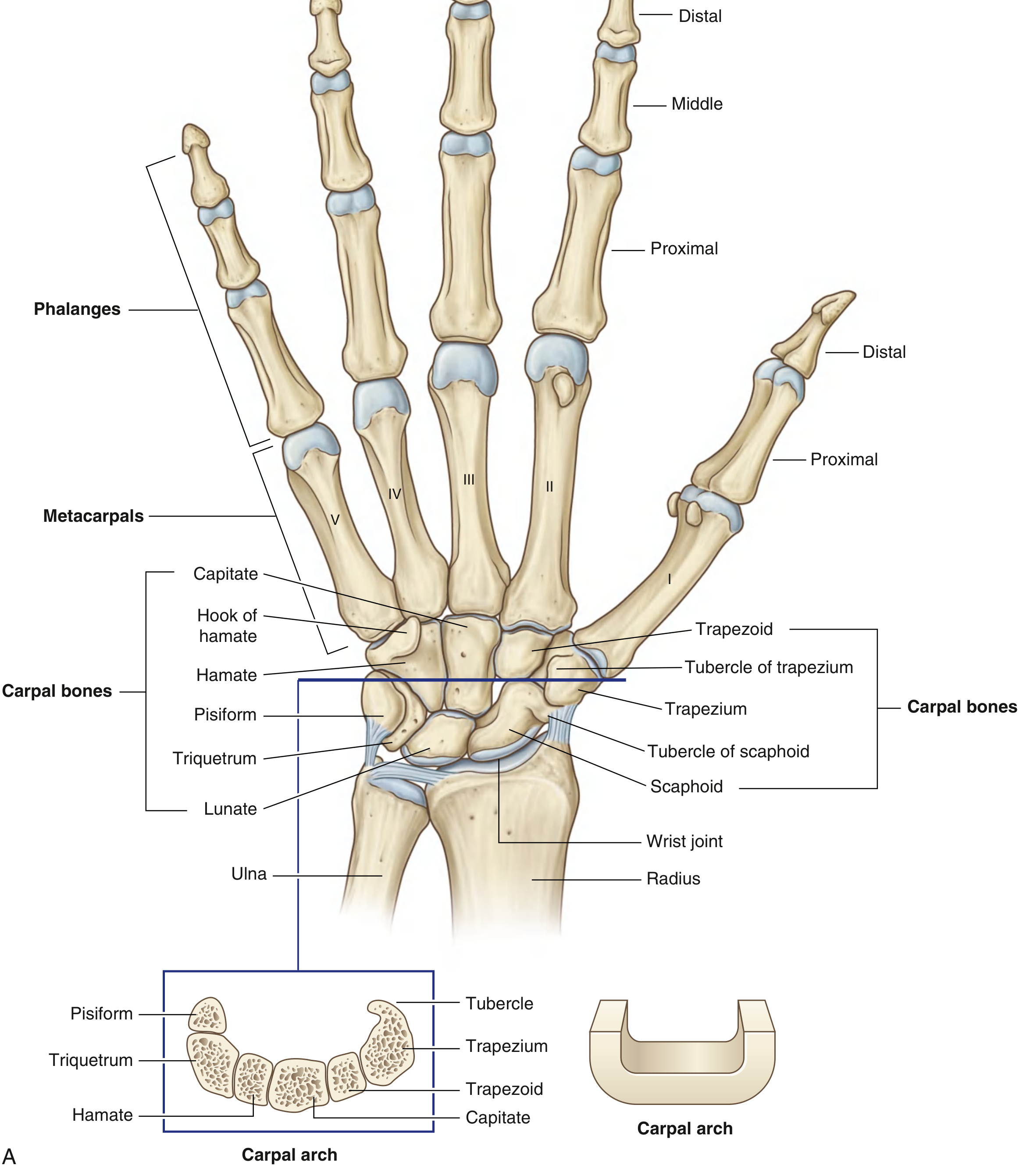
8. Carpal Bones
Fig. 7.94A - Right Hand and Wrist showing all 8 carpal bones with carpal arch cross-section
Note: Moore's Clinically Oriented Anatomy is not currently in our textbook library. These diagrams are from Gray's Anatomy for Students, which covers the same bones with equivalent detail and labeling. The diagrams include multiple views, cross-sections, muscle attachment sites, and articular surface labels for each bone.
This is a shared conversation. Sign in to Orris to start your own chat.