


+16 more
Give me detailed summary of this uploaded images in easy way so that can explain and give oral survey to my proffessor i m a 5th yr medical student
I can see all 10 pages from your textbook - these cover Stroke I, Stroke II, Stroke III, TIA & Prevention, and Subarachnoid Haemorrhage. Let me analyze all of them in detail and build you a structured oral survey guide.
analyse_media
analyse_media
analyse_media
analyse_media
analyse_media
I've now extracted detailed content from all 10 pages. Here is your complete oral survey guide, written in a clear, easy-to-explain format for your professor:
🧠 COMPLETE ORAL SURVEY GUIDE: STROKE, TIA & SUBARACHNOID HAEMORRHAGE
PART 1 - STROKE I: Definition, Epidemiology & Pathogenesis
What is a Stroke?
Stroke is damage to the brain caused by abnormalities of blood supply, presenting as a rapidly developing focal neurological deficit that may progress to coma or death. If the deficit lasts less than 24 hours, it is called a TIA (Transient Ischaemic Attack).
Epidemiology (Numbers to Remember)
- 3rd most common cause of death in developed countries
- Incidence of first stroke: 2 per 1000 per year
- Rare below age 45; rises to 30 per 1000/year in those over 80
- 25% die within 6 months (most within 1 month)
- 40% of survivors are dependent at 6 months
Types of Stroke (The 80-15-5 Rule)
| Type | Percentage |
|---|---|
| Cerebral infarction (ischaemic) | 80% |
| Primary intracerebral haemorrhage | 15% |
| Subarachnoid haemorrhage | 5% |
Causes of Cerebral Infarction
| Cause | % of strokes |
|---|---|
| Emboli | 30% |
| Thrombosis | 30% |
| Small vessel disease | 20% |
Atheroma - The Key Villain
Atheroma (fatty plaque build-up) is the most important underlying cause. It commonly forms at artery junctions - carotid bifurcation, vertebrobasilar junction. It causes ischaemia in 3 ways:
- Stenosis not compensated by anastomoses
- Plaque ulcerates → fragments become emboli
- Ulcerated base acts as focus for thrombus
Risk Factors for Atheroma (Relative Risk of Stroke)
| Risk Factor | Relative Risk |
|---|---|
| Hypertension | 5x (most important!) |
| Smoking | 3x |
| Diabetes | 2x |
| Excess alcohol | 1-4x |
| Family history, Cholesterol | Present |
Embolic Strokes - Sources
Cardiac sources (10% of all strokes):
- Atrial fibrillation (most common cardiac source)
- Mural thrombus post-MI
- Left ventricular dilatation
- Rare: bacterial endocarditis vegetations, atrial myxoma
Artery-to-artery emboli: From atheromatous plaque in carotid/vertebral vessels
Thrombotic Strokes - 3 Mechanisms
- Vessel wall abnormality - atheroma causes irregular surface → thrombus
- Hypercoagulable states - polycythaemia, sickle cell, protein C/S deficiency, antiphospholipid syndrome
- Stasis - severe stenosis or arterial dissection (commonest identifiable cause in young adults)
Small Vessel Disease (Lacunar strokes)
- Process of lipohyalinosis / microatheroma in small penetrating arteries
- Leads to occlusion → lacunar infarcts
Intracerebral Haemorrhage
- Usually due to hypertension
- Mechanism: Charcot-Bouchard microaneurysms rupture
- Locations: Basal ganglia 50%, Lobar white matter 20%, Pons 10%, Cerebellum 10%
- Other causes: AVM, anticoagulants, ruptured saccular aneurysm
Ischaemic Penumbra (Important Concept!)
- After infarction, the core dies but the penumbra (surrounding area) may be saved
- Cytotoxic oedema peaks at 5 days then gradually clears
- By 3 weeks: gliosis begins, infarct shrinks
- In small vessel strokes: gliosis creates fluid-filled pockets called "lacunes"
PART 2 - STROKE II: Clinical Features & Classification
Key Clinical Feature: ONSET
The hallmark of stroke is sudden onset - seconds to hours. This differentiates it from:
- Space-occupying lesions: weeks to months
- MS plaques: days to weeks
Stroke Classification (TACS/PACS/POCS/Lacunar)
1. Total Anterior Circulation Stroke (TACS)
Classic triad:
- Dense flaccid hemiparesis (arm + face + leg)
- Homonymous hemianopia
- Higher cortical dysfunction (aphasia if dominant, neglect if non-dominant)
- Eyes deviate away from hemiparetic side
- High mortality, severe morbidity
2. Partial Anterior Circulation Stroke (PACS)
- Only some features of TACS
- Depends on which MCA branch:
- Inferior MCA: Hemianopia + Wernicke's aphasia (or constructional apraxia)
- Superior MCA: Hemiparesis + Broca's aphasia (or neglect)
- ACA infarct: Leg > arm weakness + apathy + incontinence
3. Posterior Circulation Stroke (POCS)
- PCA: Contralateral homonymous hemianopia + hemisensory loss + cortical blindness
- Basilar occlusion: "Locked-in syndrome" - conscious but can't move (only eye movements may remain)
- Lateral Medullary Syndrome (Wallenberg's):
- Ipsilateral: facial pain/temp loss, Horner's, cerebellar ataxia, vocal cord paralysis
- Contralateral: body pain/temp loss
- Note: crossed signs = brain stem lesion
4. Lacunar Strokes (5 Pure Syndromes to Remember)
| Syndrome | Features | Site |
|---|---|---|
| Pure motor hemiparesis | Face + arm + leg weakness, NO sensory | Internal capsule |
| Ataxic hemiparesis | Motor + cerebellar ataxia same side | Post. internal capsule/pons |
| Dysarthria/clumsy hand | Dysarthria + hand clumsiness | Pons |
| Pure sensory stroke | Hemisensory loss only | Thalamus |
| Sensorimotor stroke | Both sensory + motor | Variable |
Differential Diagnosis of Stroke (Box 1)
- Space-occupying lesion (tumour, abscess, bleed into tumour)
- Subdural haematoma (slower onset, confused patient)
- Multiple sclerosis (younger patients)
- Head injury
- Hypoglycaemia (always check BSL first!)
- Todd's paralysis (post-seizure weakness mimicking stroke)
Important Clinical Pearls
- Infarction vs haemorrhage CANNOT be distinguished clinically - need CT/MRI
- Haemorrhage more likely if: severe headache + coma at onset
- Horner's syndrome + anterior circulation stroke = think carotid dissection
- Multi-infarct dementia: multiple small deep infarcts → dementia + small-stepped gait
PART 3 - STROKE III: Investigations & Treatment
Investigations - Answer 4 Questions
- Is it a stroke? → CT/MRI (DWI MRI best for early ischaemia)
- What type? → Imaging differentiates haemorrhagic from ischaemic
- Why did it occur? → Risk factor screen, cardiac workup, vascular imaging
- What could make it worse? → Metabolic screen (glucose, electrolytes, O2)
Tests Done in EVERY Patient (*)
- BP, blood glucose*, cholesterol*, thyroid function*, ECG*, full blood count*, ESR/plasma viscosity*
Additional Tests Based on Clinical Picture
- Cardiac source suspected: Echo, 24h tape, blood cultures
- Young patient, no atheroma risk factors: Thrombophilia screen (protein C/S, lupus anticoagulant), ANA, anticardiolipin Ab, syphilis serology, temporal artery biopsy
- Anterior circulation stroke: Carotid Doppler (looking for operable stenosis)
- Haemorrhagic, young, non-hypertensive: Cerebral angiography (AVM/aneurysm)
Treatment Goals (5 Aims)
- General medical support
- Minimize stroke size
- Prevent complications
- Optimize recovery
- Prevent recurrence
Key Treatments
Thrombolysis (rTPA):
- Given within 3 hours of onset
- Must do CT first to exclude haemorrhage
- Reduces stroke size and improves outcome
- Contraindications: no motor deficit, onset >3h, impaired consciousness, recent seizure, ICH history, pregnancy, recent surgery, BP >185/110, coagulopathy (INR >1.5)
Aspirin: Reduces 30-day stroke mortality
BP Management:
- Elevated in >80% of acute strokes
- Usually manage conservatively (don't lower aggressively - cerebral perfusion at risk)
- Only treat if diastolic >120 or hypertensive encephalopathy
Complications to Prevent (Box 3):
- Acute: Raised ICP + herniation, aspiration pneumonia
- Immobility: DVT, pneumonia, contractures, bed sores, UTI, constipation
- Later: Depression (50%!), epilepsy (5%), thalamic pain, social problems
Recovery (Multidisciplinary Team):
- Nurses, physiotherapists, OT, speech therapist, dietician, psychologist, social worker
- Stroke unit care reduces mortality by 25% and improves functional recovery
Prognosis
| Stroke Type | Mortality | Dependence at 6 months |
|---|---|---|
| Primary ICH | 50% | 50% |
| TACS | Similar to ICH | 90% |
| PACS/Lacunar/POCS | 10-15% | 20-40% |
PART 4 - TIA AND PREVENTION OF STROKE
What is a TIA?
- Acute loss of neurological function due to ischaemia lasting less than 24 hours, with complete recovery
- Occurs in 10% of patients prior to a stroke
- Annual incidence: 30 per 100,000
- Cause: artery-to-artery emboli or cardiac emboli (same as embolic stroke)
Clinical Features by Circulation
Anterior Circulation TIAs:
- Amaurosis fugax - fleeting monocular blindness ("like a shutter coming down") - classic!
- Aphasia, dyslexia, dysgraphia
Posterior Circulation TIAs:
- Homonymous visual field loss
- Dysarthria
- Vertigo + diplopia + dysphagia (only TIA if combined, not in isolation)
- Bilateral weakness/sensory loss
Either Circulation:
- Unilateral weakness (face/arm/leg)
- Unilateral sensory loss
TIA vs Mimics
| Condition | Distinguishing Feature |
|---|---|
| Migraine | Slower progression (15-30 min), positive symptoms (flashing lights), headache follows |
| Partial seizures | Very short (seconds to minutes), stereotyped |
| Transient global amnesia | Profound anterograde amnesia, hours duration, normal physical function |
| Hypoglycaemia | Low blood sugar, resolves with glucose |
| Todd's paralysis | Post-seizure weakness |
Secondary Prevention of Stroke
Risk is highest immediately after TIA - rapid treatment essential.
Control risk factors: Treat hypertension, diabetes, hypercholesterolaemia, stop smoking, reduce alcohol
Aspirin: Reduces stroke recurrence from 10% to 8%/year (dose: 75-300 mg/day)
Other antiplatelets: Dipyridamole (with aspirin) or Clopidogrel (aspirin-intolerant)
Anticoagulation (Warfarin):
- For AF - reduces risk from 12% to 4%/year (INR 2-3)
- Also in mural thrombus, LV dilatation
Carotid Endarterectomy:
- For symptomatic stenosis >70% (especially >80%)
- Reduces recurrent stroke risk significantly (NASCET/ECST trials)
- Requires surgical morbidity/mortality <7% to be worthwhile
- Occluded carotid = no further embolic risk, so surgery not indicated
Primary prevention: Control hypertension reduces stroke rate by 40% regardless of age
PART 5 - SUBARACHNOID HAEMORRHAGE (SAH)
What is SAH?
Bleeding into the subarachnoid space (between pia mater and arachnoid/dura mater). Incidence: 6-20 per 100,000/year. Peak age: 40-60 years.
Pathology
- Most SAH = ruptured intracranial aneurysm (usually saccular/berry aneurysm on Circle of Willis)
- 5% caused by AVM (arteriovenous malformation)
- Aneurysm distribution (where they occur most):
- Anterior communicating artery: 40%
- Posterior communicating artery: 30%
- Middle cerebral artery: 20%
- Basilar artery: 10%
Classical Presentation (4 Features)
- Sudden severe headache - "thunderclap", "worst headache of my life", "as if hit by a bat"
- Transient loss of consciousness
- Vomiting
- Neck stiffness + sometimes focal neurological signs
Key point: A 3rd nerve palsy with pupillary involvement = posterior communicating artery aneurysm - urgent neurosurgical referral!
Important Clinical Alert
- 1/6 patients die before reaching hospital
- On arrival: 1/3 drowsy, 20% stuporous or in coma
- 50% are initially misdiagnosed - always think SAH in sudden severe headache
- About 1/3 have had a warning "sentinel headache" in the weeks before - a smaller bleed they recovered from
Investigations
Step 1 - Prove SAH:
- CT brain: positive in 95% within 24h, only 67% by 3 days
- If CT negative: Lumbar puncture (wait at least 6 hours after onset) - look for xanthochromia (yellow CSF = blood breakdown products - confirmed by spectrophotometry)
Step 2 - Find the source:
- Four-vessel cerebral angiogram (gold standard) - Fig 4a in images
- CT angiography increasingly used, less invasive - Fig 4b
- 1 in 10 patients with definite SAH will have no source found (low recurrence risk)
Complications (The Big 3)
- Rebleeding from ruptured aneurysm
- Cerebral vasospasm → ischaemia (reduced by nimodipine + hypervolaemia)
- Hydrocephalus (CSF reabsorption blocked → needs drainage)
Other: Hyponatraemia, neurogenic pulmonary oedema, cardiac arrhythmias, DVT, aspiration pneumonia
Treatment
- Transfer to neurosurgical centre
- Monitor: pulse, BP, Glasgow Coma Scale
- Prevent rebleeding: Coiling (endovascular - preferred if technically possible) or surgical clipping
- Reduce vasospasm: Nimodipine (calcium antagonist) + sustained hypervolaemia (3 litres saline/day)
- Avoid antihypertensives (worsen vasospasm/ischaemia)
- Treat hydrocephalus with drainage
Prognosis
| Consciousness at Arrival | Outcome |
|---|---|
| Alert | 75% make full recovery, 10% mortality |
| Comatose | 75% die, 10% severe disability, only 10% good recovery |
QUICK REVISION CHEAT-SHEET FOR YOUR ORAL
Must-Know Numbers:
- Stroke types: 80% infarction / 15% ICH / 5% SAH
- Infarction causes: Emboli 30% / Thrombosis 30% / Small vessel 20%
- Hypertension = relative risk 5x for stroke (biggest modifiable risk factor)
- TIA precedes 10% of strokes; risk highest immediately after TIA
- rTPA window: 3 hours from onset
- SAH CT: positive 95% in 24h; LP if CT negative (after 6h)
- SAH aneurysm sites: Anterior comm 40%, Post comm 30%, MCA 20%, Basilar 10%
- Carotid endarterectomy: indicated for symptomatic stenosis >70%
- Aspirin reduces recurrence from 10% → 8%/year
- Anticoagulation in AF: reduces stroke risk from 12% → 4%/year
Key Distinctions Professors Love to Ask:
- Infarct vs haemorrhage: cannot be distinguished clinically - need CT
- TACS vs PACS: TACS has all 3 features (hemiparesis + hemianopia + cortical dysfunction)
- Lacunar vs cortical: Lacunar has NO cortical features (no aphasia, no neglect, no hemianopia)
- Wallenberg's (lateral medullary): crossed signs - ipsilateral face + contralateral body
- Locked-in syndrome: basilar artery occlusion - conscious but paralysed, only eye movement preserved
- SAH headache vs migraine: SAH is instantaneous "thunderclap"; migraine builds up over 15-30 min
This covers everything in your 10 uploaded pages. Practice explaining the classification tree (TACS → PACS → POCS → Lacunar) and the SAH investigation algorithm - professors commonly target these in oral surveys. Good luck! 🎓
Explain Thalamic stroke
A thalamic stroke is a type of lacunar/small vessel stroke affecting the thalamus - a deep brain structure that acts as the main relay station for sensory, motor, and consciousness signals. Let me pull up detailed textbook content on this.
Searching sources...
Finding Sources
Finding Sources
Searching the Web
thalamic stroke clinical features syndromes anatomy 2024
Found an excellent dedicated chapter in Bradley & Daroff's Neurology. Let me pull the full content now.
Reading File
Reading File
Reading File
Reading File
Reading File
Excellent - I now have all the detailed textbook content. Let me fetch the thalamic territory diagram too.
Loading Image
Here is your complete, easy-to-explain guide on thalamic stroke:
🧠 THALAMIC STROKE - Complete Easy Guide for Oral Survey
What is the Thalamus? (Start Here)
Think of the thalamus as the "relay station" or "switchboard" of the brain. It sits deep in the centre of the brain and:
- Relays ALL sensory information (touch, pain, temperature, vision, hearing, taste) to the cortex
- Helps regulate consciousness and alertness
- Plays a role in memory, emotions, and executive function
- Controls the sleep-wake cycle
So when it gets damaged by a stroke, you get a wide and sometimes confusing mix of symptoms - sensory loss, consciousness changes, memory problems, eye movement issues - all from one small lesion.
Blood Supply of the Thalamus (Important!)
The thalamus gets its blood from 4 arteries, all branches of the posterior cerebral artery (PCA) and basilar artery (i.e., posterior circulation):
| Artery | Territory |
|---|---|
| Polar artery (tuberothalamic) | Anterolateral thalamus |
| Paramedian artery (thalamic perforators) | Medial/paramedian thalamus |
| Geniculothalamic artery (inferolateral) | Posterolateral thalamus |
| Posterior choroidal artery | Posterior/dorsal thalamus |
Key fact: There is a special anatomical variant called the Artery of Percheron - a single artery that supplies BOTH paramedian thalami. If this one artery is occluded → bilateral thalamic infarction → sudden coma + memory loss + vertical gaze palsy. Classic exam question!
4 Types of Thalamic Stroke (by vascular territory)
1. ANTEROLATERAL (Polar/Tuberothalamic Artery)
Think: Neuropsychiatric + cognitive + sensorimotor mix
- Contralateral weakness, hemisensory loss, hemiataxia
- Homonymous hemianopia
- Language disturbance / aphasia (if left-sided)
- Visuospatial problems and neglect (if right-sided)
- Confusion and disorientation
2. MEDIAL/PARAMEDIAN (Paramedian Artery or Artery of Percheron)
Think: Consciousness + memory + eye movements
This is the most dramatic presentation:
- Sudden decreased consciousness - disorientation, stupor, coma
- Memory loss (amnesia - both anterograde and retrograde)
- Vertical gaze palsy - patient can't look up or down
- Loss of pupillary reflexes
- Loss of convergence
- Neuropsychiatric disturbances
If the Artery of Percheron is involved (bilateral): patient suddenly becomes deeply comatose with vertical gaze palsy - a pattern that can be mistaken for a midbrain lesion or metabolic coma.
3. POSTEROLATERAL (Geniculothalamic / Inferolateral Artery)
Think: Sensory + Dejerine-Roussy syndrome
This is the most important and classic thalamic territory:
- Contralateral loss of touch, pain, temperature, and vibration (ALL modalities)
- Hemiparesis (in some)
- Hemiataxia
- Homonymous hemianopia
- Left hemispatial neglect
- Poor attention span
Classic syndrome here = Dejerine-Roussy Syndrome (see below)
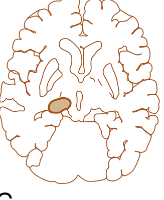
4. POSTERIOR (Posterior Choroidal Artery)
Think: Visual field defects + pulvinar involvement
- Homonymous quadrantanopia or horizontal sectoranopsia
- Involvement of the pulvinar nucleus → thalamic aphasia (if left side)
- Memory impairment
The 3 Classic Clinical Syndromes of Thalamic Stroke
These arise mainly from posterolateral infarction and are the most commonly tested:
A. Pure Sensory Stroke
- Hemisensory loss ONLY - no motor, no vision, no cognitive deficits
- Contralateral face + arm + leg
- All modalities affected (touch, pain, temperature, proprioception)
- Lesion: VPL nucleus (ventral posterolateral nucleus) of thalamus
- This is a classic lacunar syndrome
B. Sensorimotor Stroke
- Sensory loss + hemiparesis combined
- Lesion extends from thalamus into posterior limb of internal capsule
C. Dejerine-Roussy Syndrome (Thalamic Pain Syndrome)
This is the most famous thalamic syndrome - named after French neurologist Dejerine (1906):
Triad:
- Moderate contralateral hemiparesis (initially, may improve)
- Hemisensory loss (hemihypesthesia)
- Severe, burning, agonizing contralateral pain that comes AFTER the stroke (central post-stroke pain)
Features of the pain:
- Develops days to weeks AFTER the stroke, not immediately
- Affects contralateral face, arm, trunk, and leg
- Burning, aching, gnawing - made WORSE by touch, cold, or even emotions
- Called "allodynia" - normal stimuli cause pain
- Hemiataxia and hemialgia (dysesthesia - perversion of sensation)
- The "thalamic hand/arm" - a dystonic posture of the hand may develop in extensive strokes
Why does this pain occur?
The thalamus normally "gates" or modulates pain. When it is damaged, inhibition is lost and pain signals become amplified and distorted.
Treatment of thalamic pain:
- Tricyclic antidepressants (amitriptyline) - first line
- Carbamazepine / anticonvulsants
- Often very difficult to treat (as mentioned in your textbook)
Thalamic Haemorrhage (vs Infarction)
- Thalamic haemorrhage usually occurs from hypertension (like other deep brain haemorrhages)
- Produces same symptoms as infarction but often more severe and sudden
- Additional feature: downward and inward deviation of the eyes (looking down at the nose - the "setting sun" sign for thalamic bleed)
- May rupture into the third ventricle → hydrocephalus
Summary Table: Quick Reference
| Feature | Details |
|---|---|
| Type of stroke | Lacunar / small vessel (posterior circulation) |
| Main artery involved | Branches of PCA (posterior cerebral artery) |
| Most classic syndrome | Dejerine-Roussy (thalamic pain syndrome) |
| Pure sensory stroke | VPL nucleus, all modalities |
| Paramedian infarct | Coma + amnesia + vertical gaze palsy |
| Bilateral thalamic | Artery of Percheron occlusion |
| Thalamic haemorrhage | Hypertension, eyes deviate downward |
| Late complication | Thalamic pain (central post-stroke pain) |
| Treatment of pain | Amitriptyline, carbamazepine |
How to Spot a Thalamic Stroke Clinically
Ask yourself: Does the patient have an unusual combination of:
- Sensory loss (especially all modalities)
- Altered consciousness or memory out of proportion to motor deficit
- Eye movement abnormalities (vertical gaze palsy)
- Later: inexplicable burning pain on one side
If yes → think thalamic stroke. The textbook notes: "in particular where there is impaired vigilance and an unusual collection of deficits difficult to explain by a single cortical lesion, thalamic disease should be considered." - Bradley & Daroff's Neurology
Imaging
- MRI (DWI) is the best modality for acute thalamic infarction - CT may miss small thalamic lesions
- MRI shows the exact thalamic nucleus/territory involved, guiding syndrome prediction
Here is the axial brain diagram showing the posterior choroidal territory (shaded region = thalamic infarct zone):
Quick Oral Talking Points for Your Professor
- "Thalamic stroke is a type of posterior circulation small vessel stroke..."
- "The 4 vascular territories give rise to 4 distinct syndromes - anterolateral, medial, posterolateral, and posterior..."
- "The most classic syndrome is Dejerine-Roussy - a triad of hemiparesis, hemisensory loss, and delayed contralateral pain..."
- "A special case is bilateral thalamic infarction from the Artery of Percheron - presenting as sudden coma + amnesia + vertical gaze palsy..."
- "Thalamic pain is central post-stroke pain treated with amitriptyline or carbamazepine..."
- "MRI-DWI is the investigation of choice as CT often misses small thalamic lesions..."
Sources: Bradley and Daroff's Neurology in Clinical Practice, 8th Ed.; Ganong's Review of Medical Physiology, 26th Ed.
This is a shared conversation. Sign in to Orris to start your own chat.