
/generatehandwrittenimages Describe the propagation of action potentials in a mixed nerve. What are the functional implications of their distinct conduction velocities and properties. - for md physiology 10 marks theory question. You can make many images and not just one for completing the answer
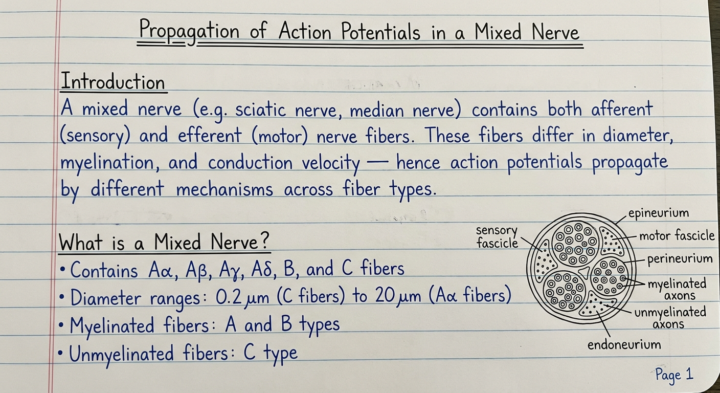
Handwritten medical exam answer on ruled notebook paper, blue ink pen, neat student handwriting. Page 1 of a 10-mark MD Physiology theory answer. Title at top (underlined twice): "Propagation of Action Potentials in a Mixed Nerve" Subheading (underlined): "Introduction" Text: "A mixed nerve (e.g. sciatic nerve, median nerve) contains both afferent (sensory) and efferent (motor) nerve fibers. These fibers differ in diameter, myelination, and conduction velocity — hence action potentials propagate by different mechanisms across fiber types." Subheading (underlined): "What is a Mixed Nerve?" Bullet points: "• Contains Aα, Aβ, Aγ, Aδ, B, and C fibers" "• Diameter ranges: 0.2 μm (C fibers) to 20 μm (Aα fibers)" "• Myelinated fibers: A and B types" "• Unmyelinated fibers: C type" Small neat hand-drawn cross-section diagram of a peripheral nerve showing fascicles labeled 'motor fascicle', 'sensory fascicle', myelinated axons (circles with rings) and unmyelinated axons (small dots) within epineurium, perineurium, endoneurium labeled. Bottom corner: "Page 1" in small writing. Lined paper background texture visible.
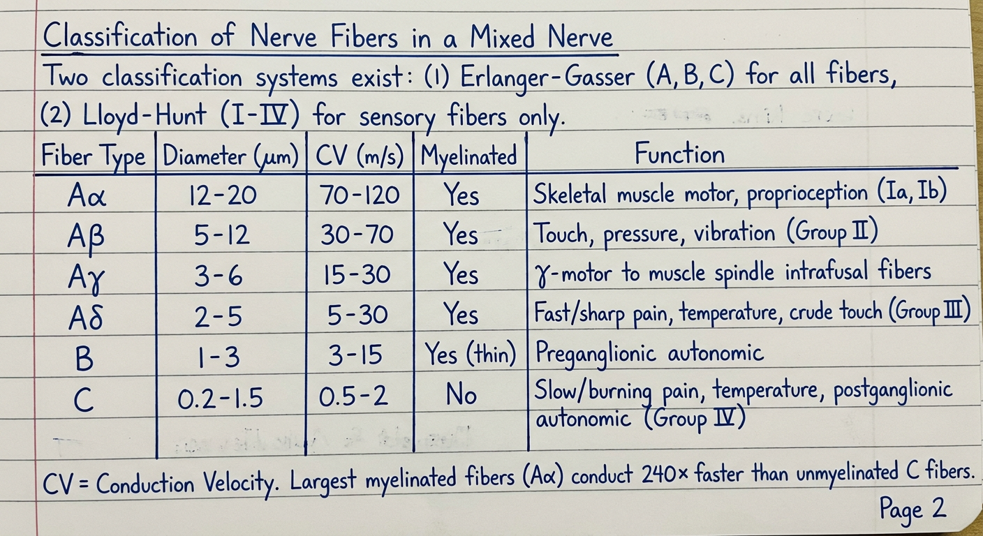
Handwritten medical exam answer on ruled notebook paper, blue ink pen, neat student handwriting. Page 2 of a 10-mark MD Physiology theory answer. Heading (underlined): "Classification of Nerve Fibers in a Mixed Nerve" Text: "Two classification systems exist: (1) Erlanger-Gasser (A, B, C) for all fibers, (2) Lloyd-Hunt (I-IV) for sensory fibers only." Neatly hand-drawn table with the following columns: Fiber Type | Diameter (μm) | CV (m/s) | Myelinated | Function Rows: "Aα | 12-20 | 70-120 | Yes | Skeletal muscle motor, proprioception (Ia, Ib)" "Aβ | 5-12 | 30-70 | Yes | Touch, pressure, vibration (Group II)" "Aγ | 3-6 | 15-30 | Yes | γ-motor to muscle spindle intrafusal fibers" "Aδ | 2-5 | 5-30 | Yes | Fast/sharp pain, temperature, crude touch (Group III)" "B | 1-3 | 3-15 | Yes (thin) | Preganglionic autonomic" "C | 0.2-1.5 | 0.5-2 | No | Slow/burning pain, temperature, postganglionic autonomic (Group IV)" Below table, small note: "CV = Conduction Velocity. Largest myelinated fibers (Aα) conduct 240× faster than unmyelinated C fibers." Bottom corner: "Page 2"
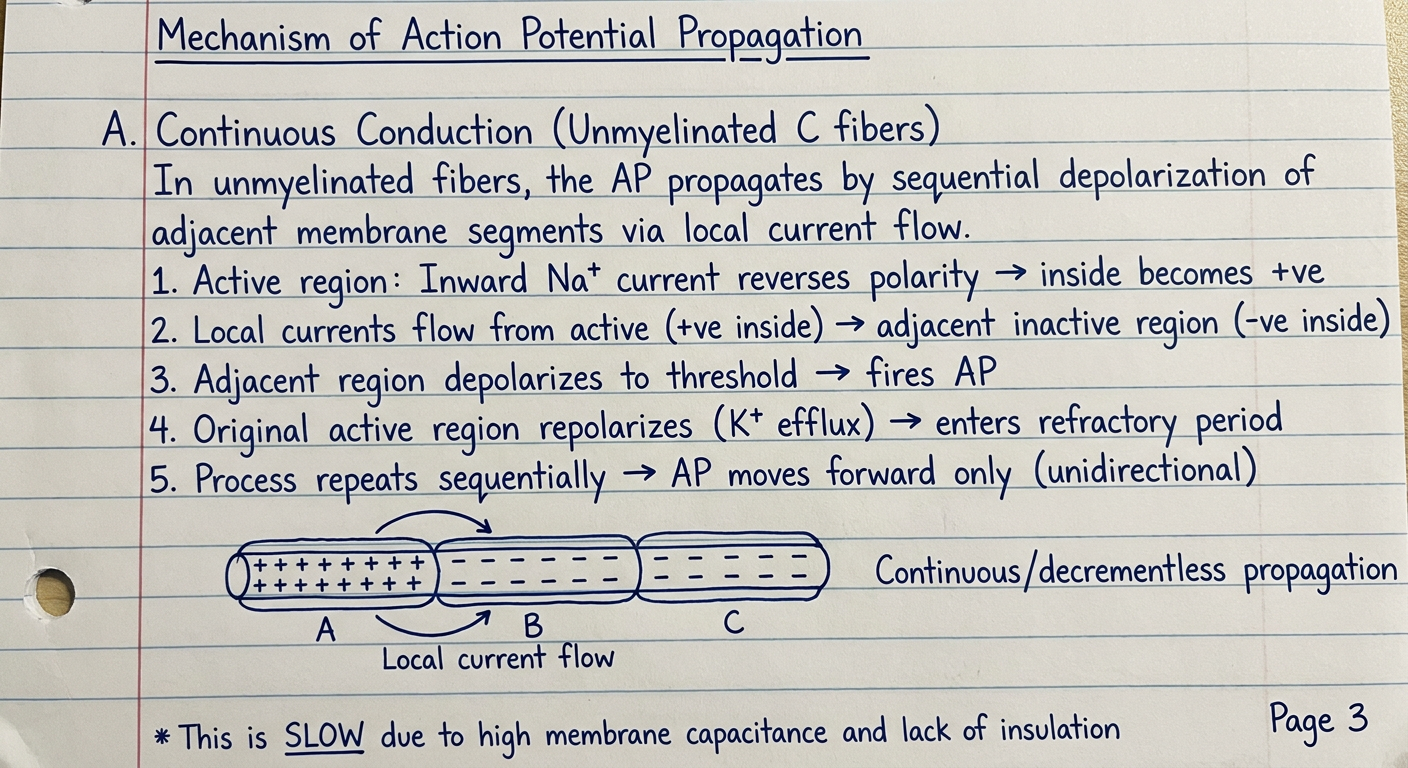
Handwritten medical exam answer on ruled notebook paper, blue ink pen, neat student handwriting. Page 3 of a 10-mark MD Physiology theory answer. Heading (underlined): "Mechanism of Action Potential Propagation" Subheading: "A. Continuous Conduction (Unmyelinated C fibers)" Explanatory text: "In unmyelinated fibers, the AP propagates by sequential depolarization of adjacent membrane segments via local current flow." Step-by-step with numbers: "1. Active region: Inward Na+ current reverses polarity → inside becomes +ve" "2. Local currents flow from active (+ve inside) → adjacent inactive region (-ve inside)" "3. Adjacent region depolarizes to threshold → fires AP" "4. Original active region repolarizes (K+ efflux) → enters refractory period" "5. Process repeats sequentially → AP moves forward only (unidirectional)" Hand-drawn diagram showing a horizontal unmyelinated axon with: - Three segments labeled A, B, C - Segment A: shows + signs inside (active, depolarized) with curved arrows showing local current flowing to B - Segment B: shows - inside (about to be depolarized) - Arrows labeled "Local current flow" - Note: "Continuous/decrementless propagation" - Small note at bottom: "This is SLOW due to high membrane capacitance and lack of insulation" Bottom corner: "Page 3"
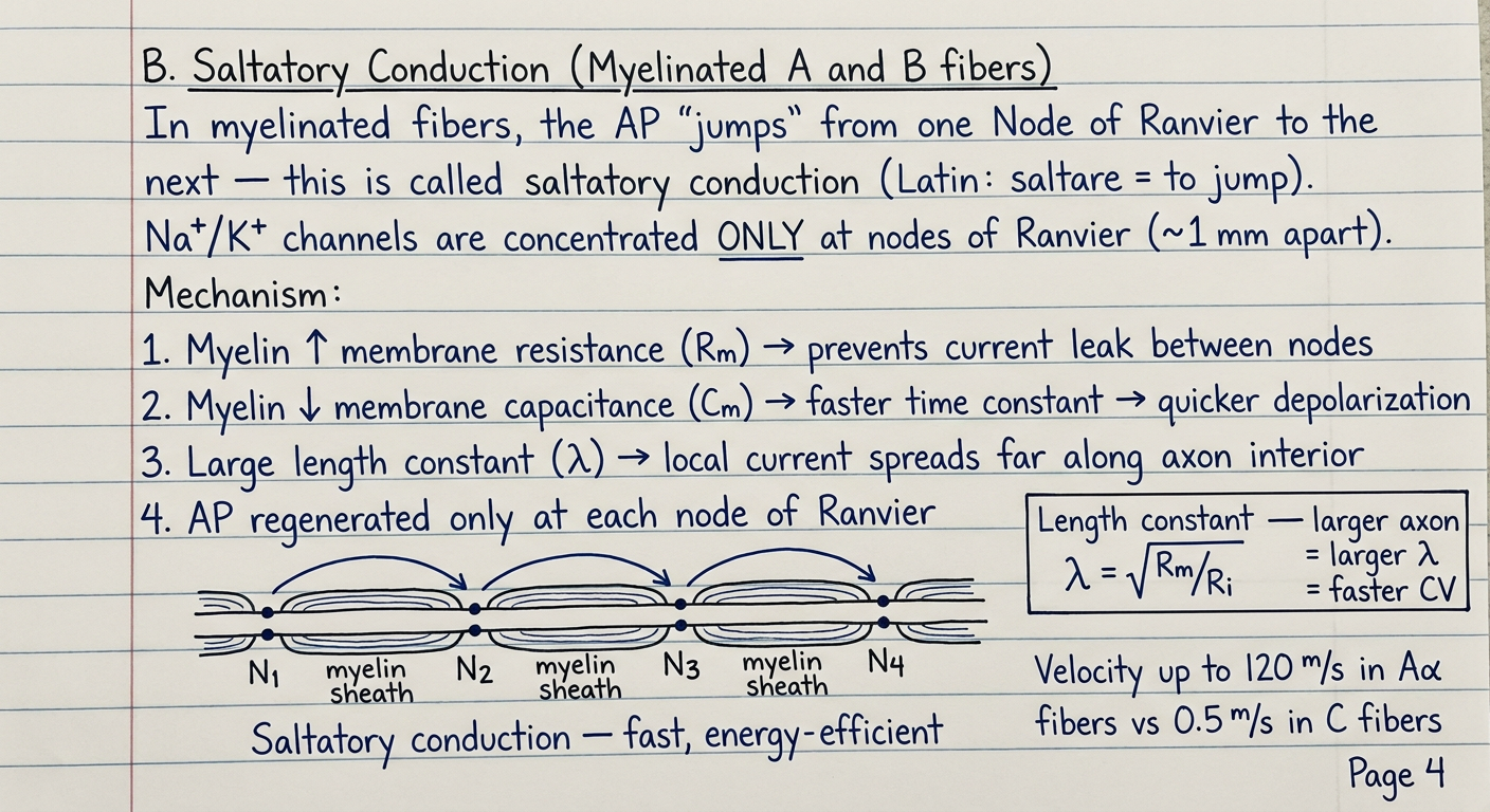
Handwritten medical exam answer on ruled notebook paper, blue ink pen, neat student handwriting. Page 4 of a 10-mark MD Physiology theory answer. Heading (underlined): "B. Saltatory Conduction (Myelinated A and B fibers)" Text paragraph: "In myelinated fibers, the AP 'jumps' from one Node of Ranvier to the next — this is called saltatory conduction (Latin: saltare = to jump). Na+/K+ channels are concentrated ONLY at nodes of Ranvier (~1 mm apart)." "Mechanism:" "1. Myelin ↑ membrane resistance (Rm) → prevents current leak between nodes" "2. Myelin ↓ membrane capacitance (Cm) → faster time constant → quicker depolarization" "3. Large length constant (λ) → local current spreads far along axon interior" "4. AP regenerated only at each node of Ranvier" Hand-drawn diagram of a myelinated axon showing: - Horizontal axon with 4 nodes of Ranvier labeled N1, N2, N3, N4 - Myelin segments between nodes labeled "myelin sheath" - Curved arrows showing current leaping from N1 → N2 → N3 (skipping the myelin) - Label: "Saltatory conduction — fast, energy-efficient" - Small inset box: "Length constant λ = √(Rm/Ri) — larger axon = larger λ = faster CV" - Note: "Velocity up to 120 m/s in Aα fibers vs 0.5 m/s in C fibers" Bottom corner: "Page 4"
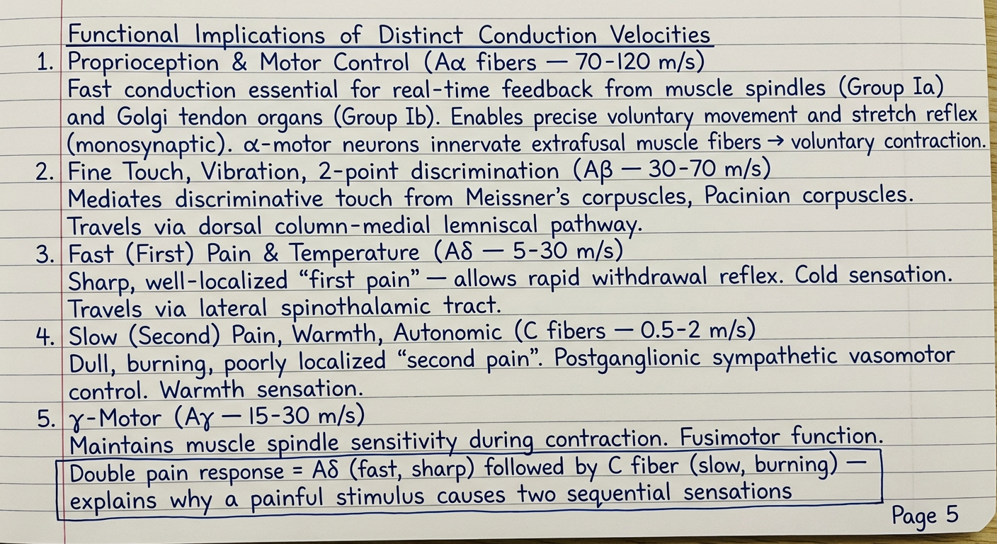
Handwritten medical exam answer on ruled notebook paper, blue ink pen, neat student handwriting. Page 5 of a 10-mark MD Physiology theory answer. Heading (underlined): "Functional Implications of Distinct Conduction Velocities" Subheading: "1. Proprioception & Motor Control (Aα fibers — 70-120 m/s)" "Fast conduction essential for real-time feedback from muscle spindles (Group Ia) and Golgi tendon organs (Group Ib). Enables precise voluntary movement and stretch reflex (monosynaptic). α-motor neurons innervate extrafusal muscle fibers → voluntary contraction." Subheading: "2. Fine Touch, Vibration, 2-point discrimination (Aβ — 30-70 m/s)" "Mediates discriminative touch from Meissner's corpuscles, Pacinian corpuscles. Travels via dorsal column-medial lemniscal pathway." Subheading: "3. Fast (First) Pain & Temperature (Aδ — 5-30 m/s)" "Sharp, well-localized 'first pain' — allows rapid withdrawal reflex. Cold sensation. Travels via lateral spinothalamic tract." Subheading: "4. Slow (Second) Pain, Warmth, Autonomic (C fibers — 0.5-2 m/s)" "Dull, burning, poorly localized 'second pain'. Postganglionic sympathetic vasomotor control. Warmth sensation." Subheading: "5. γ-Motor (Aγ — 15-30 m/s)" "Maintains muscle spindle sensitivity during contraction. Fusimotor function." Box/note: "Double pain response = Aδ (fast, sharp) followed by C fiber (slow, burning) — explains why a painful stimulus causes two sequential sensations" Bottom corner: "Page 5"
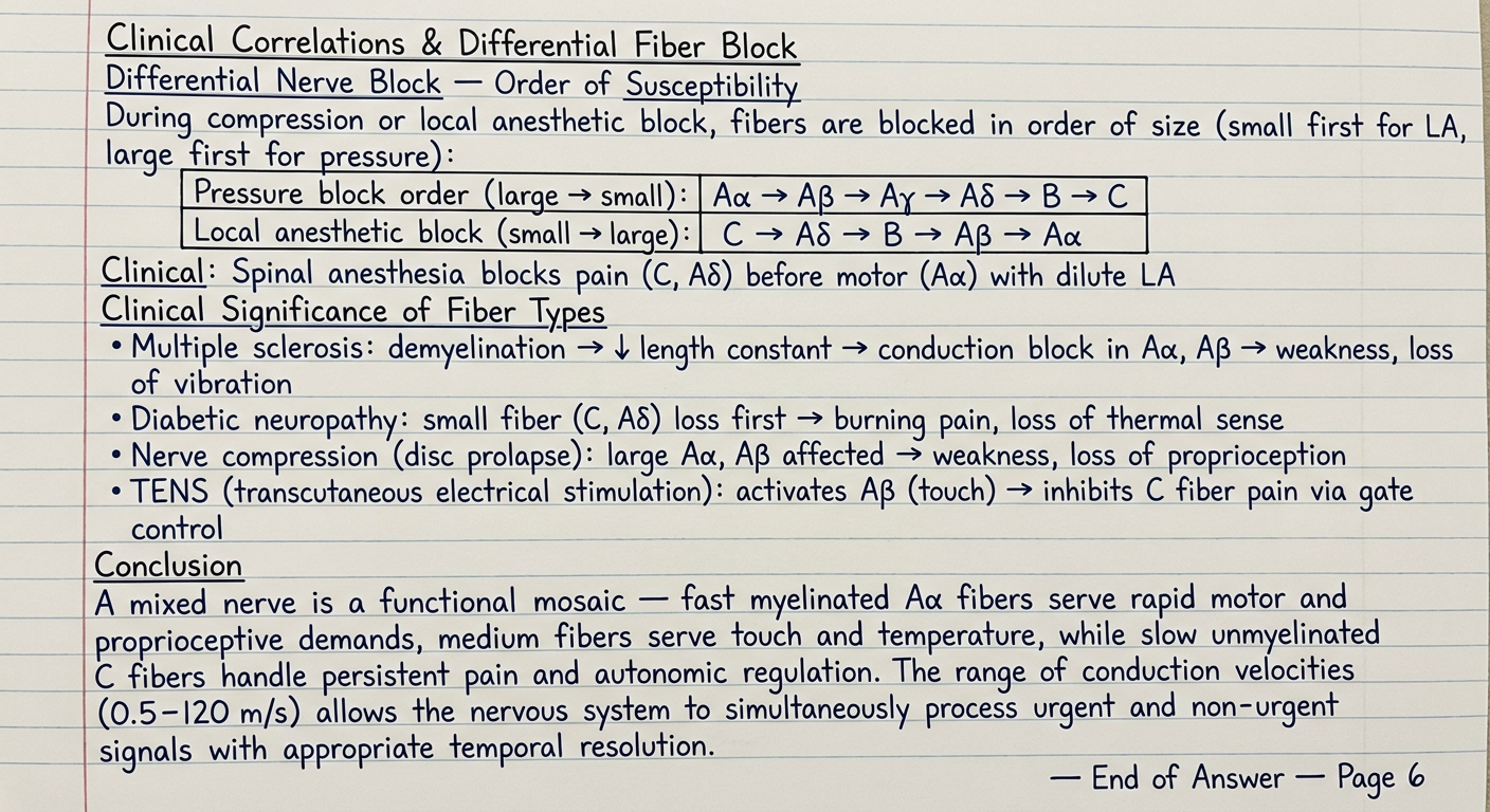
Handwritten medical exam answer on ruled notebook paper, blue ink pen, neat student handwriting. Page 6 (final page) of a 10-mark MD Physiology theory answer. Heading (underlined): "Clinical Correlations & Differential Fiber Block" Subheading: "Differential Nerve Block — Order of Susceptibility" Text: "During compression or local anesthetic block, fibers are blocked in order of size (small first for LA, large first for pressure):" Small hand-drawn table: "Pressure block order (large → small): Aα → Aβ → Aγ → Aδ → B → C" "Local anesthetic block (small → large): C → Aδ → B → Aβ → Aα" "Clinical: Spinal anesthesia blocks pain (C, Aδ) before motor (Aα) with dilute LA" Subheading: "Clinical Significance of Fiber Types" Bullet list: "• Multiple sclerosis: demyelination → ↓ length constant → conduction block in Aα, Aβ → weakness, loss of vibration" "• Diabetic neuropathy: small fiber (C, Aδ) loss first → burning pain, loss of thermal sense" "• Nerve compression (disc prolapse): large Aα, Aβ affected → weakness, loss of proprioception" "• TENS (transcutaneous electrical stimulation): activates Aβ (touch) → inhibits C fiber pain via gate control" Heading (underlined): "Conclusion" "A mixed nerve is a functional mosaic — fast myelinated Aα fibers serve rapid motor and proprioceptive demands, medium fibers serve touch and temperature, while slow unmyelinated C fibers handle persistent pain and autonomic regulation. The range of conduction velocities (0.5–120 m/s) allows the nervous system to simultaneously process urgent and non-urgent signals with appropriate temporal resolution." Bottom: "— End of Answer — Page 6"
Page 1 - Introduction & Structure of a Mixed Nerve
Page 2 - Classification of Nerve Fibers (Erlanger-Gasser & Lloyd-Hunt)
Page 3 - Continuous Conduction (Unmyelinated C Fibers)
Page 4 - Saltatory Conduction (Myelinated Fibers) + Cable Properties
Page 5 - Functional Implications of Distinct Conduction Velocities
Page 6 - Clinical Correlations, Differential Fiber Block & Conclusion
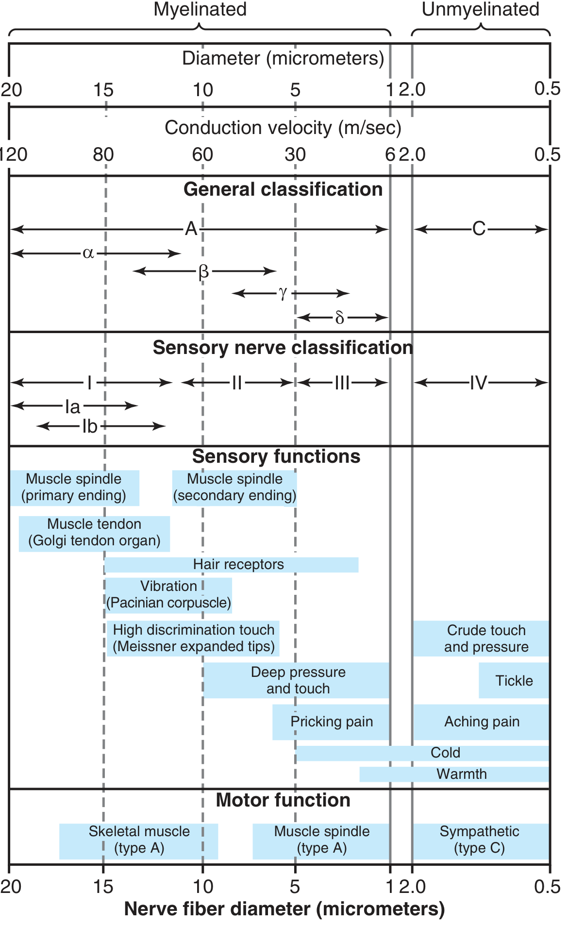
Reference Chart (from Guyton & Hall)
Answer Summary (What's covered across 6 pages)
| Page | Content |
|---|---|
| 1 | Introduction, definition of mixed nerve, fiber composition, nerve cross-section diagram |
| 2 | Full classification table (Erlanger-Gasser A/B/C + Lloyd-Hunt I-IV) with diameter, CV, myelination, function |
| 3 | Continuous conduction in unmyelinated C fibers - local current spread, step-by-step mechanism, diagram |
| 4 | Saltatory conduction in myelinated fibers, nodes of Ranvier, cable properties (λ, τ), energy efficiency |
| 5 | Functional implications - proprioception (Aα), touch (Aβ), fast pain (Aδ), slow pain/autonomic (C), double pain response |
| 6 | Differential nerve block, clinical diseases (MS, diabetic neuropathy, TENS), conclusion |
- Aα (12-20 μm, 70-120 m/s) - fastest, proprioception and somatic motor
- Aδ vs C fibers explain "double pain" - sharp first then burning second
- Saltatory conduction is ~50x faster than continuous conduction AND more energy-efficient (Na+/K+ ATPase only works at nodes)
- Local anesthetics block small fibers first (C > Aδ > Aβ > Aα) - this is the basis of selective spinal analgesia
/generatehandwrittenimages Explain the gibbs donnan effect and its role in determining the distribution of ions and water across capillary walls. How do changes in plasma protein concentration, capillary permeability and lymphatic drainage impact gibbs Donnan equilibrium and fluid balance in body - for md physiology 10 marks theory question. You can make many images and not just one for completing the answer
Handwritten medical exam answer on ruled notebook paper, blue ink pen, neat student handwriting. Page 1 of a 10-mark MD Physiology theory answer. Title (double underlined): "Gibbs-Donnan Effect & Fluid Balance Across Capillary Walls" Subheading (underlined): "Introduction" Text: "The Gibbs-Donnan equilibrium (Donnan 1911) describes the unequal distribution of diffusible ions across a semipermeable membrane when one side contains non-diffusible, charged macromolecules (e.g. plasma proteins). In capillaries, plasma proteins (mainly albumin, MW 69,000 Da) are confined to the intravascular space and cannot cross the capillary wall. This creates a Donnan effect that governs ion and water distribution between plasma and interstitial fluid." Subheading (underlined): "Basic Principle of Gibbs-Donnan Equilibrium" Text: "When a membrane separates two compartments — one containing impermeant anions (proteins) and one protein-free — the diffusible ions redistribute to satisfy TWO conditions simultaneously:" Numbered list: "1. Electrical neutrality must be maintained in each compartment" "2. Electrochemical equilibrium: [C+]plasma × [A-]plasma = [C+]ISF × [A-]ISF (Donnan equation)" Small hand-drawn diagram: Two boxes separated by a vertical line (membrane). Left box labeled 'PLASMA' with large oval 'Pr-' (protein, negatively charged), several Na+ and fewer Cl-. Right box labeled 'INTERSTITIAL FLUID (ISF)' with more Cl- and fewer Na+ and no protein. Arrow on membrane indicating 'impermeable to protein'. Label: 'Donnan ratio = [Na+]ISF/[Na+]plasma = [Cl-]plasma/[Cl-]ISF = 0.95' Bottom: "Page 1"
Handwritten medical exam answer on ruled notebook paper, blue ink pen, neat student handwriting. Page 2 of a 10-mark MD Physiology theory answer. Heading (underlined): "Ion Distribution: The Donnan Ratio at Capillary Wall" Text paragraph: "Because plasma proteins (Pr-) are negatively charged and confined to plasma, they attract cations and repel anions across the capillary wall:" Neat hand-drawn table with 3 columns: Ion | Plasma Conc. (mEq/L) | ISF Conc. (mEq/L) Rows: "Na+ | 153 | 145 (plasma > ISF)" "K+ | 4.2 | 4.0 (plasma > ISF)" "Ca2+ | 5.4 | 5.0 (plasma > ISF)" "Cl- | 110 | 116 (ISF > plasma)" "HCO3- | 27 | 28.5 (ISF > plasma)" Note below table: "Cations: [plasma] > [ISF] by factor ~1.05" "Anions: [ISF] > [plasma] by factor ~1.05" "Gibbs-Donnan ratio = 0.95 for all monovalent ions" Text: "The Donnan effect causes plasma to have a HIGHER total osmolality than ISF (by ~2 mOsm/L). This creates a small additional osmotic gradient — the oncotic contribution of the Donnan effect — which acts ALONGSIDE the direct colloid osmotic pressure (COP) of proteins to retain water in the capillary." Formula box: "COP (oncotic pressure) of plasma ≈ 25–28 mmHg" "Of which, ~8 mmHg is the Donnan effect component (due to retained small cations)" "~17 mmHg is the direct protein osmotic effect" Bottom: "Page 2"
Handwritten medical exam answer on ruled notebook paper, blue ink pen, neat student handwriting. Page 3 of a 10-mark MD Physiology theory answer. Heading (underlined): "Role of Gibbs-Donnan Effect in Capillary Fluid Exchange — Starling's Law" Text paragraph: "Fluid movement across the capillary wall is governed by the Starling-Landis equation, in which the Donnan-driven colloid osmotic pressure (COP/oncotic pressure) is the critical inward-pulling force:" Large formula box with clear writing: "Net filtration pressure (NFP) = (Pc - Pi) - (πp - πi)" "Where:" "Pc = capillary hydrostatic pressure (pushes fluid OUT)" "Pi = interstitial hydrostatic pressure (opposes filtration)" "πp = plasma colloid osmotic pressure (pulls fluid IN) ← Donnan-dependent" "πi = interstitial COP (pulls fluid OUT)" Text: "Normal values at arteriolar end:" "Pc = 32 mmHg, Pi = -3 mmHg, πp = 28 mmHg, πi = 8 mmHg" "NFP = (32 - (-3)) - (28 - 8) = 35 - 20 = +15 mmHg → filtration" "At venular end: Pc = 15 mmHg → NFP = (15+3) - (28-8) = 18 - 20 = -2 mmHg → absorption" Hand-drawn capillary diagram: Horizontal tube labeled 'Capillary' with arteriolar end (left) and venular end (right). Arrows pointing outward (filtration) at arteriolar end. Arrows pointing inward (absorption) at venular end. Small arrows labeled 'To lymphatics' exiting from midpoint. Label: 'Net filtration > absorption; excess returned by lymphatics' Bottom: "Page 3"
Handwritten medical exam answer on ruled notebook paper, blue ink pen, neat student handwriting. Page 4 of a 10-mark MD Physiology theory answer. Heading (underlined): "Effect of Changes in Plasma Protein Concentration" Subheading: "A. Decreased Plasma Proteins (Hypoproteinemia)" Causes listed in small text: "Nephrotic syndrome (urinary protein loss), liver cirrhosis (↓ albumin synthesis), malnutrition (kwashiorkor), protein-losing enteropathy, severe burns" Pathophysiology chain (with arrows): "↓ Plasma albumin → ↓ COP (oncotic pressure) → ↓ Donnan effect → ↓ inward osmotic force → NFP becomes positive at venular end → excess filtration > reabsorption → ↑ Interstitial fluid accumulation → EDEMA" "At plasma protein < 2.5 g/dL → severe generalized edema (Guyton & Hall)" Subheading: "B. Increased Plasma Proteins (Hyperproteinemia/Dehydration)" Text: "↑ Plasma protein → ↑ COP → ↑ inward osmotic pull → net absorption from ISF → ↓ interstitial volume → reduced skin turgor, sunken eyes in dehydration" Neat table summarizing: "Condition | Albumin | COP | NFP | Effect" "Nephrotic | ↓↓ | ↓↓ | ↑ filtration | Generalized edema" "Liver cirrhosis | ↓↓ | ↓ | ↑ filtration | Ascites + edema" "Malnutrition | ↓ | ↓ | ↑ filtration | Peripheral edema" "Dehydration | ↑ | ↑ | ↑ absorption | ↓ skin turgor" Bottom: "Page 4"
Error calling model 'gemini-3-pro-image' (RESOURCE_EXHAUSTED): 429 RESOURCE_EXHAUSTED. {'error': {'code': 429, 'message': 'Resource has been exhausted (e.g. check quota).', 'status': 'RESOURCE_EXHAUSTED'}}
We encountered an error while processing your request. Please try again or reach out to us for help.
Retry
An error occurred (ValidationException) when calling the ConverseStream operation: Expected toolResult blocks at messages.54.content for the following Ids: tooluse_MksH530vVDVpj7FBvdgMwS
We encountered an error while processing your request. Please try again or reach out to us for help.
/generatehandwrittenimages Explain the gibbs donnan effect and its role in determining the distribution of ions and water across capillary walls. How do changes in plasma protein concentration, capillary permeability and lymphatic drainage impact gibbs Donnan equilibrium and fluid balance in body - for md physiology 10 marks theory question. You can make many images and not just one for completing the answer
An error occurred (ValidationException) when calling the ConverseStream operation: Expected toolResult blocks at messages.54.content for the following Ids: tooluse_MksH530vVDVpj7FBvdgMwS
We encountered an error while processing your request. Please try again or reach out to us for help.