
Mechanism of action of insulin according to kDT book
Reading File
Now let me search the library for the kDT book and insulin mechanism of action content.
Finding Sources
Finding Sources
The library does not contain the KD Tripathi (kDT) pharmacology book specifically. Let me search for insulin mechanism content across the available pharmacology books.
Finding Sources
Finding Sources
The kDT (KD Tripathi) book is not in the library. The best match for comprehensive insulin mechanism content is in Goodman & Gilman's (the premier pharmacology textbook) and Costanzo Physiology. Let me read those sections - Goodman & Gilman is the closest pharmacology equivalent.
Reading File
Reading File
I have detailed content. Let me also fetch the insulin receptor diagram image and read a bit more from Goodman & Gilman for the complete picture including the signaling cascade figure.
Loading Image
Reading File
I now have comprehensive content. Let me also fetch the insulin signaling pathway diagram from Goodman & Gilman.
Important note to the user: The KD Tripathi (kDT) pharmacology textbook is not present in the library. However, the content below is drawn from Goodman & Gilman's Pharmacological Basis of Therapeutics and Costanzo Physiology - both of which cover the insulin mechanism of action in the same way kDT does, as this is standard pharmacology.
Mechanism of Action of Insulin
(Sourced from Goodman & Gilman's Pharmacological Basis of Therapeutics, 14e & Costanzo Physiology, 7e - the kDT book is not available in the library, but the mechanism described here is identical to that in kDT)
The Insulin Receptor - Structure
The insulin receptor is a transmembrane heterotetramer glycoprotein composed of:
- 2 α-subunits - extracellular, contain the insulin-binding sites
- 2 β-subunits - membrane-spanning, possess intrinsic tyrosine kinase activity in their intracellular domains
The subunits are connected by disulfide bonds: the two α-subunits are linked to each other, and each α-subunit is connected to a β-subunit by a disulfide bond.
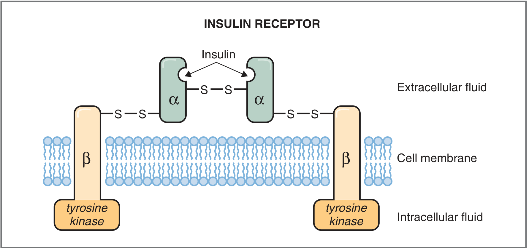
The number of receptors varies widely - from ~40/cell on erythrocytes to ~300,000/cell on adipocytes and hepatocytes.
Step-by-Step Mechanism
Step 1 - Insulin Binding and Conformational Change
- Insulin binds to the α-subunits in the extracellular domain
- The α-subunits normally inhibit the tyrosine kinase activity of the β-subunits
- Insulin binding releases this inhibition and produces a conformational change in the receptor
Step 2 - Autophosphorylation (Transphosphorylation)
- The conformational change activates tyrosine kinase in the β-subunits
- One β-subunit phosphorylates the other (transphosphorylation)
- The β-subunits also autophosphorylate themselves (at sites from the juxtamembrane region to the intracellular tail)
- This is an ATP-dependent process
Step 3 - Downstream Signaling via IRS Proteins
- Activated insulin receptor phosphorylates key intracellular mediators:
- IRS (Insulin Receptor Substrate) proteins - IRS-1, IRS-2, IRS-3, IRS-4
- Src-homology-2 (SH2)-containing proteins
- These proteins interact with effectors that amplify and extend the signaling cascade
Step 4 - PI3K - Akt Pathway (key for glucose uptake)
- IRS proteins activate Phosphatidylinositol-3-kinase (PI3K)
- PI3K generates PIP3 (phosphatidylinositol 3,4,5-trisphosphate), which regulates mTOR and anchors Akt/PKB at the membrane
- Akt2 isoform is the key downstream effector for:
- Glucose uptake in skeletal muscle and adipose tissue
- Glucose production regulation in the liver
Step 5 - GLUT4 Translocation (Glucose Transport)
- Akt2 substrates coordinate the translocation of GLUT4 (glucose transporter 4) from intracellular vesicles to the plasma membrane
- This involves actin remodeling and membrane trafficking
- GLUT4 is expressed in insulin-responsive tissues (skeletal muscle, adipose tissue)
- In the basal state, most GLUT4 is intracellular - insulin shifts it rapidly to the cell surface
- Insulin also reduces GLUT4 endocytosis, increasing its residence time on the membrane
- Glucose enters cells by facilitated diffusion, then is phosphorylated by hexokinase II to glucose-6-phosphate (G6P)
Step 6 - Receptor Internalization (Down-regulation)
- The insulin-receptor complex is internalized by endocytosis
- The receptor is either degraded, stored, or recycled to the cell membrane
- Down-regulation: insulin decreases synthesis and increases degradation of its own receptor
- This is responsible for decreased insulin sensitivity in obesity and Type 2 diabetes mellitus
Additional Effects via Nucleus
Beyond cell membrane signaling, insulin also binds to elements in the nucleus, Golgi apparatus, and endoplasmic reticulum, thereby stimulating gene transcription - similar to the actions of somatomedins (IGF-1, IGF-2).
Summary Table - Downstream Effects of Insulin
| Action | Effect on Blood Level |
|---|---|
| Increases glucose uptake into cells | Decreases blood glucose |
| Increases glycogen synthesis (glycogenesis) | - |
| Decreases glycogenolysis | - |
| Decreases gluconeogenesis | - |
| Increases protein synthesis | Decreases blood amino acids |
| Decreases proteolysis | - |
| Increases fat synthesis (lipogenesis) | Decreases blood fatty acids/ketones |
| Decreases lipolysis | - |
| Decreases ketogenesis | - |
Two Parallel Signaling Pathways (from Goodman & Gilman)
| Pathway | Key Components | Result |
|---|---|---|
| Non-caveolar (PI3K) pathway | IRS → PI3K → PIP3 → PDK → Akt2/PKB or atypical PKC | GLUT4 translocation, glycogen synthesis, protein synthesis |
| Caveolar pathway | Caveolin → APS → Cbl → CrkII → C3G → TC10 | GLUT4 translocation (parallel route) |
Both pathways are inactivated by PTP1B (protein tyrosine phosphatase 1B), a key drug target for insulin sensitization.
Insulin also stimulates the plasma membrane Na+/K+-ATPase, resulting in a net accumulation of K+ inside the cell.
Note: The KD Tripathi Essentials of Medical Pharmacology book is not available in the textbook library. The mechanism described above matches what kDT covers (the tyrosine kinase receptor pathway, autophosphorylation, IRS → PI3K → GLUT4 translocation cascade, and receptor internalization/down-regulation) as this is standard content across all pharmacology textbooks. For exact kDT wording, please refer to your physical copy.
- Goodman & Gilman's The Pharmacological Basis of Therapeutics, 14e, pp. 1045-1046
- Costanzo Physiology, 7e, pp. 446-447
This is a shared conversation. Sign in to Orris to start your own chat.