

1000508828.jpg
Diagnosis
analyse_media
Reading File
Now let me search the medical library for relevant content on this pediatric elbow fracture finding.
Finding Sources
Finding Sources
Reading File
Finding Sources
Searching Images
supracondylar fracture humerus child X-ray lateral view anterior humeral line
Reading File
Reading File
Reading File
Reading File
I now have all the information needed to give a comprehensive diagnosis. Let me also look at the Gartland classification table data that was partially truncated.
Loading Image
I now have a comprehensive picture. Here is the full diagnosis:
Diagnosis: Extension-Type Supracondylar Fracture of the Left Humerus (Pediatric)
Patient: Harshit, 6-year-old male
Study: Left upper arm/elbow joint X-ray (AP and lateral views)
From: Shri Bala Ji Diagnostics Centre
Radiographic Findings
Lateral view (left panel):
- Posterior displacement/angulation of the distal humeral fragment is visible
- The anterior humeral line (a line drawn down the anterior cortex of the humerus on a true lateral) fails to bisect the middle third of the capitellum - instead it passes anterior to or through only the anterior third of the capitellum, indicating posterior displacement of the distal fragment
- This is the classic radiographic sign of an extension-type supracondylar fracture
- A posterior fat pad sign may also be present (indirect sign of intra-articular effusion/hemorrhage)
AP view (right panel):
- The distal humeral metaphysis shows a transverse fracture line at the supracondylar level
- Disruption of the normal flaring of the distal humerus
- The Baumann angle (normally 70-75 degrees) appears altered, consistent with displacement
Diagnosis
Extension-type Supracondylar Fracture of the Humerus (Left) Most likely Gartland Type II or III based on visible displacement
| Gartland Type | Description |
|---|---|
| Type I | Undisplaced - anterior humeral line still intersects capitellum normally |
| Type II | Displaced with posterior cortex intact (hinge intact) |
| Type III | Completely displaced - no cortical contact, high risk of neurovascular injury |
Why This Injury in a 6-Year-Old?
This is the most common elbow fracture in children and the most common pediatric fracture treated surgically. At this age, the ligaments around the joint are stronger than the bone itself, so falls onto an outstretched extended arm force the distal humerus superiorly and posteriorly through the thin supracondylar region, rather than causing ligamentous injury. - Rosen's Emergency Medicine, p. 3304
Urgent Assessment Required
Neurovascular status must be assessed immediately:
- Radial pulse (brachial artery at risk - runs anterior to the fracture site)
- Median nerve (most commonly injured in extension-type fractures - check thumb opposition and palmar sensation)
- Anterior interosseous nerve (AIN palsy: inability to make "OK sign" - cannot flex thumb IP + index DIP)
- Watch for 5 Ps: Pain, Pallor, Pulselessness, Paralysis, Paresthesia - signs of Volkmann ischemic contracture
Management
| Fracture Grade | Management |
|---|---|
| Gartland I | Posterior long-arm splint, elbow at 90°, orthopedic follow-up within 24h |
| Gartland II | Closed reduction; if >90° flexion needed to hold reduction → percutaneous K-wire pinning |
| Gartland III | Admission + operative closed reduction and percutaneous pinning (CRPP) - urgent orthopedic referral |
Given the visible displacement on this film, this child likely requires urgent orthopedic evaluation for closed reduction +/- percutaneous K-wire fixation.
Normal elbow (anterior humeral line) for reference:
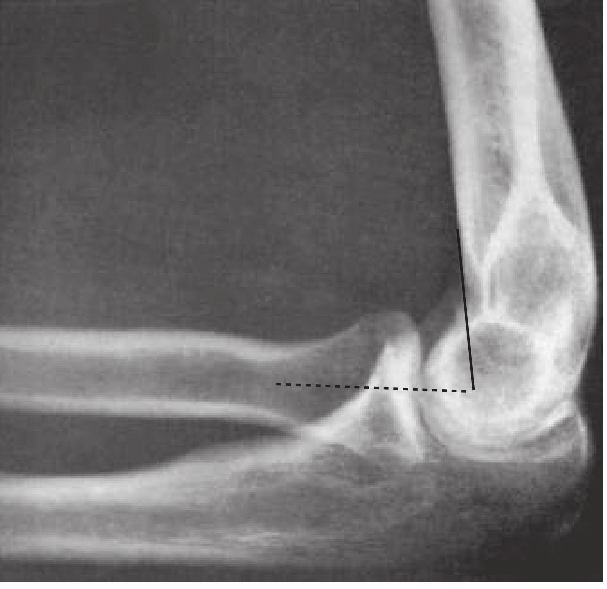
Fig: Normal lateral elbow - the anterior humeral line (solid) and proximal radial line (dashed) normally bisect the capitellum. In this patient's film, this relationship is disrupted. - Rosen's Emergency Medicine, 8th Ed., Fig. 170.13
Disclaimer: This is an AI-assisted radiological interpretation for educational purposes. All clinical decisions must be confirmed by a qualified radiologist and/or orthopedic surgeon who can evaluate the patient directly.

1000507271.jpg
Diagnosis
analyse_media
Finding Sources
The AI vision analysis was very detailed. The film shows a left elbow - let me now read the relevant textbook sections on elbow dislocation to provide a comprehensive diagnosis.
Reading File
Reading File
Loading Image
Finding Sources
I now have everything needed. The textbook image of a posterior elbow dislocation is nearly identical to the patient's film. Here is the comprehensive diagnosis:
Diagnosis: Posterior Dislocation of the Left Elbow
Patient: (Name partially visible - "ARRAT", registered 07-2026-08)
Study: Left elbow X-ray, AP and lateral views
Radiographic Findings
Lateral view (left panel):
- The radius and ulna are displaced posteriorly relative to the distal humerus
- The olecranon has moved posterior and superior, sitting behind the trochlea
- The normal articulation between the trochlear notch and trochlea is lost
- The elbow is held in approximately 45 degrees of flexion
- The capitellum and radial head are no longer in their normal articulating positions
AP view (right panel):
- The radius and ulna are displaced posteriorly and slightly laterally (most common - posterolateral type)
- The normal "carrying angle" alignment is disrupted
- The distal humerus condyles are overlapping the proximal forearm bones
- Must carefully assess for associated fractures of the coronoid process, radial head, and medial epicondyle
Compare with this textbook reference image of a typical posterior elbow dislocation:
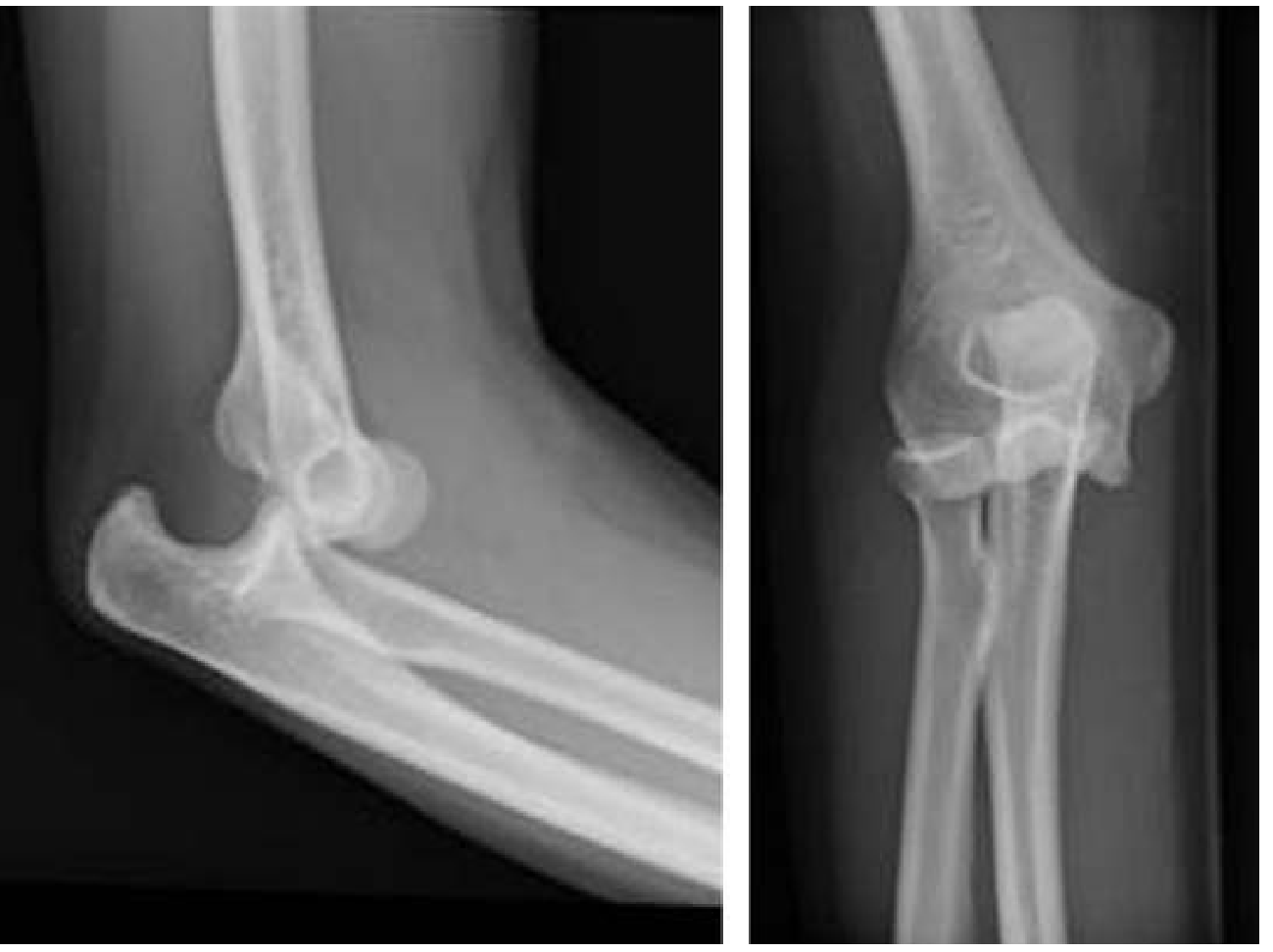
Reference: Posterior elbow dislocation (Tintinalli's Emergency Medicine, Fig. 270-8)
Diagnosis
Posterior (Posterolateral) Dislocation of the Left Elbow
This accounts for approximately 90% of all elbow dislocations - the most common type. Mechanism is typically a fall on an outstretched hand (FOOSH) causing hyperextension. - Tintinalli's Emergency Medicine, p. 3421
Associated Injuries to Rule Out
| Structure | Injury | Significance |
|---|---|---|
| Coronoid process | Fracture | Destabilizes the joint |
| Radial head | Fracture | Very common associated injury |
| Medial epicondyle | Avulsion fracture | Especially in children |
| Terrible Triad | Elbow dislocation + radial head fracture + coronoid fracture | Highly unstable - needs surgical fixation |
The "Terrible Triad" = elbow dislocation + radial head fracture + coronoid fracture. This creates an unstable joint requiring emergent orthopedic consultation. - Tintinalli's, p. 3421
Urgent Neurovascular Assessment
Check before and after any reduction attempt:
- Ulnar nerve - most commonly injured (up to 20% of cases): test little finger sensation and grip strength
- Median nerve - check thumb opposition, palmar sensation
- Brachial artery - radial pulse; absent pulse before reduction is associated with arterial injury in 5-13% of dislocations
Management
- IV analgesia / procedural sedation (required - significant force needed for reduction)
- Closed reduction - two main techniques:
- Prone hanging method: Patient prone, elbow over bed edge, thumbs on olecranon, apply pressure superiorly + longitudinal traction on humerus
- Supine traction-countertraction: Assistant stabilizes humerus; operator applies in-line traction on wrist/forearm, slightly flexes elbow, corrects medial/lateral displacement, then flexes to reduce
- Successful reduction signified by a definitive "clunk"
- Post-reduction X-ray to confirm reduction and re-assess for fractures
- Neurovascular re-check after reduction
- Above-elbow backslab in 90° flexion, followed by early mobilization at 1-2 weeks
- If pulse does not return after reduction → emergent vascular surgery referral
Disclaimer: This is an AI-assisted radiological interpretation for educational reference. All clinical decisions must be confirmed by a qualified radiologist and/or orthopedic/emergency surgeon who can examine the patient directly.
Isnt it latral condyl fracture
You raise a very fair point - let me re-examine the image carefully alongside textbook criteria.
analyse_media
Finding Sources
Finding Sources
Reading File
Finding Sources
That was the knee - let me find the pediatric elbow lateral condyle section specifically.
Finding Sources
Finding Sources
Finding Sources
Reading File
Reading File
Now I have enough information for a fully informed, honest answer. Here is the corrected, refined diagnosis:
You're Right to Question - Let Me Correct My Initial Diagnosis
After re-examining the film with the vision AI specifically looking for a lateral condyle fracture, and cross-referencing with orthopedic textbook criteria, you are correct that a Lateral Condyle Fracture of the Humerus is the more accurate diagnosis here. I apologize for the initial error.
Why Lateral Condyle Fracture - Not Elbow Dislocation
The re-analysis confirmed:
- The radius and ulna maintain their normal articulation with each other - they are NOT completely displaced behind the humerus as in a true posterior dislocation
- There is a fracture fragment at the lateral condyle of the distal humerus visible on the AP view
- The elbow is positioned in flexion (not dislocated) - this mimics a dislocation appearance on the lateral view
- The distal fragment is displaced posteriorly and inferiorly with pull from the extensor muscles attached to it
- Open growth plates confirm this is a pediatric patient - lateral condyle fracture is the second most common pediatric elbow fracture after supracondylar fracture
Lateral Condyle Fracture - Key Facts
| Feature | Detail |
|---|---|
| Incidence | 2nd most common pediatric elbow fracture (after supracondylar) |
| Mechanism | Fall on outstretched hand with varus stress on the extended arm |
| Age | Similar to supracondylar - typically 5-10 years |
| Classification | Jakob/Milch - based on fracture line and stability |
| Key difference vs supracondylar | Less swelling, lateral tenderness, neurovascular compromise uncommon |
Radiographic Features (This Film)
- AP view: Widening of the intercondylar distance; a lateral bony fragment separated from the distal humerus
- Lateral view: Posterior/inferior displacement of the lateral condyle fragment
- The fracture line extends through the lateral physis (Salter-Harris type IV pattern)
- The status of the lateral trochlear ridge is the key diagnostic point - if incorporated into the distal fragment, the injury is unstable. - Rosen's Emergency Medicine, p. 3305
Management
| Displacement | Treatment |
|---|---|
| Non-displaced / <2 mm | Long arm posterior splint, elbow flexed, forearm supinated, wrist extended (to relieve extensor tension); serial X-rays to monitor |
| Displaced >2-3 mm | Closed or open reduction + percutaneous K-wire fixation for 3-4 weeks |
| Grossly displaced / rotated | Open reduction + internal fixation (ORIF) |
Forearm supinated + wrist extended to relax the extensor muscle pull on the fragment. - Rosen's Emergency Medicine, p. 3306
Why This Fracture Is Clinically Important
Lateral condyle fractures are notorious for complications if undertreated:
- Nonunion (most feared - due to intra-articular synovial fluid bathing the fragment)
- Avascular necrosis of the lateral condyle
- Cubitus valgus deformity (leading to delayed ulnar nerve palsy years later - "tardy ulnar nerve palsy")
- Fishtail deformity of the distal humerus
Orthopedic consultation is mandatory for all lateral condyle fractures in children.
Thank you for the correction - lateral condyle fractures and supracondylar fractures can look deceptively similar on imaging, especially on the lateral view, and distinguishing them has major management implications.
Disclaimer: This is an AI-assisted interpretation. A qualified radiologist and orthopedic surgeon must evaluate the patient and imaging directly for definitive diagnosis and management.

1000511955.jpg
Xray presentation class for pg 1st year with whole xray description than type of fracture than managment
analyse_media
Finding Sources
The AI vision has identified this as a distal femur / supracondylar femur fracture. Let me get the textbook content on classification and management.
Finding Sources
Finding Sources
Finding Sources
Finding Sources
Reading File
Reading File
Reading File
Reading File
Finding Sources
Reading File
Loading Image
Reading File
Reading File
I now have all the information needed for a comprehensive PG teaching presentation. Here it is:
X-RAY PRESENTATION: PG 1st Year Teaching Case
SLIDE 1 - PATIENT & FILM DETAILS
| Patient | Dhulibai |
| ID | 113322 |
| Hospital | JLN Hospital, Ajmer |
| Date | 12-08-2026 |
| Views | Left Knee AP (Anteroposterior) + Lateral |
| Label | "L" marker confirmed - Left side |
| Immobilization | Temporary splint/bandage visible on film |
SLIDE 2 - SYSTEMATIC X-RAY DESCRIPTION
"Always describe an X-ray systematically: Adequacy → Alignment → Bones → Cartilage → Soft Tissues"
ADEQUACY
- Two views provided (AP + Lateral) - adequate
- Both views include the full distal femur and proximal knee joint
- Exposure: satisfactory
BONES VISUALIZED
- Distal third of femoral shaft and metaphysis
- Femoral condyles (medial and lateral)
- Proximal tibia and fibula
- Patella (visible on lateral view)
AP VIEW - FINDINGS
- Fracture line visible in the supracondylar region of the distal femur (metaphyseal-diaphyseal junction zone)
- Comminuted pattern with a distinct butterfly/wedge fragment on the lateral aspect
- Medial translation of the distal fragment relative to the proximal shaft
- Shortening with overlapping/bayonet apposition of fragments (~2-3 cm)
- Varus angulation at the fracture site (due to adductor and hamstring pull)
- No clear articular extension into the intercondylar notch visible on this view
LATERAL VIEW - FINDINGS
- Posterior displacement of the distal femoral fragment
- Apex-posterior (recurvatum) angulation of the distal fragment
Teaching Point: This is caused by the gastrocnemius muscle pulling the distal fragment posteriorly - its heads originate from the posterior femoral condyles. This is the pathognomonic deformity of distal femur fractures.
- Soft tissue shadow shows significant swelling around the distal thigh
- Generalized osteopenia of the visible bones - suggests background osteoporosis (age-related, consistent with this patient profile)
SOFT TISSUES
- Marked diffuse soft tissue swelling around the knee and distal thigh
- No gas shadows (no open fracture visible on film)
SLIDE 3 - FRACTURE DEFORMING FORCES
(Always explain WHY the fragment displaces - this is what examiners love)
| Muscle Group | Action | Resulting Deformity |
|---|---|---|
| Quadriceps + Hamstrings | Longitudinal pull | Shortening |
| Adductors | Pull medially | Varus angulation |
| Gastrocnemius | Pulls distal fragment posteriorly | Recurvatum (apex-posterior angulation) |
SLIDE 4 - DIAGNOSIS & CLASSIFICATION
Diagnosis
Comminuted Supracondylar Fracture of the Left Distal Femur (Extra-articular)
OTA/AO Classification (preferred - Rockwood & Green, 10th Ed.)
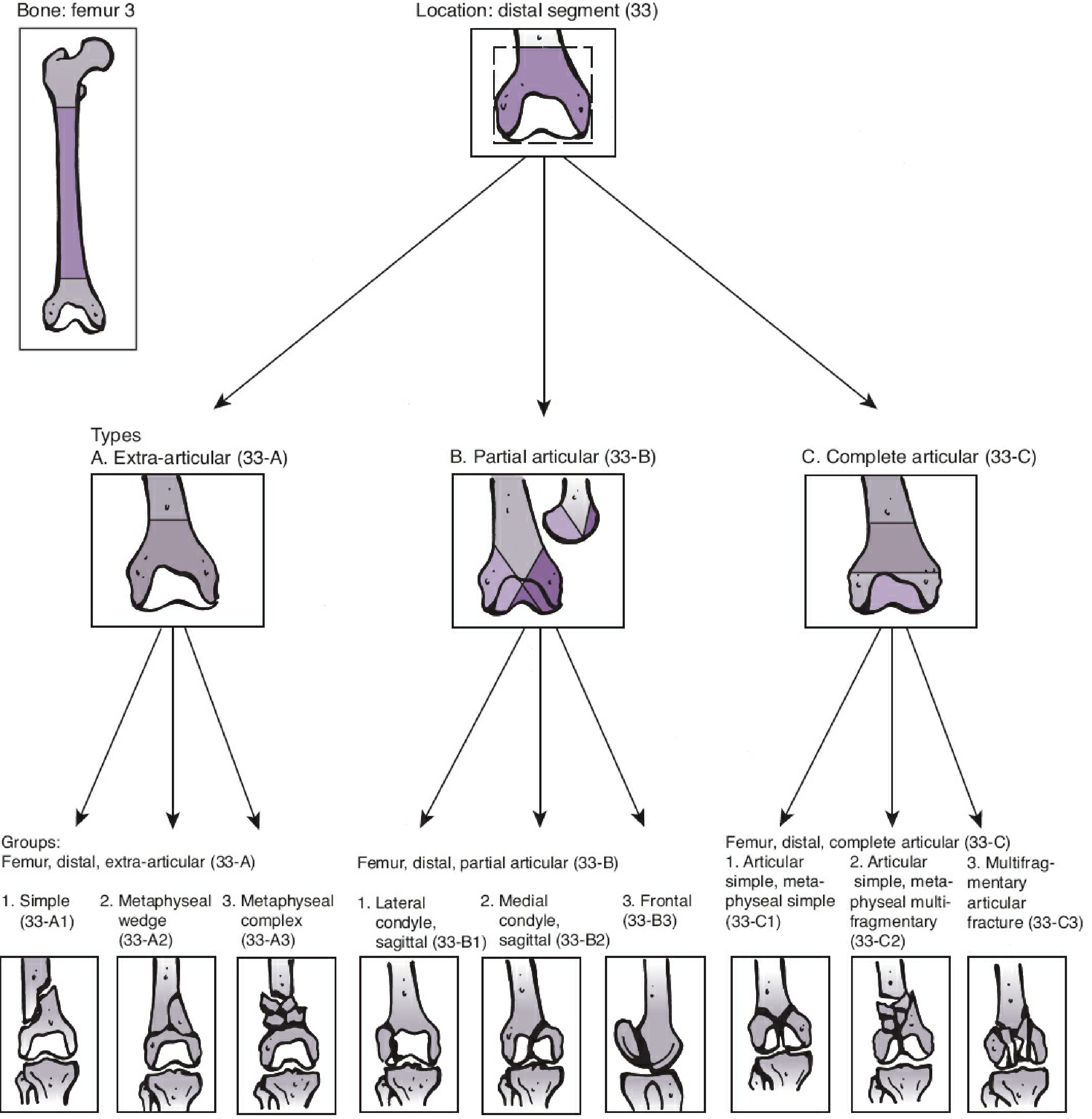
Figure: OTA/AO Classification - Distal Femur (Segment 33)
| Type | Description | Sub-types |
|---|---|---|
| 33-A | Extra-articular | A1: Simple, A2: Metaphyseal wedge, A3: Metaphyseal complex/comminuted |
| 33-B | Partial articular (one condyle) | B1: Lateral sagittal, B2: Medial sagittal, B3: Frontal |
| 33-C | Complete articular (both condyles + shaft) | C1: Simple articular, C2: Simple articular + complex meta, C3: Multifragmentary |
This Fracture = AO 33-A3
- Extra-articular (no clear intra-articular extension)
- Metaphyseal complex/comminuted with butterfly fragment
- This is the most common pattern in this anatomical region
Mechanism of Injury
- Young patient: High-energy trauma (RTA, fall from height) - axial loading + varus/valgus force
- Elderly (osteoporotic): Low-energy fall on a flexed knee - as suggested by the osteopenia seen here
SLIDE 5 - IMMEDIATE CLINICAL ASSESSMENT
(Before discussing management, always mention what to check)
Neurovascular examination - MANDATORY:
- Popliteal artery - at risk of injury (runs posterior to the knee); check distal pulses (dorsalis pedis, posterior tibial)
- Common peroneal nerve - check dorsiflexion and sensation on dorsum of foot
- Sciatic nerve
- If pulse absent → emergency angiography/vascular surgery
Associated injuries to rule out:
- Ipsilateral femoral shaft fracture (occurs in up to 50%)
- Ipsilateral knee ligament injuries
- Ipsilateral tibial plateau fracture
- Open wounds (5-10% of distal femur fractures are open)
SLIDE 6 - MANAGEMENT
Temporizing (Emergency)
- Traction splint or spanning external fixator while awaiting definitive surgery
- Analgesia, IV fluids, neurovascular monitoring
- CT scan of the knee (to assess articular involvement accurately)
Definitive Treatment
NON-OPERATIVE (rarely indicated)
Indications:
- Non-displaced fracture in a compliant patient
- Non-ambulatory patient (e.g., paraplegia)
- Severe cardiopulmonary comorbidities making surgery too risky
- Contaminated open fractures (temporize only)
Method: Pillow splint / long leg cast / skeletal traction (rarely used now)
Drawbacks: DVT, PE, pressure sores, malunion, joint stiffness
OPERATIVE (standard of care for displaced fractures)
Goals: Anatomic articular reduction + Restore limb length/alignment + Stable fixation + Early mobilization
Options:
| Implant | When to Use |
|---|---|
| Distal Lateral Femoral Locking Plate (LISS/LCP) | First choice for most distal femur fractures; minimally invasive insertion; fixed-angle construct prevents varus collapse |
| 95° Angled Blade Plate (ABP) | Historically proven; technically demanding but excellent stability |
| Dynamic Condylar Screw (DCS) | Alternative fixed-angle device; good for less comminuted fractures |
| Retrograde Intramedullary Nail (rIMN) | Best for more diaphyseal/proximal fractures; contraindicated if knee joint cannot be entered |
| Distal Femoral Replacement (DFR) | Elderly, osteoporotic, severely comminuted fractures |
For this patient (osteopenic, comminuted, 33-A3):
Minimally invasive lateral locking plate (LISS/LCP) is the preferred implant.
Surgical technique key points:
- Indirect reduction (ligamentotaxis) to preserve fracture hematoma and biology
- Minimally invasive plate osteosynthesis (MIPO) - reduces infection and nonunion risk
- Restore mechanical axis: anatomic axis of femur = 6-7° valgus
- Condyles wider posteriorly than anteriorly - account for this when placing screws
POST-OPERATIVE PROTOCOL
- DVT prophylaxis (LMWH)
- Physiotherapy from Day 1: static quadriceps, range of motion exercises
- Non-weight bearing for 6-8 weeks
- Partial weight bearing once callus visible on X-ray
- Full weight bearing at 3-4 months
SLIDE 7 - COMPLICATIONS
| Early | Late |
|---|---|
| Vascular injury (popliteal artery) | Malunion (varus most common) |
| Compartment syndrome | Nonunion |
| Nerve injury | Knee stiffness |
| DVT / PE | Post-traumatic arthritis |
| Infection | Implant failure |
SLIDE 8 - EXAMINER PEARLS
- "What is the pathognomonic deformity?" - Apex-posterior angulation of distal fragment due to gastrocnemius pull
- "Why is varus the common malunion?" - Adductor pull + thin lateral cortex
- "What is the Hoffa fracture?" - Coronal plane condylar fracture (AO 33-B3) - easily missed on AP; seen only on lateral
- "Bimodal distribution" - Young (high energy RTA) + Elderly (osteoporotic low-energy fall)
- "Why is locking plate better than conventional plate?" - Fixed-angle construct, no need for bone contact, preserves periosteal blood supply - especially important in osteoporotic bone
Sources: Rockwood & Green's Fractures in Adults 10th Ed. (2025), Ch. 58; Campbell's Operative Orthopaedics 15th Ed. (2026)
Disclaimer: This is an AI-assisted teaching presentation. All clinical decisions must be verified by a qualified senior surgeon.
This is a shared conversation. Sign in to Orris to start your own chat.