
maternal and fetal complication of htn in pregnancy
"hypertensive disorders pregnancy" AND complications
complications of hypertension in pregnancy preeclampsia maternal fetal
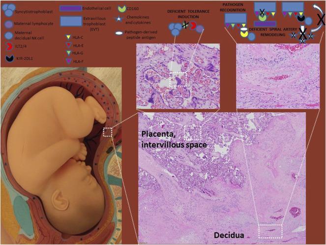
This composite educational infographic illustrates the pathophysiology of preeclampsia at the maternal-fetal interface. It combines a gross anatomical model of a fetus in utero with H&E-stained histopathological sections and a schematic immunological diagram. The histology displays the placental intervillous space, characterized by branched chorionic villi and syncytiotrophoblasts, adjacent to the maternal decidua. A schematic legend identifies key immune mediators, including HLA-C, HLA-E, HLA-G, and HLA-F, alongside maternal lymphocytes and decidual NK cells (dNK). The diagram contrasts normal pregnancy physiology with pathological states in preeclampsia, specifically highlighting 'Deficient Tolerance Induction' and 'Deficient Spiral Artery Remodeling.' It depicts how impaired interactions between extravillous trophoblasts (EVTs) and maternal immune cells—mediated by specific HLA-receptor bindings like HLA-G with ILT2/4 and HLA-C with KIR-2DL1—contribute to shallow trophoblast invasion and failed vascular transformation of decidual spiral arteries. Black 'X' marks denote inhibited pathways, signaling failures in pathogen recognition and chemokine/cytokine release necessary for healthy placental development.
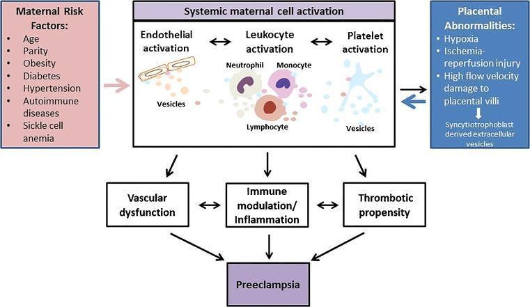
A pathophysiology diagram illustrating the integrated role of extracellular vesicles (EVs) in the pathogenesis of preeclampsia. The flowchart identifies two primary triggers: 'Maternal Risk Factors' (including age, obesity, diabetes, and hypertension) and 'Placental Abnormalities' (such as hypoxia, ischemia-reperfusion injury, and syncytiotrophoblast-derived EV release). These triggers lead to 'Systemic maternal cell activation,' which is depicted as a central mechanism involving three main pathways: 1) Endothelial activation, 2) Leukocyte activation (neutrophils, monocytes, and lymphocytes), and 3) Platelet activation. Each cell type is shown releasing vesicles that mediate cross-talk between systems. The diagram shows how these cellular activations result in three downstream clinical consequences: vascular dysfunction, immune modulation/inflammation, and increased thrombotic propensity. These pathological processes collectively culminate in the clinical manifestation of Preeclampsia. This diagram serves as an educational summary of how maternal-placental interactions through EVs drive systemic inflammation and vascular damage in pregnancy-induced hypertension disorders.
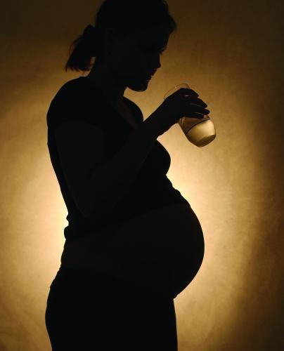
A clinical educational photograph utilizing a silhouette technique to represent a pregnant woman in her second or third trimester. The individual is shown in profile, with a visibly enlarged abdomen indicating advanced pregnancy. She is holding a glass of liquid to her mouth, symbolizing the ingestion of water or other substances. From a medical and public health perspective, this image serves as a conceptual illustration of prenatal environmental exposures. It is used to teach concepts related to maternal-fetal health, toxicology, and environmental health risks, specifically the impact of contaminated drinking water (e.g., perfluorooctanoic acid or PFOA) on gestational outcomes such as pregnancy-induced hypertension or preeclampsia. The educational focus is on the vulnerability of the maternal-fetal unit to ingested environmental pollutants and the importance of safe hydration during pregnancy.
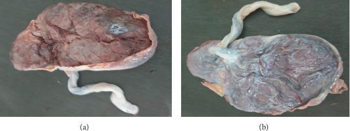
Two gross clinical photographs displaying the maternal and fetal surfaces of a human placenta, specifically illustrating morphological changes associated with pregnancy-induced hypertension (PIH). Panel (a) shows the maternal surface, which exhibits a reddish-brown, mottled texture with cotyledons. Areas of darker discoloration and a notable grayish-white patch are visible, potentially indicating infarction or calcification. The umbilical cord is shown with an eccentric insertion. Panel (b) depicts the fetal surface, characterized by a smooth, translucent amnion with a distinct bluish-purple hue. Branching chorionic vessels are visible radiating from the site of umbilical cord insertion, which is more centrally located compared to panel (a). The umbilical cords in both specimens appear thick, whitish, and demonstrate typical helical coiling. These images serve as educational examples of gross placental pathology, highlighting how systemic maternal conditions like hypertension can manifest in placental morphology and cord insertion patterns.
Maternal and Fetal Complications of Hypertension in Pregnancy
Classification (Brief Review)
| Category | Definition |
|---|---|
| Gestational HTN | New BP ≥ 140/90 mmHg after 20 weeks; resolves postpartum; no proteinuria |
| Preeclampsia | Gestational HTN + proteinuria (>300 mg/24 hr) or end-organ damage |
| Eclampsia | Seizures or coma in the setting of preeclampsia |
| HELLP Syndrome | Hemolysis + Elevated Liver enzymes + Low Platelets (<100,000/mL) |
| Chronic HTN | Present before pregnancy or persists >6 weeks postpartum |
| Superimposed preeclampsia | Chronic HTN + new-onset preeclampsia features |
Pathophysiology at a Glance
- Increased thromboxane / decreased prostacyclin (pro-vasoconstriction)
- Loss of normal angiotensin resistance
- Decreased VEGF and PlGF; increased antiangiogenic sFlt-1 and sEng
- Systemic endothelial dysfunction and vasospasm affecting multiple organs
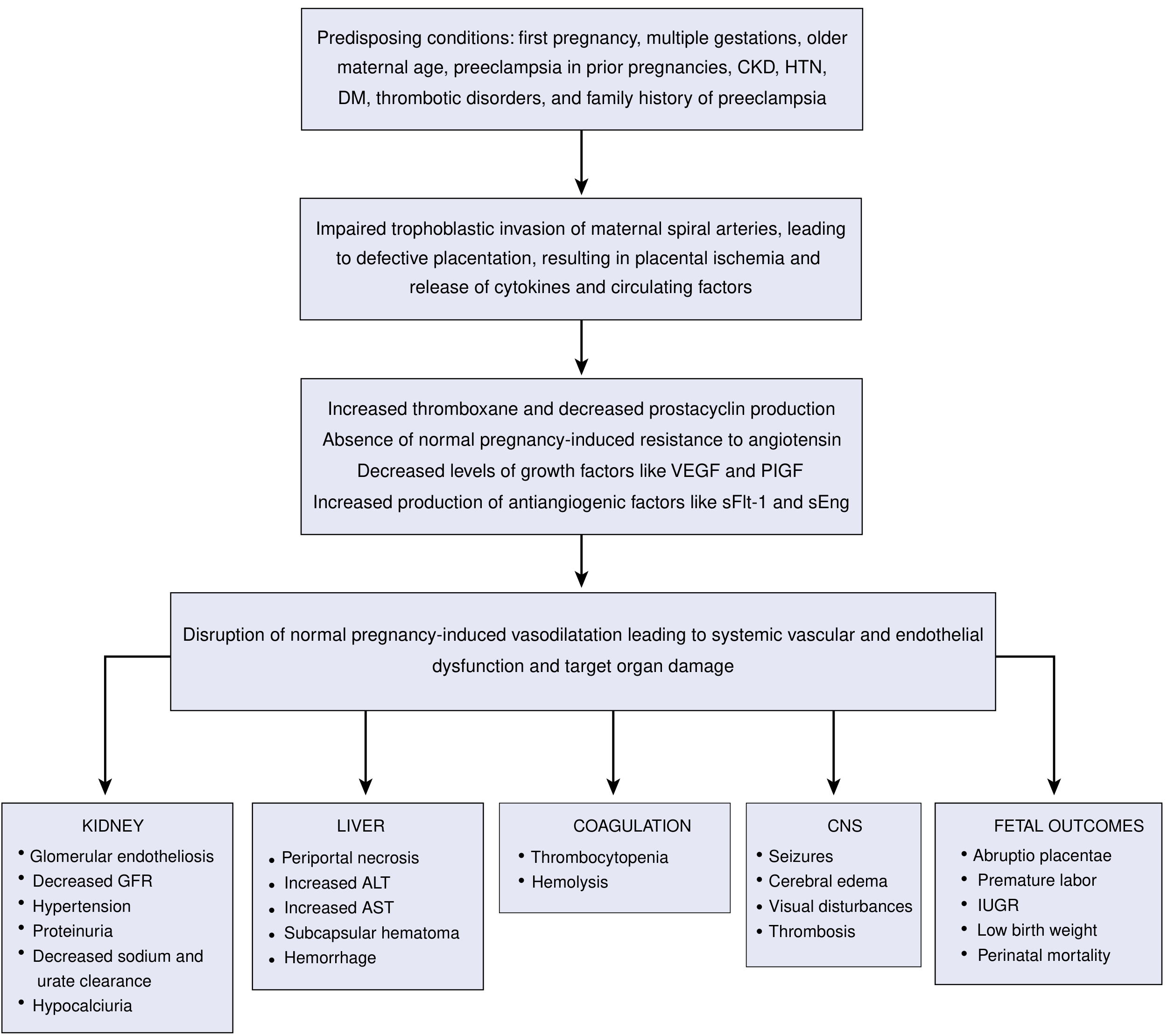
MATERNAL COMPLICATIONS
1. Eclampsia
- Can develop abruptly before 32 weeks, sometimes without prior edema or proteinuria
- Postpartum eclampsia occurs in >55% of cases who had no prior preeclampsia diagnosis - often within 48 hours but up to 6-12 weeks after delivery
- CT may show patchy cortical hemorrhage, microinfarcts, or diffuse cerebral edema in 50% of patients
- Maternal mortality has declined to <1% with modern management
- Permanent CNS damage, renal insufficiency, and intracranial hemorrhage are serious sequelae
- ROSEN's Emergency Medicine, p. 3357
2. HELLP Syndrome
- H - Hemolysis (microangiopathic hemolytic anemia)
- EL - Elevated liver enzymes (ALT and AST > 70 U/L)
- LP - Low platelets (<100,000/mL)
- PT, PTT, and fibrinogen are typically normal
- Presents at older maternal age compared to preeclampsia
- ROSEN's, p. 3357; Textbook of Family Medicine 9e, p. 3025
3. Renal Complications
- Decreased GFR
- Proteinuria (ranging from mild to nephrotic-range)
- Oliguria (in severe disease)
- Decreased sodium and urate clearance, hypocalciuria
- NKF Primer on Kidney Diseases, Fig. 47.2
4. Hepatic Complications
- Periportal necrosis from hepatocellular ischemia
- Elevated ALT/AST
- Subcapsular hematoma
- Spontaneous hepatic and splenic hemorrhage - rare but life-threatening
- ROSEN's, p. 3357
5. Neurological Complications
- Severe headache and hyperreflexia
- Visual disturbances (blurred vision, scotoma, cortical blindness)
- Cerebral edema and thrombosis
- Intracerebral hemorrhage
- ROSEN's, p. 3358
6. Coagulation / DIC
- Thrombocytopenia (from microangiopathic process)
- Microangiopathic hemolytic anemia
- In severe cases, disseminated intravascular coagulation (DIC)
- NKF Primer, Fig. 47.2
7. Placental Abruption
8. Cardiovascular Complications
9. Caesarean Section Risk
10. Long-Term Cardiovascular Risk (Post-Preeclampsia)
| Outcome | Relative Risk |
|---|---|
| Chronic hypertension | RR 3.6-3.7 |
| Cardiovascular disease | RR 2.2 |
| Stroke | RR 1.8-2.0 |
| Chronic kidney failure | RR 4.7-16.0 |
- NKF Primer on Kidney Diseases, p. 492
FETAL / NEONATAL COMPLICATIONS
1. Intrauterine Growth Restriction (IUGR)
2. Premature Birth / Preterm Labour
3. Low Birth Weight
4. Placental Infarction and Abruption
5. Fetal Hypoxia
6. Perinatal Mortality
- Perinatal mortality remains 4-8% even with modern management
- Causes: placental infarcts, IUGR, abruptio placentae, and complications of prematurity
- Fetal hypoxia from maternal seizures contributes significantly
- ROSEN's Emergency Medicine, p. 3358
7. Neonatal Complications of Prematurity
Summary Table
| System | Maternal Complications | Fetal/Neonatal Complications |
|---|---|---|
| CNS | Eclampsia, cerebral edema, ICH, visual disturbances | Fetal hypoxia from maternal seizures |
| Liver | HELLP, subcapsular hematoma, spontaneous rupture | - |
| Renal | Proteinuria, decreased GFR, oliguria, AKI | - |
| Coagulation | Thrombocytopenia, hemolysis, DIC | - |
| Placenta | Abruption, infarction | Abruption, IUGR, fetal distress |
| Cardiovascular | Pulmonary edema, long-term CVD/stroke/CKD | - |
| Fetal growth | - | IUGR, SGA, low birth weight |
| Delivery | Caesarean section | Prematurity, perinatal mortality |
Pathogenesis Diagram (Preeclampsia)