attachment
attachment

Convert these into diagrams

analyse_media
Now I'll generate two clear diagrams - one for Class I modifications and one for Class III modifications of Dewey.
Generating Image

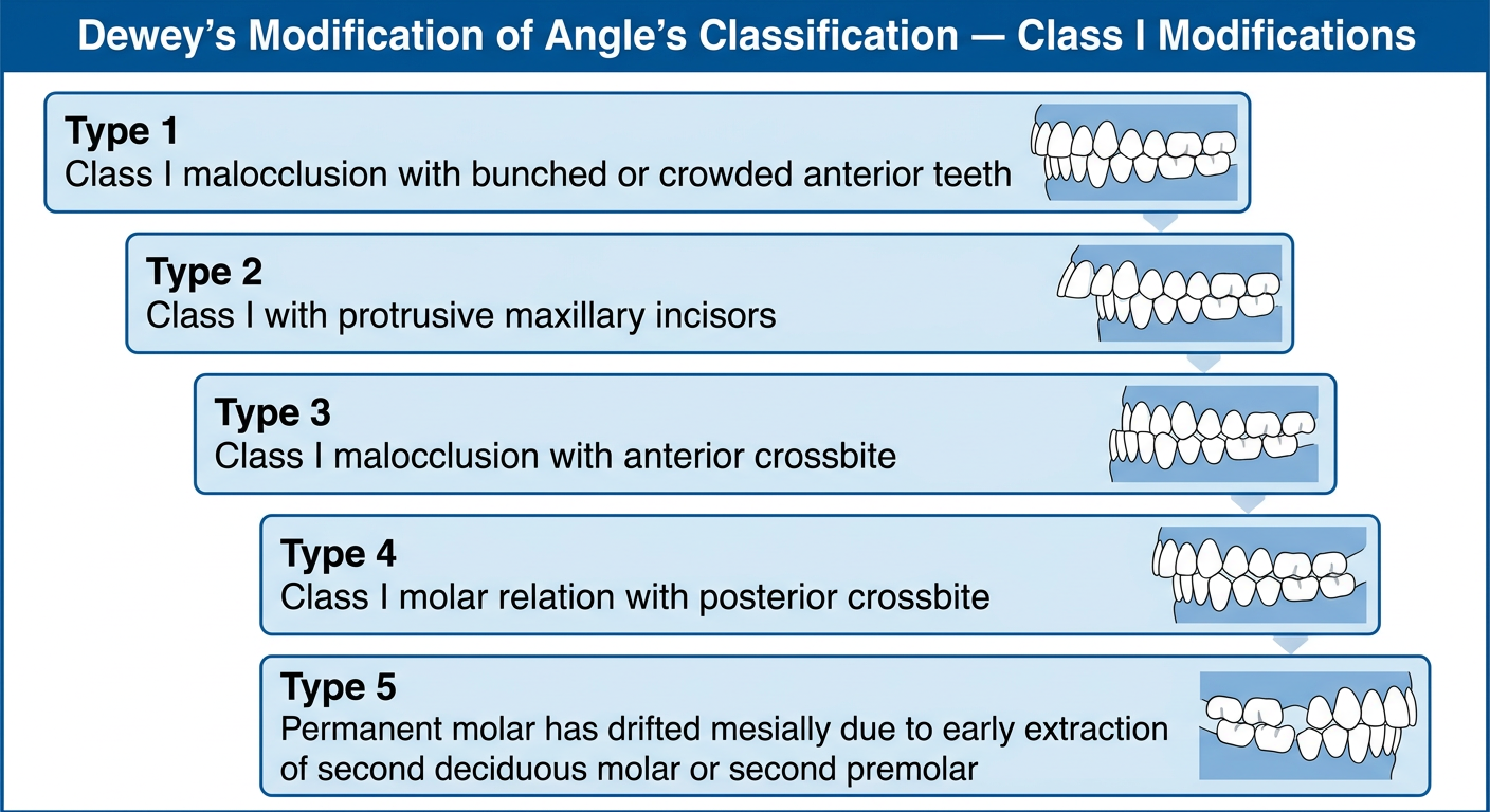
Clean educational diagram titled "Dewey's Modification of Angle's Classification — Class I Modifications" with a dark blue header. Below the title, show 5 labeled boxes arranged vertically or in a structured flow, each with a bold "Type" label and a short description: Type 1: Class I malocclusion with bunched or crowded anterior teeth Type 2: Class I with protrusive maxillary incisors Type 3: Class I malocclusion with anterior crossbite Type 4: Class I molar relation with posterior crossbite Type 5: Permanent molar has drifted mesially due to early extraction of second deciduous molar or second premolar Each box should have a light blue background with bold type number and clear description text. White background overall. Medical/dental textbook style infographic.

Generating Image

Clean educational diagram titled "Dewey's Modification of Angle's Classification — Class III Modifications" with a dark teal header. Below the title, show 3 labeled boxes arranged vertically, each with a bold "Type" label and a short description: Type 1: Upper and lower dental arches when viewed separately are in normal alignment. When made to occlude, patient shows edge-to-edge incisor alignment. Type 2: Mandibular incisors are crowded and are in lingual relation to the maxillary incisors. Type 3: Maxillary incisors are crowded and are in crossbite in relation to the mandibular anteriors. Each box should have a light teal/green background with bold type number and clear description text. White background overall. Medical/dental textbook style infographic.

Here are your two diagrams based on Dewey's Modification of Angle's Classification of Malocclusion:

Class I Modifications of Dewey

Class I Modifications of Dewey

Class III Modifications of Dewey
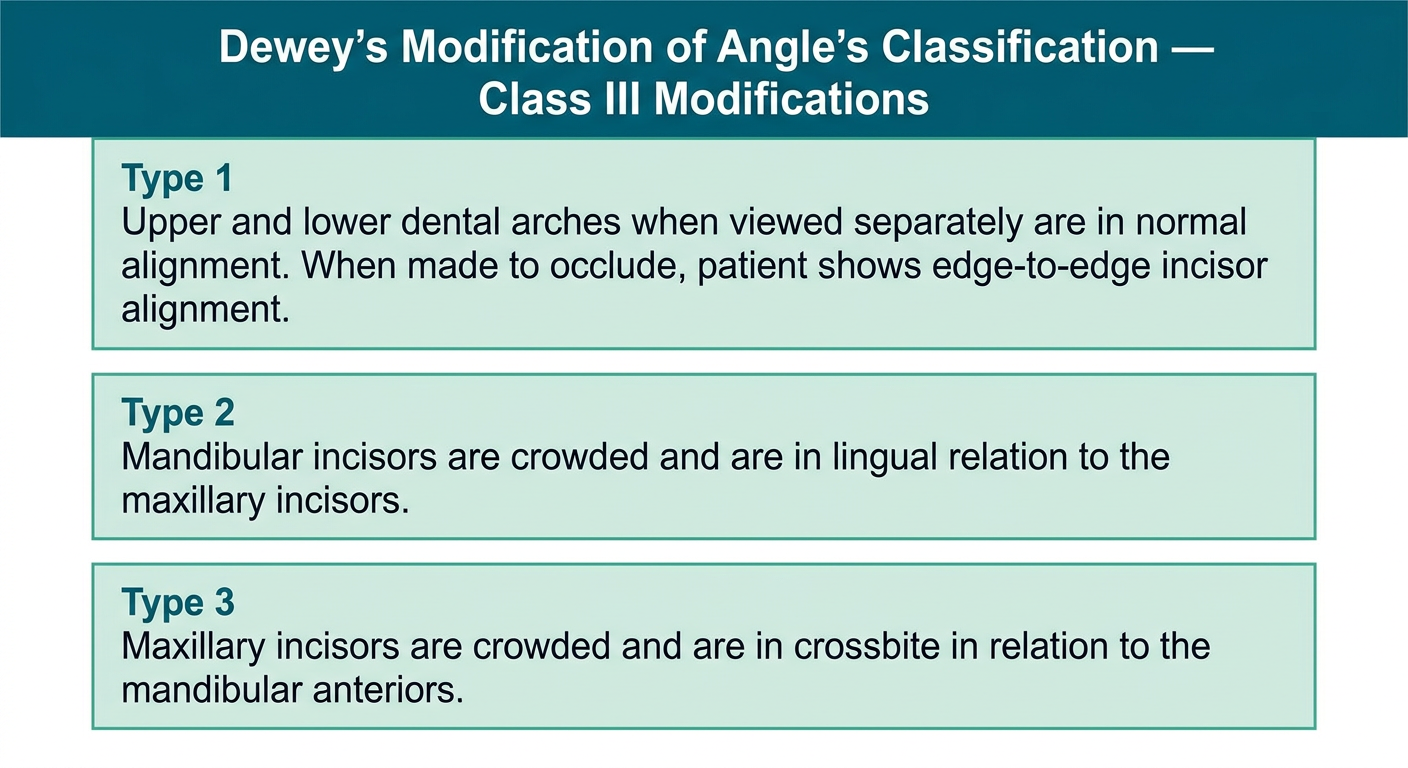
Class III Modifications of Dewey

Quick reference summary from your textbook (Orthodontics: The Art and Science):
Class I - 5 types:
  • Type 1: Crowded/bunched anterior teeth
  • Type 2: Protrusive maxillary incisors
  • Type 3: Anterior crossbite
  • Type 4: Posterior crossbite with Class I molar relation
  • Type 5: Mesial drift of permanent molar (post-early extraction)
Class III - 3 types:
  • Type 1: Normal arch form separately; edge-to-edge incisors on occlusion
  • Type 2: Crowded mandibular incisors in lingual relation to maxillary incisors
  • Type 3: Crowded maxillary incisors in crossbite with mandibular anteriors
attachment
attachment
attachment
attachment

Explain in easy language

analyse_media
analyse_media
Here is the full content explained in simple, easy language:

Prenatal Growth of Maxilla (How the Upper Jaw Develops Before Birth)

What does "prenatal growth" mean?

Prenatal = before birth. This section explains how your upper jaw (maxilla) and the middle part of your face form while you are still a baby inside the womb.

Step-by-Step Story of How the Face Forms

🟠 Week 4 of pregnancy - The story begins

1. A big bulge appears on the baby At 4 weeks in the womb, the baby (embryo) has a large bump on its front side. This bump is the developing brain. The maxilla's growth follows the same growth pattern as the brain (called the neural growth curve in Scammon's curves - basically it grows fast early on, just like the brain does).
2. A hollow dent below the bulge Just below that brain bulge, there is a small shallow dip/depression. This dip is going to become the mouth. It's called the stomodeum (think of it as the "primitive mouth" - the very first version of your mouth).
3. The floor of that primitive mouth The bottom of this stomodeum is sealed off by a thin membrane called the buccopharyngeal (or oropharyngeal) membrane. This acts like a wall separating the future mouth from the gut (foregut). Later, this wall breaks down to open up the mouth passage.

🟠 Still Week 4 - Five arches appear

4. Five branchial arches form Around the same time, 5 curved ridges called branchial arches form in the region of the future head and neck. Think of them like 5 building blocks for the face, head, and neck.
Each arch contributes to:
  • Muscles
  • Blood vessels
  • Bones and cartilage
  • Nerves
5. The 1st arch is most important for us The first branchial arch is called the mandibular arch. It plays the biggest role in forming the maxilla (upper jaw) and the entire middle part of the face (nasomaxillary region).

🟠 The face starts taking shape

6. Frontonasal process forms The tissue (mesoderm) covering the developing brain grows downward and forms a flap that hangs over the top of the primitive mouth. This downward flap is called the frontonasal process (fronto = forehead, nasal = nose area). It covers the upper part of the stomodeum.
7. Mandibular arches form the sides and bottom of the mouth
  • The mandibular arches from both sides grow and form the side walls of the primitive mouth.
  • Below the mouth, two mandibular processes fuse together in the middle to form the lower lip and lower jaw.
  • The mandibular arch also gives off a little bud from its back end called the maxillary process.
8. The maxillary process This bud (maxillary process) grows upward and toward the middle. It will eventually become the upper jaw (maxilla) and cheek regions.
At this stage, the primitive mouth (stomodeum) is surrounded by:
  • Above - frontonasal process
  • Below - mandibular process
  • On each side - maxillary process
Think of it like a box being built around the mouth opening from all 4 sides.

🟠 The nose forms

9. Nasal placodes appear On the frontonasal process, the surface cells thicken at two spots to form two oval patches called nasal placodes (placode = thickened plate of tissue). These are the very beginning of the nostrils.
10. Nasal pits form These placode patches then sink inward to form two pits called nasal pits - these become the nostrils/nasal cavities.
11. The frontonasal process splits into two parts Because the nasal pits form in the middle of the frontonasal process, they divide it into:
  • Medial nasal process (the part between the two pits - forms the middle of the nose and philtrum)
  • Lateral nasal process (the outer parts - forms the sides/wings of the nose)

🟠 Final fusion steps

12. Everything fuses together
  • As the maxillary process grows, the frontonasal process gets narrower, and the two nasal pits come closer together.
  • The maxillary process fuses with the medial nasal process. The line of fusion between these two becomes the nasolacrimal duct (the small tube that drains tears from your eye into your nose - that's why your nose runs when you cry!).

Simple Summary Table

StructureWhat it becomes
StomodeumPrimitive mouth
Frontonasal processForehead, nose, middle of upper lip
Maxillary processUpper jaw (maxilla), cheeks
Mandibular processLower jaw, lower lip
Medial nasal processMiddle of nose, philtrum (groove above lip)
Lateral nasal processSides/wings of the nose
Nasal placodes → pitsNostrils/nasal cavities
Fusion line (maxillary + medial nasal)Nasolacrimal duct

The One-Line Takeaway

By the end of week 4-6, the baby's face is basically formed by 5 processes growing toward each other and fusing - like puzzle pieces coming together to make the face. Any failure to fuse properly results in cleft lip or cleft palate.

Sentences by sentence

Here is every sentence from your notes explained one by one, in simple language:

Prenatal Growth of Maxilla


"Growth of maxilla closely follows the neural growth curve in the Scammon's curves." šŸ‘‰ The upper jaw grows in the same pattern as the brain - meaning it grows very fast in early life and then slows down. Scammon made a chart of 4 growth patterns, and the brain/neural one is the fastest early on. The maxilla follows this same fast-early pattern.

"During the fourth week of intrauterine life, prominent bulge is seen on ventral aspect of embryo which corresponds to the developing brain." šŸ‘‰ At just 4 weeks inside the womb, if you look at the front side of the tiny baby (embryo), you can see a big round bump. That bump is simply the baby's brain growing under the skin. The face will form just below this bump.

"Below the bulge there is a shallow depression, which corresponds to primitive mouth and is known as stomodeum." šŸ‘‰ Just under that brain bump, there is a small dent or hollow area. This dent is where the mouth will eventually form. Right now it's just a dip, and it's called the stomodeum - basically the very first, most primitive version of the mouth.

"Floor of the stomodeum is formed by buccopharyngeal membrane or oropharyngeal membrane which separates stomodeum from foregut." šŸ‘‰ The bottom of this primitive mouth-dent is covered by a thin wall called the buccopharyngeal membrane. This membrane acts like a closed door separating the mouth area from the baby's gut/throat below. Later it breaks open to connect them.

"By around 4th week of intrauterine life, five branchial arches form in region of future head and neck." šŸ‘‰ Also around week 4, five curved, ridge-like structures appear in the area that will become the baby's head and neck. These are called branchial arches - think of them as 5 building blocks or Lego pieces that will be used to construct the face, jaws, and neck.

"Each of these arches gives origin to muscles, connective tissue, vasculature, skeletal components and neural components of future face." šŸ‘‰ Every one of those 5 arches contributes different things to the face:
  • Muscles (so you can chew, smile, etc.)
  • Connective tissue (the support/filler tissue)
  • Blood vessels (vasculature)
  • Bones and cartilage (skeletal)
  • Nerves (neural) Each arch makes a specific set of these structures.

"First branchial arch is known as mandibular arch and it plays an important role in development of nasomaxillary region." šŸ‘‰ The first and most important branchial arch is called the mandibular arch. It is the main one responsible for building the upper jaw (maxilla), nose, and the middle part of the face - the whole nasomaxillary region.

"Mesoderm covering the developing forebrain proliferates and forms downward projection which overlaps upper part of stomodeum." šŸ‘‰ The tissue (called mesoderm) that is sitting on top of the developing brain starts multiplying and grows downward, forming a flap that hangs over the top of the primitive mouth. Imagine a curtain coming down from above the mouth opening.

"Downward projection is known as frontonasal process. Stomodeum is thus overlapped superiorly by frontonasal process." šŸ‘‰ This downward-growing flap is named the frontonasal process (fronto = forehead region, nasal = nose region). So now the top of the primitive mouth is covered by this flap from above.

"Mandibular arches of both sides form the lateral walls of stomodeum." šŸ‘‰ The mandibular arches grow in from both the left and right sides and form the side walls of the primitive mouth. So now the mouth has walls on both sides too.

"Mandibular arch gives off a bud from dorsal end known as maxillary process. Maxillary process grows ventro-medial-cranial to main part of the mandibular arch which is now known as mandibular process." šŸ‘‰ The mandibular arch sprouts a small bud from its back (dorsal) end. This bud is called the maxillary process - it will grow upward and inward to become the upper jaw. Meanwhile, the main body of the mandibular arch is now called the mandibular process - it will become the lower jaw.

"At this stage primitive mouth or stomodeum is overlapped above by frontonasal process, below by mandibular process and on either side by maxillary process." šŸ‘‰ Now the primitive mouth is completely surrounded on all sides:
  • Top = frontonasal process
  • Bottom = mandibular process
  • Left & right sides = maxillary processes Like a frame around a picture.

"Ectoderm which overlying the frontonasal process demonstrate bilateral localized thickening above the stomodeum. These are known as nasal placodes." šŸ‘‰ On the surface of the frontonasal process (the top flap), the outer skin layer (ectoderm) thickens at two specific spots - one on the left and one on the right. These two thickened oval patches are called nasal placodes - they are the very first sign that the nose is beginning to form.

"Such placodes soon sink and they form the nasal pits." šŸ‘‰ Those two thickened patches then push inward (sink into the tissue) and create two small holes/pits. These are called nasal pits and they will eventually become your nostrils and the inside of your nose.

"Formation of such nasal pits divides frontonasal process into two parts i.e. 1. Medial nasal process, 2. Lateral nasal process." šŸ‘‰ Because those two pits form on the frontonasal process, they split it into two parts:
  • Medial nasal process - the strip between the two pits (becomes the middle/tip of the nose and the philtrum - the groove above your lips)
  • Lateral nasal process - the parts on the outer sides (become the wings/sides of the nose)

"Two of the mandibular processes grow medially and fuse to form lower lip and the lower jaw." šŸ‘‰ The two mandibular processes (one from each side) grow toward each other and join in the middle. When they fuse, they form the lower lip and the lower jaw (mandible).

"As maxillary process undergoes growth, frontonasal process becomes narrow, so that two of the nasal pits come close." šŸ‘‰ As the maxillary processes grow bigger and push inward, they squeeze the frontonasal process, making it narrower. This brings the two nasal pits closer together - which is why both nostrils end up right next to each other in the middle of the face.

"Line of the fusion of maxillary process and medial nasal process correspond to nasolacrimal duct." šŸ‘‰ When the maxillary process and the medial nasal process join/fuse together, the line where they meet becomes the nasolacrimal duct - the tiny tube in the corner of your eye that drains tears into your nose. That is why your nose runs when you cry - the tears travel down this duct!

Quick Visual Summary

Week 4 Embryo Face:

         [Frontonasal Process]
               ↓
    [Max. P] [MOUTH] [Max. P]
               ↑
        [Mandibular Process]

Nasal pits form on Frontonasal Process
→ splits into Medial + Lateral nasal process

All fuse together → Face is complete!

Summarise this process in short form and also easy language

Prenatal Growth of Maxilla - Short Summary


The One-Paragraph Version

At week 4 in the womb, a baby's face begins forming around a small dent called the stomodeum (primitive mouth). Five branchial arches appear and act as building blocks for the face. A flap called the frontonasal process covers the mouth from above, the mandibular process covers it from below, and two maxillary processes cover it from the sides. On the frontonasal process, two spots thicken, sink in, and form nasal pits (future nostrils), splitting it into medial and lateral nasal processes. Everything then grows toward the center and fuses together to complete the face. The fusion line of the maxillary and medial nasal process becomes the nasolacrimal duct.

Super Short Point Form

WeekWhat Happens
Week 4Brain bulge visible on embryo
Week 4Stomodeum (primitive mouth dent) forms below the bulge
Week 45 branchial arches appear
Week 4Frontonasal process grows DOWN from above
Week 4Maxillary process buds off - grows to form upper jaw
Week 4Mandibular process forms lower jaw + lower lip
LaterNasal placodes → sink in → nasal pits form
LaterFrontonasal process splits into medial + lateral nasal process
LaterEverything fuses → face is complete
Fusion lineMaxillary + medial nasal process = nasolacrimal duct

The Simplest Possible Version (Like a Story)

šŸ—ļø Think of building a face like building a house from 4 walls.
  • The top wall = frontonasal process (becomes forehead + nose)
  • The bottom wall = mandibular process (becomes lower jaw + lower lip)
  • The two side walls = maxillary processes (become upper jaw + cheeks)
The nose is made by two holes (nasal pits) punching into the top wall and splitting it into two pieces.
All 4 walls then grow inward and stick together to complete the face.
Where the side wall and top wall join = the nasolacrimal duct (tear duct).

Memory Trick

"Front Covers Top, Max Covers Sides, Mand Covers Bottom → Pits Split Front → All Fuse = Face Done!"
This is a shared conversation. Sign in to Orris to start your own chat.