
Sealability nd biocompatabililty of various root canal sealers 10 marks ntruhs university exm like answer . Give me from contents to reference everything . If possible hand drawn notes and diagrams
root canal sealers sealability biocompatibility types zinc oxide eugenol calcium hydroxide resin NTRUHS exam
https://johs.com.sa/published/article/6a29e64b284fbd8a7a57fa…
root canal sealer biocompatibility sealability
root canal sealer types classification diagram endodontics
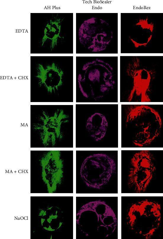
This Comparison Chart displays representative Confocal Laser Scanning Microscopy (CLSM) images at 10x magnification, illustrating endodontic sealant penetration into dentinal tubules. The grid compares three types of root canal sealers (AH Plus in green, Tech BioSealer Endo in purple, and EndoRez in red) against five different irrigation protocols: EDTA, EDTA + Chlorhexidine (CHX), Maleic Acid (MA), MA + CHX, and Sodium Hypochlorite (NaOCl). The images demonstrate transverse sections of the coronal third of the root canal. Key visual elements include radial 'tag-like' projections extending from the canal lumen into the pericanalar dentin, representing sealer penetration. Variations in the density, depth, and uniformity of these projections are visible across the protocols. For example, EDTA and MA treatments generally show more extensive radial tag formation compared to the patchy, less uniform distribution seen with NaOCl. This image is used in dental research and endodontics to evaluate how different chemical irrigation regimens affect the sealing ability and adaptation of resin-based and bioceramic sealers to the root canal wall by removing the smear layer.
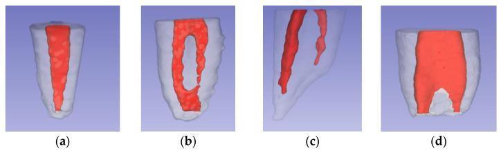
This anatomical diagram displays four three-dimensional reconstructions (labeled a-d) illustrating Weine’s classification of root canal morphology, a critical concept in endodontics. The images use color-coding where the translucent grey/white outer structure represents the tooth root dentin and the solid red internal structure represents the pulp canal system. Panel (a) shows Type I: a single continuous canal extending from the pulp chamber to one apical foramen. Panel (b) depicts Type II: two distinct canals that originate separately in the coronal portion but converge and fuse to exit through a single apical foramen. Panel (c) illustrates Type III: two independent canals that remain separate throughout the length of the root, terminating in two distinct apical foramina. Panel (d) shows Type IV: a single coronal canal that bifurcates in the middle or apical third, ending in two separate apical foramina. This visual comparison serves as an educational tool for identifying variations in root anatomy, informing diagnostic and treatment protocols during root canal therapy.
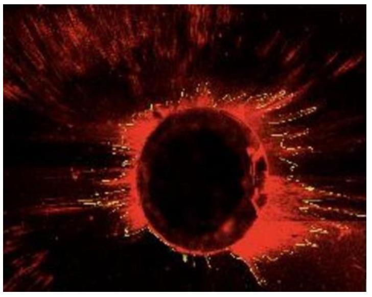
This diagnostic image is a Confocal Laser Scanning Microscopy (CLSM) micrograph showing a cross-section of a human tooth root canal. The central dark circular area represents the lumen of the root canal. Surrounding this central void is a prominent, irregular ring of bright red fluorescence, which identifies the penetration of endodontic sealer into the pericanal dentin. Radiating outward from this primary ring are fine, fluorescent red streaks and tag-like structures, illustrating the penetration of the sealer material into the dentinal tubules. The distribution and depth of these streaks are used in endodontics to evaluate the efficiency of different irrigation protocols (such as NaOCl or CHX) and sealing techniques. The image highlights the interface between the root canal wall and the dentinal substrate, emphasizing the adaptation and tubular penetration of dental biomaterials necessary for a fluid-tight apical seal.
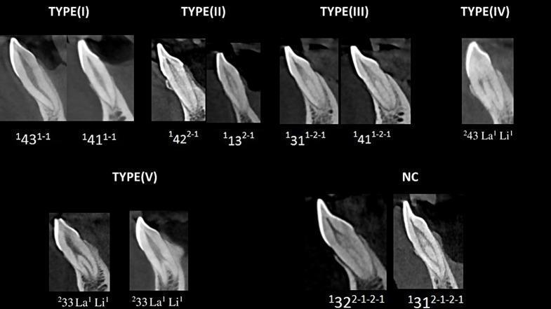
This diagnostic composite image presents sagittal Cone Beam Computed Tomography (CBCT) scans of human mandibular anterior teeth, illustrating root canal morphology variations. The visual is categorized according to the Vertucci classification (Types I, II, III, IV, and V) and the Ahmed classification system. Type I shows a single canal from pulp chamber to apex (1-1 configuration). Type II shows two canals merging into one (2-1). Type III depicts a single canal bifurcating into two then rejoining as one (1-2-1). Type IV demonstrates two independent canals from chamber to apex (2-2). Type V illustrates a single canal dividing near the apex into two distinct foramina (1-2). An additional 'NC' (Non-Classifiable) section displays complex variations like the 2-1-2-1 configuration. These images serve as educational resources for endodontics, highlighting the anatomical complexity of root canal systems necessary for successful dental treatment planning and biomechanical preparation.
endodontic sealer biocompatibility review
Hand-drawn style dental education diagram showing classification of root canal sealers with branches: 1) Zinc Oxide Eugenol (Rickert's, Grossman's, Tubliseal), 2) Calcium Hydroxide (CRCS, Apexit), 3) Resin-based (AH26, AH Plus, Diaket), 4) Glass Ionomer (Ketac-Endo), 5) Calcium Silicate/MTA-based (MTA Fillapex, iRoot SP), 6) Silicon-based (Lee Endo-fill), 7) Gutta Percha-based (Kloropercha). With properties listed: sealability, biocompatibility, radiopacity, solubility. Black ink on white background, like medical notes.
Hand-drawn medical notes style diagram comparing biocompatibility and sealability of root canal sealers in a table format. Columns: Sealer Type, Sealability (Excellent/Good/Fair/Poor), Biocompatibility (Excellent/Good/Fair/Poor), Cytotoxicity, Examples. Rows: ZOE sealers, Calcium Hydroxide, Epoxy Resin (AH Plus), Glass Ionomer, Calcium Silicate/MTA, Bioceramic. Include a small cross-section diagram of tooth root showing sealer filling dentinal tubules. Black pen on white paper appearance, educational dental notes style.
https://pmc.ncbi.nlm.nih.gov/articles/PMC4379664
Hand-drawn pencil sketch notes for dental exam showing: cross-section of tooth root with labels - dentin, sealer layer filling irregularities, gutta percha core, dentinal tubules showing sealer penetration. Arrows showing where leakage occurs: sealer-dentin interface, sealer-GP interface, through sealer itself. Title: "SEALABILITY - Mechanism of Sealing". Black on white, medical student notes style, rough sketch aesthetic.
Hand-drawn dental exam notes flowchart: "Ideal Properties of Root Canal Sealer" in center circle with branches - Adhesion to canal walls, Dimensional stability after setting, Radiopaque, Insoluble in tissue fluids, Non-irritating (biocompatible), Antibacterial, Easy to manipulate, Hermetic seal, Non-staining, Slow setting, Resorbable if extruded. Black ink pen on white paper, student notes style with underlines and circles.
Error calling model 'gemini-3-pro-image' (RESOURCE_EXHAUSTED): 429 RESOURCE_EXHAUSTED. {'error': {'code': 429, 'message': 'Resource has been exhausted (e.g. check quota).', 'status': 'RESOURCE_EXHAUSTED'}}
We encountered an error while processing your request. Please try again or reach out to us for help.