
Left eye esotropia what could be the possible muscles affected
- Overacting left medial rectus (most common - pulls eye inward)
- Underacting left lateral rectus (fails to abduct the eye)
esotropia eye muscles anatomy diagram

This medical illustration features a semi-transparent schematic overlay of facial musculature onto a monochrome clinical photograph of a human face. The primary focus is the orbicularis oculi muscle, depicted with a distinctive circular, striated fiber pattern that surrounds the orbital rim and extends from the eyebrow to the upper cheek and lower eyelid. The diagram highlights the muscle's superficial position in the periocular region. Adjacent facial muscles, including the zygomaticus major and minor and levator labii superioris, are shown extending across the midface and cheek area, illustrating the complex, overlapping anatomical relationships between the periocular and midfacial soft tissues. This visual is designed for educational instruction on facial anatomy, specifically addressing the soft tissue components involved in eye movement, wrinkle formation, and clinical conditions such as periocular dark circles (dark eye circles) and eyelid edema. It demonstrates the anatomical basis for cosmetic and therapeutic interventions targeting the facial nerve branches and orbicularis oculi.
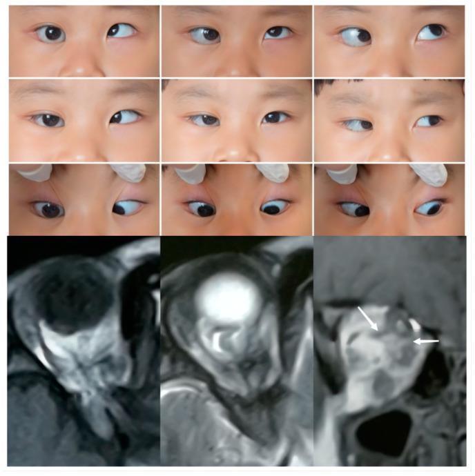
Educational composite image illustrating Case 2 of supernumerary extraocular muscles (SEOM). The upper panel consists of nine clinical gaze photographs of a pediatric patient, demonstrating a large-angle right esotropia (approx. 30 prism diopters) and a severe abduction limitation (-4) of the right eye. The primary position and multiple directions of gaze show restrictive motility, along with eyelid entropion and an enlarged palpebral aperture in the right eye. The bottom panel displays diagnostic MRI scans, including T1 axial, T2 axial, and T2 coronal sections. These images reveal anatomical abnormalities in the right orbit, characterized by anomalous extraocular muscle (EOM) bands that extend from the orbital apex and connect multiple rectus muscles (SR, IR, LR, and MR). White arrows on the coronal section highlight these atypical muscular connections. Additionally, the right optic nerve is shown to be maldeveloped (hypoplastic) with an indistinct course, representing a Type 2 SEOM classification where anomalous tissue surrounds the optic nerve and disrupts normal ocular motility.
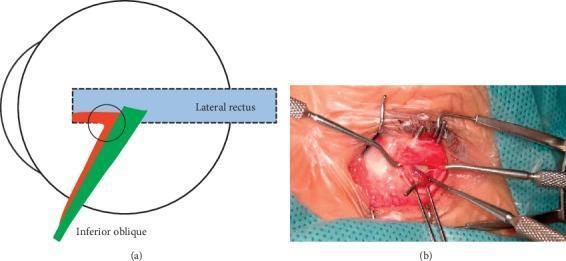
This composite educational material consists of an anatomical diagram and a clinical photograph detailing Type-beta inferior oblique muscle enlargement (IO-E). Part (a) is a schematic illustration of the lateral aspect of the eye globe. It depicts the lateral rectus muscle (blue) and the inferior oblique muscle bifurcating into two bundles: a posterior bundle (green) that inserts into the conventional scleral position, and an anterior bundle (red) that loosely connects at the conventional insertion (purple circle) before coursing anteriorly beneath the lateral rectus to fuse with its insertion point. Part (b) is an intraoperative clinical photograph providing a surgical view of the ocular surface during strabismus surgery. The white sclera is exposed, and various surgical instruments, including muscle hooks, forceps, and a lid speculum, are used to manipulate the extraocular tissues and muscles for visualization of anomalous attachments. This content is intended for ophthalmic specialty training, focusing on surgical anatomy and the diagnosis of extraocular muscle variations related to vertical strabismus and V-pattern exotropia.
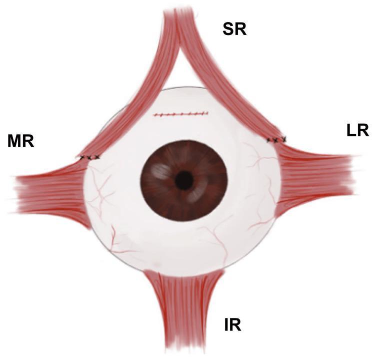
This medical illustration depicts a surgical procedure on the extraocular muscles of the left eye, identified as the Sesma-AlGhazal procedure. The anatomical diagram shows the eyeball with four rectus muscles: Superior Rectus (SR), Medial Rectus (MR), Lateral Rectus (LR), and Inferior Rectus (IR). The SR muscle is demonstrated as longitudinally split into two distinct halves. The medial half of the SR has been transposed and sutured anterior to the original insertion of the MR. Simultaneously, the lateral half of the SR has been transposed and sutured superior to the insertion point of the LR. Black surgical sutures are visible at both new insertion points and at the original superior scleral site where the SR was detached. This muscle transpositioning technique is used in ophthalmology to correct specific forms of strabismus, such as hypotropia, by redistributing the mechanical forces of the SR to assist adjacent muscles. The diagram serves as an educational tool for ophthalmologists and medical students to understand ocular motility surgery and muscle transposition geometry.
Left Eye Esotropia - Muscles Affected
PRIMARY MUSCLES INVOLVED
| Muscle | Role in Esotropia | Mechanism |
|---|---|---|
| Left Medial Rectus (MR) | Overacting / spastic | Adducts the eye - pulls it nasally inward |
| Left Lateral Rectus (LR) | Underacting / paretic | Fails to abduct the eye outward |
MECHANISM - HOW EACH MUSCLE CONTRIBUTES
1. Left Lateral Rectus Weakness (Most neurologically significant)
- Innervated by CN VI (Abducens nerve)
- Paresis/palsy of the left CN VI = left LR cannot abduct the eye
- Results in incomitant esotropia that worsens on left lateral gaze (toward the weak muscle)
- The angle of deviation increases when looking left and decreases when looking right
- Per Bradley & Daroff's Neurology: "a right lateral rectus palsy will cause esotropia that increases upon looking to the right... Similarly, with a right medial rectus weakness, an exotropia will be present"
2. Left Medial Rectus Overaction / Contracture
- Innervated by CN III (Oculomotor nerve)
- Can be primary (overactive pull) or secondary (contracture of the antagonist during recovery from LR palsy)
- Per Bradley & Daroff's: "Abducens palsy with contracture of antagonist (ipsilateral medial rectus) during recovery" is a distinct cause
- Also seen in: thyroid eye disease (restrictive fibrosis of MR), orbital pseudotumor, medial orbital wall fracture with entrapment
CLASSIFICATION OF ESOTROPIA BY CAUSE (from Kanski's Clinical Ophthalmology)
- Early-onset (infantile) esotropia - large angle (>30Δ), typically with cross-fixation pattern
- Accommodative esotropia - driven by excessive convergence with accommodation; MR overacts in response to hypermetropia
- Refractive (fully/partially accommodative)
- Non-refractive (convergence excess type - MR dominates for near)
- Basic esotropia - equal deviation at distance and near
- Divergence insufficiency/paralysis - bilateral LR underaction
- Convergence spasm - intermittent MR spasm (accompanied by miosis and pseudomyopia)
- Sensory esotropia - poor vision in one eye removes fusion stimulus; MR tone dominates
| Cause | Muscle Affected |
|---|---|
| CN VI palsy | Left Lateral Rectus paresis |
| Thyroid eye disease | Left MR fibrosis/restriction |
| Duane syndrome (Type 1) | Abnormal LR innervation by CN III |
| Myasthenia gravis | LR fatigue (variable) |
| Medial orbital wall fracture | MR entrapment |
| CN III palsy (rare - ocular neuromytonia) | MR spasm |
| Dorsal midbrain syndrome | Convergence/tonic MR spasm |
SURGICAL TREATMENT - WHICH MUSCLES ARE OPERATED ON
- Recession of both medial rectus muscles (weakening procedure) - for concomitant esotropia
- Unilateral MR recession + LR resection (weakening MR, strengthening LR) - for smaller/asymmetric deviations
- Angles >50Δ may require surgery on more than 2 muscles
QUICK SUMMARY
- Left MR (adductor) > Left LR (abductor)
Can muscles affected in right eye cause left eye esotropia
Can Right Eye Muscles Cause Left Eye Esotropia?
The Two Laws That Govern This
1. Hering's Law of Equal Innervation
"During any conjugate eye movement, equal and simultaneous innervation flows to the yoke muscles."
- Kanski's Clinical Ophthalmology, 10th ed.
2. Sherrington's Law of Reciprocal Innervation
Yoke Muscle Pairs for Horizontal Gaze
| Movement | Right Eye Muscle | Left Eye Muscle (Yoke) |
|---|---|---|
| Dextroversion (gaze right) | Right Lateral Rectus | Left Medial Rectus |
| Levoversion (gaze left) | Right Medial Rectus | Left Lateral Rectus |
The Classic Scenario: Right Lateral Rectus Palsy Causing Left Esotropia
- Right LR is paretic - it cannot fully abduct
- The brain sends normal innervation to right LR
- Equal innervation flows to its yoke: Left MR
- Left MR pulls the left eye slightly inward
- Result: small left esotropia = PRIMARY DEVIATION
- Brain sends EXTRA innervation to the paretic right LR to force fixation
- By Hering's Law, this same extra signal flows to the Left MR (yoke)
- Left MR is perfectly normal - it OVER-adducts
- Result: large left esotropia = SECONDARY DEVIATION
"Secondary deviation is always greater than primary deviation in incomitant strabismus because of the Hering law of dual innervation; it may mislead the examiner to believe that the eye with the greater deviation is the weak one."
- Bradley & Daroff's Neurology in Clinical Practice
Full Cascade of Muscle Sequelae from One Paretic Muscle
| Stage | Muscle | Eye | Mechanism |
|---|---|---|---|
| 1. Primary underaction | Right LR | Right | Directly paretic |
| 2. Secondary overaction | Left MR | Left | Hering's Law (yoke) - causes left esotropia |
| 3. Secondary overaction + contracture | Right MR | Right | Sherrington's Law (ipsilateral antagonist, unopposed) |
| 4. Secondary inhibition | Left LR | Left | Hering + Sherrington combined |
Other Right Eye Scenarios That Can Cause Left Esotropia
| Right Eye Problem | How it Causes Left Esotropia |
|---|---|
| Right CN VI palsy (right LR paresis) | Extra innervation to right LR overdrives left MR via Hering's Law |
| Right MR contracture (e.g. thyroid eye disease) | Mechanically pulls right eye into adduction; brain compensates with more LR signal; reduces innervation to left MR's antagonist (left LR) |
| Right CN III palsy (if patient fixates with right eye) | Brain overdrive of right medial rectus overdrives left lateral rectus - this would cause exotropia; but if the convergence tone increases to compensate, secondary esotropia of the left eye can occur |
| Duane syndrome right eye Type 1 | Restricted right abduction mimics right LR paresis - same Hering cascade applies |
Clinical Pearl - The Diagnostic Trap
- If the esotropia worsens on right gaze → right LR is the weak muscle
- If the esotropia is equal in all gaze positions (comitant) → the cause is central or longstanding (spread of comitance)
How to evaluate & come to know which muscle is affected inna case of left eye esotropia
Evaluating Which Muscle Is Affected in Left Eye Esotropia - A Step-by-Step Approach
STEP 1 - HISTORY
| Question | What it tells you |
|---|---|
| Age of onset? | Infantile (congenital) vs acquired esotropia |
| Sudden or gradual onset? | Sudden = neurogenic (CN VI palsy); gradual = accommodative or restrictive |
| Constant or intermittent? | Intermittent = accommodative, convergence spasm |
| Diplopia present? | Adults with acquired paresis; absent in long-standing childhood squints (suppression) |
| Diplopia worse in which gaze direction? | Points to the weak muscle |
| Associated ptosis, pupil changes? | CN III palsy |
| Thyroid disease, myasthenia, trauma? | Restrictive or neuromuscular causes |
| Family history, birth history? | Congenital/hereditary forms |
STEP 2 - VISUAL ACUITY (Both Eyes Separately)
- Test each eye independently
- A big difference between eyes raises the possibility of amblyopia (lazy eye) or sensory esotropia (poor vision in left eye → loss of fusion → left MR dominates)
- Use age-appropriate charts: Snellen, Cardiff acuity cards (infants), preferential looking
STEP 3 - CORNEAL REFLEX TESTS (Objective - works in all ages)
Hirschberg Test
- In left esotropia: left eye reflex is displaced temporally (laterally)
- Each 1 mm of decentration ≈ 7° (14 prism dioptres)
- Limbus = ~45°; mid-pupil edge = ~15°
Krimsky Test
STEP 4 - COVER TESTS (The Gold Standard)
A. Cover-Uncover Test (detects heterotropia vs heterophoria)
-
Cover the right eye - watch the left eye:
- If left eye moves inward to take up fixation → left esotropia confirmed (left eye was turned in)
- No movement → left eye was already fixating (consider right eye problem)
-
Uncover the right eye - watch for recovery:
- Immediate re-fusion = good binocular potential
- No fusion = long-standing deviation or amblyopia
B. Alternate Cover Test (measures total deviation including phoria)
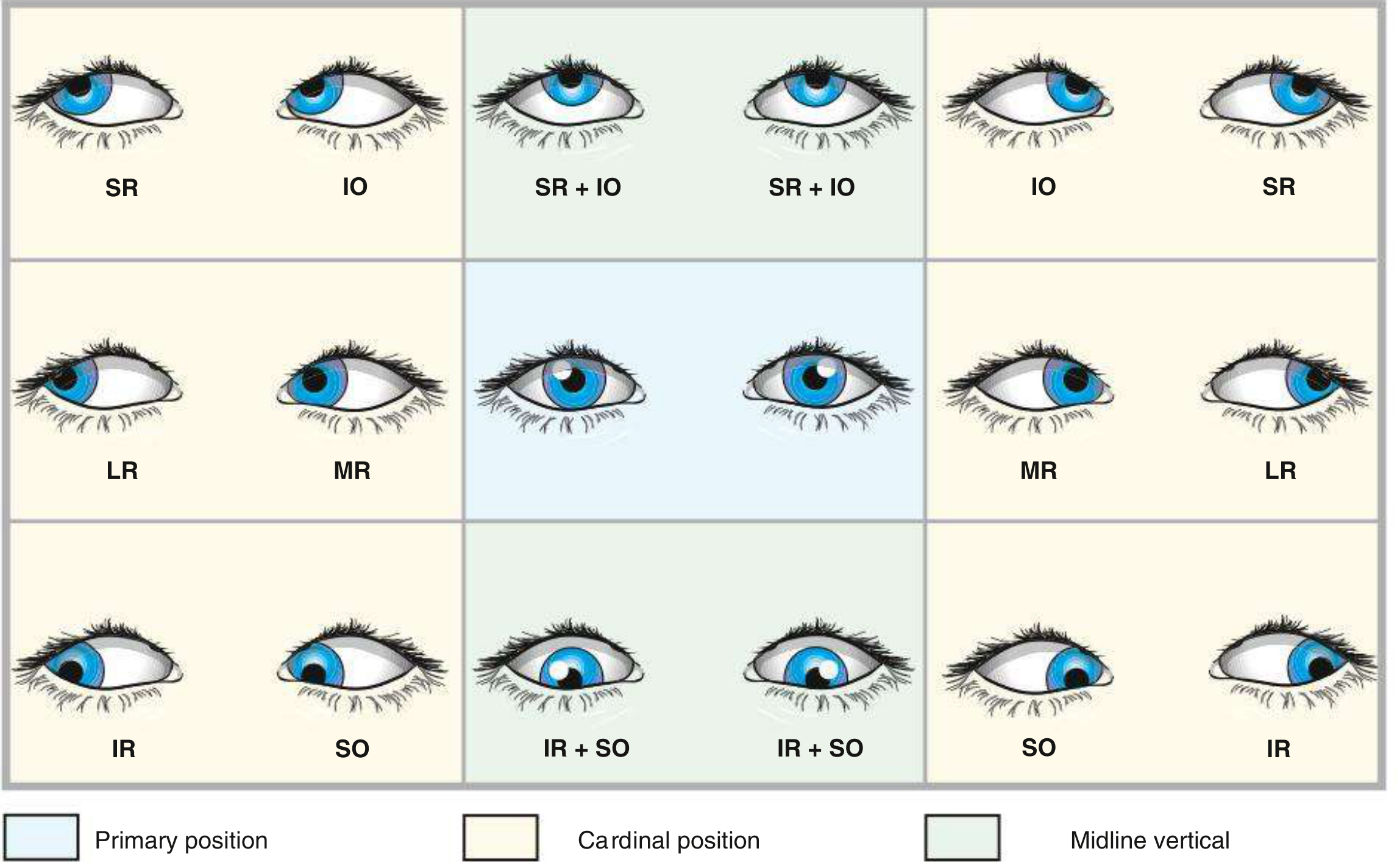
STEP 5 - COMITANCE ASSESSMENT (Key to Identifying the Weak Muscle)
- Comitant (same angle everywhere) = congenital, accommodative, or longstanding (spread of comitance)
- Incomitant (angle varies) = paretic or restrictive - tells you exactly which muscle is weak
| Angle worsens in... | Muscle at fault | Nerve |
|---|---|---|
| Left lateral gaze (looking left) | Left Lateral Rectus (weak, can't abduct) | CN VI (left abducens) |
| Right lateral gaze (looking right) | Right Medial Rectus (weak = secondary deviation) | CN III (right oculomotor) |
| Up-and-left gaze | Left Superior Rectus or Left Inferior Oblique | CN III |
| Down-and-left gaze | Left Inferior Rectus or Left Superior Oblique | CN III / CN IV |
Primary vs Secondary Deviation Rule
- Cover left eye → right eye fixates → measure left eye deviation = Primary deviation
- Cover right eye → left eye fixates → measure right eye deviation = Secondary deviation
- Secondary > Primary → confirms paretic squint (Hering's Law)
- The eye with the smaller field on Hess chart = the paretic eye
STEP 6 - DUCTIONS AND VERSIONS
- Reduced left eye abduction → Left LR weakness (CN VI palsy)
- Normal ductions but abnormal versions → consider Duane syndrome or restrictive myopathy
- Restriction on duction → mechanical/restrictive cause (thyroid, fracture, fibrosis)
STEP 7 - HESS CHART (The Most Specific Test for Paretic Muscle)
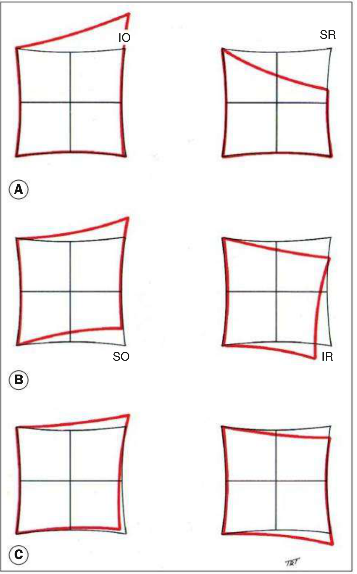
- Smaller field = paretic eye (underaction)
- Larger field = fellow eye (overaction of yoke muscles by Hering's Law)
- The plot shows exactly which gaze position is most restricted → directly names the paretic muscle
- Left chart (left eye): small field, shrunken in the lateral direction
- Right chart (right eye): large field, expanded medially (right MR overaction = yoke)
STEP 8 - FORCED DUCTION TEST (FDT)
- Grasp the conjunctiva/sclera with forceps and mechanically move the eye
- Resistance to abduction of left eye → Restrictive cause (e.g., left MR fibrosis in thyroid eye disease, orbital wall fracture with MR entrapment)
- No resistance → Paretic cause (CN VI palsy, neuromuscular disease)
STEP 9 - FURTHER INVESTIGATIONS (Based on Findings)
| Finding | Investigation |
|---|---|
| Incomitant, CN VI palsy pattern | MRI brain (Wernicke's, tumour, raised ICP, demyelination) |
| Variable, fatigable esotropia | Tensilon/ice pack test, ACh receptor antibodies (Myasthenia gravis) |
| Thyroid signs | TFTs, TSH receptor antibodies, orbital MRI |
| Trauma | CT orbit (blowout fracture, MR entrapment) |
| Accommodative component suspected | Cycloplegic refraction (atropine/cyclopentolate) to reveal hypermetropia |
| Amblyopia | Detailed VA testing, visual evoked potentials |
SUMMARY FLOWCHART
LEFT ESOTROPIA FOUND
|
Comitant?
/ \
YES NO (incomitant)
| |
Accommodative Worsens on LEFT gaze?
or congenital | |
YES NO (worsens on RIGHT gaze)
| |
Left LR weakness Right MR weakness
→ CN VI palsy → Hering's secondary deviation
→ FDT: no resistance from RIGHT eye problem
= paretic
→ FDT: resistance
= restrictive
(thyroid, fracture)
Left esotropia with positive cover test & improving with left gaze & worsening with right gaze which muscle is affected & kindly give an explanation
Left Esotropia: Improves on Left Gaze, Worsens on Right Gaze
Direct Answer: RIGHT LATERAL RECTUS is the primary affected muscle
The Logical Explanation - Step by Step
First, Understand the Anatomy
| Gaze Direction | Right Eye Muscle Used | Left Eye Yoke Muscle Used |
|---|---|---|
| Looking Right (dextroversion) | Right Lateral Rectus (abducts RE) | Left Medial Rectus (adducts LE) |
| Looking Left (levoversion) | Right Medial Rectus (adducts RE) | Left Lateral Rectus (abducts LE) |
Now Apply This to Your Case
IN RIGHT GAZE (deviation WORSENS)
- Right LR must maximally contract to abduct the right eye - but it is weak/paretic, so it cannot pull the right eye out
- By Hering's Law, that same excessive innervation signal flows simultaneously to its yoke muscle: the Left Medial Rectus
- The left MR is perfectly normal - it responds fully to that strong signal and over-adducts the left eye hard to the right (nasally)
- Result: Left esotropia is at its MAXIMUM in right gaze
"A right lateral rectus palsy will cause an esotropia that increases upon looking to the right, the side of the weak muscle."
- Bradley & Daroff's Neurology in Clinical Practice
IN PRIMARY POSITION (straight ahead)
- Some extra innervation still going to paretic right LR (and thus to left MR)
- Moderate left esotropia visible - this is what you see on the cover test in primary position
IN LEFT GAZE (deviation IMPROVES / disappears)
- Right Medial Rectus does the work for the right eye (adducts) - this is completely unaffected
- The yoke of the right MR is the Left Lateral Rectus - signal goes to LEFT LR to abduct the left eye
- The left LR is normal - it pulls the left eye outward
- The right LR (the weak one) is not being asked to work at all in this direction
- Result: The esotropia improves or fully resolves in left gaze because the paretic right LR is now out of its field of action
The Visual Summary
LEFT GAZE PRIMARY POSITION RIGHT GAZE
(looking left) (straight ahead) (looking right)
Right LR role: Not needed Moderate effort MAXIMUM effort
← PARETIC = WEAK
Left MR Normal signal Moderate signal OVER-DRIVEN signal
(yoke of RLR): → pulls LE inward → LE slightly in → LE maximally in
slightly or = moderate ET = MAXIMUM ET
neutralized
ESOTROPIA: IMPROVES / NIL MODERATE WORSENS / MAX
Why the Cover Test Is Positive
- If left eye was already fixating, no movement
- If right eye was the fixating eye: the left eye moves outward (abducts) to take up fixation = confirming esotropia of the left eye
Primary vs Secondary Deviation Confirmation
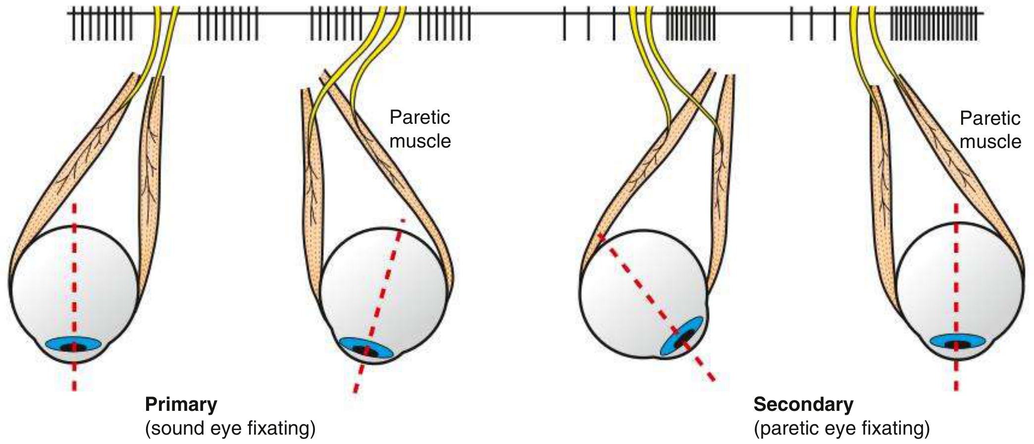
| Fixating Eye | Deviation Seen | Name | Size |
|---|---|---|---|
| Left eye fixes (right eye covered) | Right eye drifts in | Primary deviation | Smaller |
| Right eye forced to fix (left eye covered) | Left eye swings inward (over-adducts via Hering) | Secondary deviation | LARGER |
What Nerve / Structure is Causing This?
| Category | Examples |
|---|---|
| Raised intracranial pressure | Tumour, hydrocephalus (CN VI is a false localising sign) |
| Vascular | Pontine infarct, microvascular ischaemia (diabetes, hypertension) |
| Demyelination | Multiple sclerosis |
| Trauma | Petrous apex fracture (Gradenigo syndrome) |
| Wernicke's encephalopathy | Bilateral CN VI palsies |
| Meningitis / inflammation | Basal meningitis |
In One Line
A patient with tectal mengioma with hydrocephalus underwent mpvp shunting & post of developed edh for which the patient was operated After this she presented with Left esotropia with positive cover test & improving with left gaze & worsening with right gaze which muscle is affected & kindly give an explanation
Clinical Analysis: Tectal Meningioma + Hydrocephalus + VP Shunt + EDH Surgery → Left Esotropia
ANSWER: Primary Affected Muscle = Right Lateral Rectus
Nerve: Right CN VI (Abducens nerve) palsy
Left Esotropia = Secondary (Hering's) deviation - the LEFT eye is innocent
- Improves on left gaze = right LR out of its field of action
- Worsens on right gaze = right LR maximally demanded but paretic
- Positive cover test = manifest incomitant esotropia
What Makes This Case Special: 4 Stacked Mechanisms
Why CN VI Is the Most Vulnerable Cranial Nerve to Raised ICP and Surgery
- Upward through the posterior fossa
- Over the tip of the petrous bone (tethered here by Gruber's/petroclinoid ligament)
- Through Dorello's canal
- Into the cavernous sinus
- Finally to the lateral rectus
"Isolated unilateral or bilateral sixth nerve palsy with global headache can be the initial manifestation of raised intracranial pressure from any source - including brain tumour, meningitis, and pseudotumor cerebri; rarely it may appear after lumbar puncture, epidural injections, or insertion of a ventricular shunt."
- Adams & Victor's Principles of Neurology, 12th ed.
Event-by-Event Analysis of Mechanisms in This Patient
EVENT 1 - Tectal Meningioma
- Tectum compression → tonic convergence spasm → bilateral or unilateral esotropia
- This is a central cause of esotropia listed in Bradley & Daroff's Box 18.3
- The tectal mass obstructs the cerebral aqueduct
- CSF backs up → raised ICP
- Raised ICP stretches/compresses CN VI over the petrous ridge (Gruber's ligament acts as a fulcrum)
- → Right CN VI palsy as a false localising sign
EVENT 2 - Hydrocephalus (Before Shunt)
- Downward displacement of the brainstem ("transtentorial shift") stretches CN VI over the petrous apex
- Even without a mass directly touching CN VI, the mechanical traction is sufficient to cause palsy
- This is the classic Cushing-era description of CN VI as a non-localising sign of raised ICP
EVENT 3 - VP Shunt Insertion (mVP Shunting)
"Rarely, [sixth nerve palsy] may appear after... insertion of a ventricular shunt."
- Adams & Victor's Principles of Neurology
| Mechanism | Explanation |
|---|---|
| Rapid ICP reduction | After shunting, sudden drop in ICP causes brain sagging/descent; CN VI is tethered at Dorello's canal and gets kinked |
| Intracranial hypotension | Overdrainage lowers CSF pressure → brain descends → CN VI stretched from above |
| Shunt catheter tract | If catheter passes near the base of the skull, local traction possible |
| Posterior fossa crowding | Cerebellar tonsillar descent after shunting can compress exiting CN VI fibres |
EVENT 4 - EDH Surgery (Craniotomy + EDH Evacuation)
| Intraoperative / Perioperative Factor | Effect on CN VI |
|---|---|
| Intraoperative brain retraction | Temporal/frontotemporal retraction stretches the CN VI at the cavernous sinus or Dorello's canal |
| Haematoma mass effect pre-op | EDH itself raises ICP acutely → CN VI compression over petrous ridge |
| Rapid decompression during surgery | Sudden ICP drop after clot evacuation = same intracranial hypotension mechanism as shunting |
| Anaesthesia + positioning | Head positioning and hypotensive anaesthesia can reduce perfusion to long CN VI |
| Surgical trauma to skull base | Temporal bone drilling or retractor placement can injure CN VI in Dorello's canal |
Putting It All Together - The Timeline
TECTAL MENINGIOMA
↓
Obstructs aqueduct → HYDROCEPHALUS → Raised ICP
↓
CN VI tethered at petrous tip → STRETCHED by raised ICP
↓
mVP SHUNT inserted → Sudden ICP drop → Brain descent
↓
CN VI kinked/stretched by intracranial hypotension
↓
EDH develops (post-shunt) → Acute ICP rise again
↓
EDH SURGERY → Retraction + acute decompression
↓
CUMULATIVE INJURY TO RIGHT CN VI
↓
Right Lateral Rectus palsy
↓
By Hering's Law → Left MR over-driven
↓
LEFT ESOTROPIA (secondary deviation)
↓
Worsens on right gaze / Improves on left gaze
Clinical Photograph of CN VI Palsy Pattern
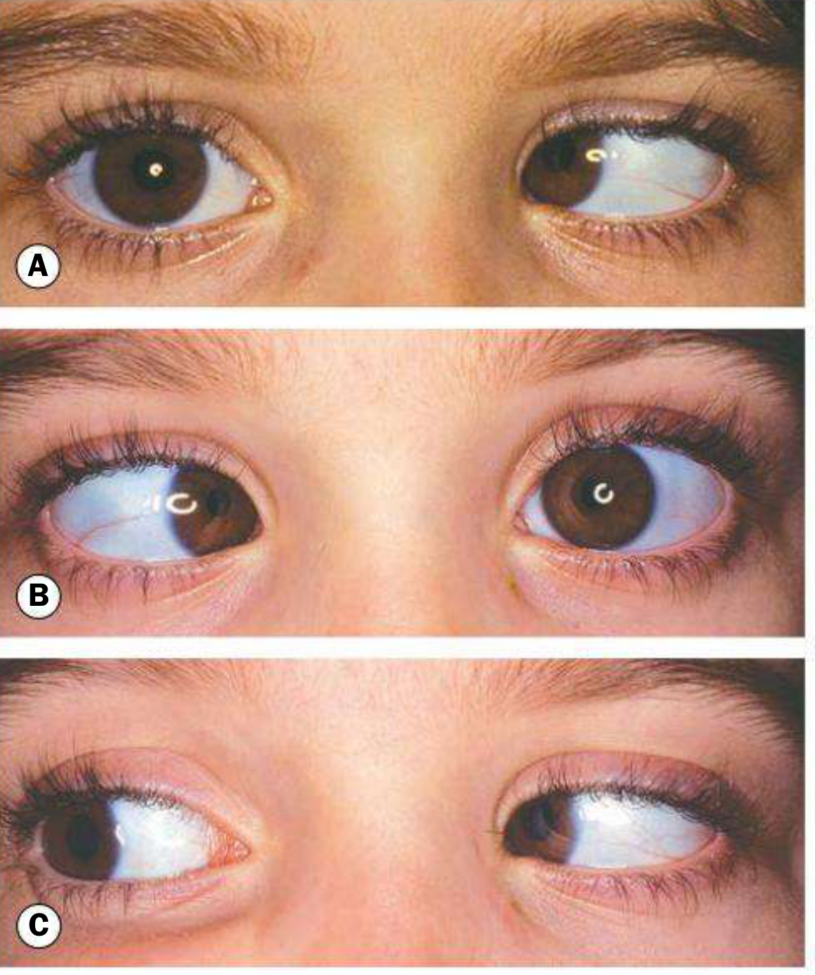
Key Bedside Signs to Confirm
| Test | Expected Finding |
|---|---|
| Cover test primary position | Positive - left esotropia manifest |
| Angle in right gaze | Maximum esotropia |
| Angle in left gaze | Minimal or absent esotropia |
| Right eye ductions | Limited abduction of right eye |
| Right eye adduction | Normal (CN III intact) |
| Primary vs Secondary deviation | Secondary (when right eye fixes) > Primary (when left eye fixes) |
| Papilledema | May be resolving post-shunt |
| Pupil | Normal bilaterally (CN III spared - no posterior communicating artery aneurysm concern) |