
Pathogenesis of thrombosis in short.
Reading File
Now let me search the medical library for thrombosis pathogenesis:
Finding Sources
Finding Sources
Robbins Basic Pathology has a dedicated thrombosis section. Let me read it:
Reading File
Reading File
Loading Image
Now let me get the core Virchow triad section and the hypercoagulability/stasis details:
Reading File
Loading Image
Reading File
I now have all the content needed. Here is the full answer:
Pathogenesis of Thrombosis
Thrombosis is the formation of a solid blood clot (thrombus) within the vascular system in a living person. The fundamental framework is Virchow's Triad, which identifies three interacting abnormalities that predispose to clot formation.
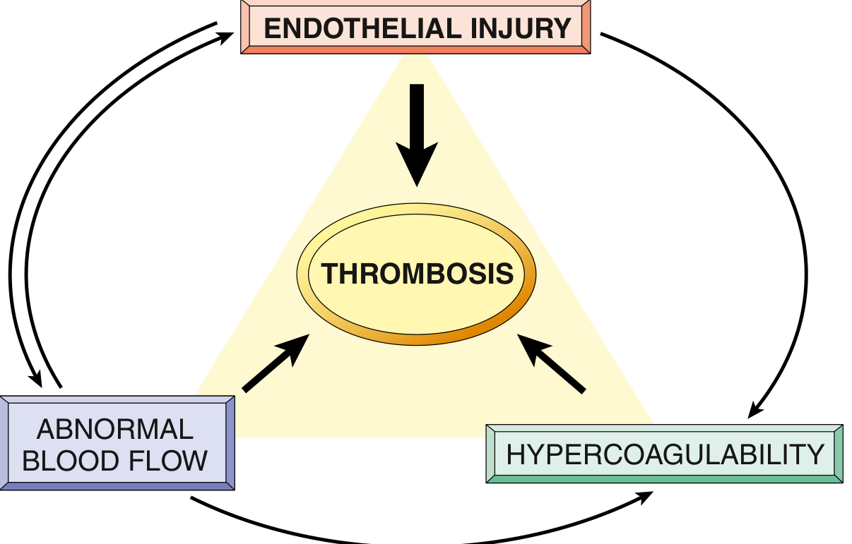
1. Endothelial Injury
Endothelial injury is the most important factor and virtually always underlies arterial and cardiac thrombosis (where high flow would otherwise prevent clotting).
- Direct injury exposes subendothelial von Willebrand factor (vWF) and tissue factor, triggering platelet adhesion and activation of the coagulation cascade.
- Endothelial activation/dysfunction (without frank injury) is triggered by: physical trauma, infectious agents, cytokines, turbulent flow, hypercholesterolemia, homocystinemia, and cigarette smoke toxins.
- Activated endothelium shifts to a prothrombotic phenotype:
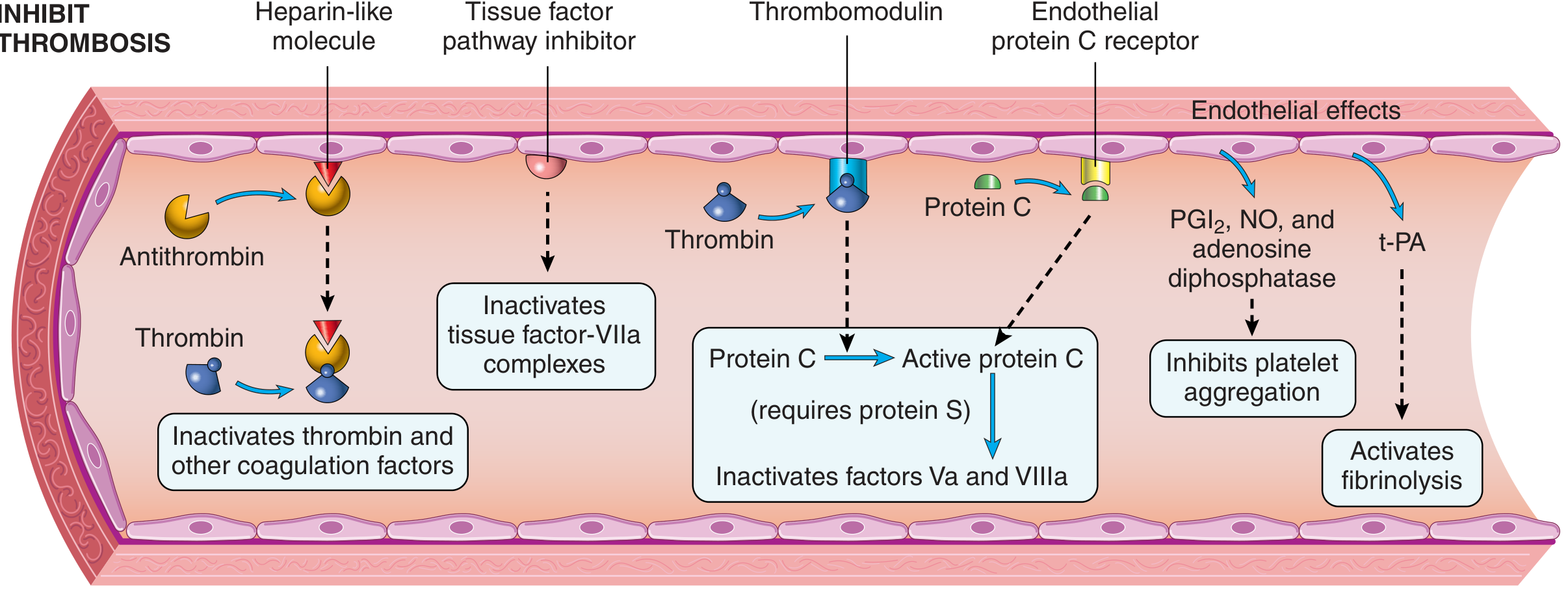
- Downregulates thrombomodulin, endothelial protein C receptor, and tissue factor pathway inhibitor (TFPI)
- Upregulates tissue factor expression
- Increases secretion of plasminogen activator inhibitors (PAI), reducing fibrinolysis
- Arterial/cardiac thrombi are platelet-rich ("white thrombi") due to the high-shear environment.
2. Abnormal Blood Flow (Stasis or Turbulence)
Turbulence contributes to arterial and cardiac thrombosis by:
- Causing endothelial injury/dysfunction
- Creating countercurrents and local pockets of stasis
Stasis is the dominant factor in venous thrombosis. It causes:
- Platelets and leukocytes to accumulate at the vessel wall (normally separated from endothelium by laminar flow)
- Slowed washout of activated clotting factors
- Impaired inflow of clotting factor inhibitors
- Activation of endothelial gene expression, promoting a procoagulant state
Clinical examples: Atrial fibrillation, mitral stenosis, ventricular aneurysm, aortic aneurysm, polycythemia vera, ulcerated atherosclerotic plaques, prolonged immobility.
3. Hypercoagulability
Abnormally high tendency of blood to clot, usually from altered coagulation factors. More important in venous thrombosis.
Primary (genetic) causes:
- Factor V Leiden mutation - renders Factor V resistant to cleavage by protein C; heterozygotes have 3-4x risk, homozygotes 25-50x risk; found in ~60% of patients with recurrent DVT
- Prothrombin G20210A mutation - 3' UTR variant causing elevated prothrombin levels; ~3x venous thrombosis risk
- Antithrombin III deficiency
- Protein C or Protein S deficiency - loss of the key anticoagulant pathway
- Elevated homocysteine (anti-phospholipid syndrome-like effects)
Secondary (acquired) causes:
- Prolonged immobility or bed rest
- Myocardial infarction, congestive heart failure
- Cancer (especially mucin-secreting adenocarcinomas - Trousseau syndrome)
- Pregnancy and the postpartum state
- Oral contraceptives/hormone replacement therapy
- Antiphospholipid antibody syndrome
- Heparin-induced thrombocytopenia (HIT)
- Disseminated intravascular coagulation (DIC)
Fate of a Thrombus
Once formed, a thrombus can undergo four outcomes:
| Outcome | Description |
|---|---|
| Propagation | Enlarges by accretion of platelets and fibrin |
| Embolization | Part/all dislodges and travels downstream |
| Dissolution | Fibrinolysis (only effective in early/fresh thrombi; older thrombi resist plasmin) |
| Organization & Recanalization | Ingrowth of endothelial cells, smooth muscle, fibroblasts; may restore partial flow |
The diagram below shows normal endothelial antithrombotic mechanisms (thrombomodulin, protein C/S, antithrombin, TFPI, PGI2/NO, t-PA) that are lost or suppressed when the endothelium is injured or activated:
- Robbins & Kumar Basic Pathology, pp. 88-91
Classification of receptor
Reading File
Receptors can be classified in multiple ways - by location, by signal transduction mechanism, or by ligand type. This is a broad pharmacology/physiology topic. Let me search the library for the best coverage.
Searching sources...
Finding Sources
Finding Sources
Finding Sources
Finding Sources
Katzung's and Guyton both look very relevant. Let me read both simultaneously:
Reading File
Reading File
Now let me get Katzung's receptor classification section and Guyton's continuation on enzyme-linked receptors:
Reading File
Reading File
Reading File
Loading Image
I have all the content needed. Here is the comprehensive answer:
Classification of Receptors
Receptors can be classified based on their location and signal transduction mechanism. The dominant pharmacological classification uses 5 major types based on how they transduce signals across the cell membrane.
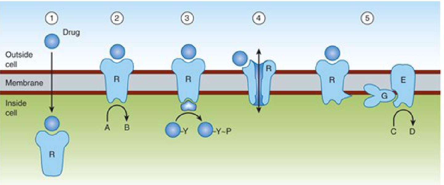
A. By Location
| Location | Receptor Type | Ligands |
|---|---|---|
| Cell membrane surface | Ion-channel, GPCR, enzyme-linked | Peptides, catecholamines, neurotransmitters |
| Cytoplasm | Steroid receptors | Corticosteroids, sex steroids, mineralocorticoids |
| Nucleus | Nuclear receptors | Thyroid hormones |
B. By Signal Transduction Mechanism (Main Classification)
Type 1 - Intracellular (Nuclear/Cytoplasmic) Receptors
- Ligand is lipid-soluble and crosses the plasma membrane directly
- Ligands: steroids (glucocorticoids, mineralocorticoids, sex hormones), vitamin D, thyroid hormone, retinoids
- Mechanism: Hormone binds receptor → receptor dissociates from hsp90 chaperone → receptor-hormone complex enters nucleus → binds DNA response elements → regulates gene transcription
- Onset of action is slow (30 min to hours) due to need for new protein synthesis
- Effects persist for hours to days even after drug is removed (slow protein turnover)
- Example: Glucocorticoid receptor, estrogen receptor, thyroid hormone receptor
Type 2 - Ligand-Gated Ion Channels (Ionotropic Receptors)
- Receptor is the ion channel
- Ligand binding directly opens or closes the channel
- Fastest signal transduction (milliseconds)
- Ions: Na⁺, K⁺, Ca²⁺, Cl⁻
- Examples:
- Nicotinic ACh receptor (Na⁺/K⁺ channel)
- GABA-A receptor (Cl⁻ channel)
- NMDA receptor (Ca²⁺/Na⁺)
- Glycine receptor (Cl⁻)
- Also called ionotropic receptors
Type 3 - G Protein-Coupled Receptors (GPCRs / Metabotropic Receptors)
- Largest family of cell-surface receptors (~1000 known)
- Have 7 transmembrane segments (heptahelical/serpentine receptors)
- Signal via heterotrimeric G proteins (α, β, γ subunits)
- Ligand binds extracellular domain → conformational change → G protein activation (GDP → GTP exchange on α subunit) → α subunit dissociates → activates/inhibits:
- Adenylyl cyclase → cAMP (via Gs or Gi)
- Phospholipase C → IP₃ + DAG (via Gq)
- Ion channels
- Gs (stimulatory) vs. Gi (inhibitory) subtypes modulate enzyme activity in opposite directions
- Signal terminated when α subunit hydrolyzes GTP → GDP; reassembles with βγ
- Examples:
- β-adrenergic receptors (Gs → ↑cAMP)
- Muscarinic M2 receptor (Gi → ↓cAMP)
- α1-adrenergic receptor (Gq → IP₃/DAG)
- All serotonin (5-HT) receptors except 5-HT3
Type 4 - Enzyme-Linked Receptors (Receptor Tyrosine Kinases and Related)
- Single transmembrane segment (single-pass, unlike GPCRs)
- Extracellular hormone-binding domain + intracellular enzyme domain
- Subtypes:
- Receptor Tyrosine Kinases (RTKs): Ligand binding causes receptor dimerization → autophosphorylation of tyrosine residues → downstream signaling cascades (Ras/MAPK, PI3K/Akt)
- Receptor Serine/Threonine Kinases: e.g., TGF-β receptor
- Receptor Guanylyl Cyclase: e.g., ANP (atrial natriuretic peptide) receptor → produces cGMP
- JAK-STAT associated receptors (cytokine receptors): lack intrinsic kinase activity; associate with JAK kinases → phosphorylate STAT proteins
- Ligands: Insulin, EGF, PDGF, VEGF, GH, leptin, prolactin, IGF-1, cytokines
Type 5 - Receptor Tyrosine Kinase-Associated Receptors
(sometimes merged with Type 4)
- Receptor binds and stimulates a separate (non-covalently attached) protein tyrosine kinase
- Does not have intrinsic enzymatic activity itself
- Example: Growth hormone receptor - associates with JAK2 kinase
Summary Table
| Type | Mechanism | Speed | Examples |
|---|---|---|---|
| Intracellular/Nuclear | Gene transcription | Hours-days | Steroid receptors, thyroid receptor |
| Ion-channel linked | Direct ion flow | Milliseconds | nAChR, GABA-A, NMDA |
| GPCR (7-TM) | Via G proteins → 2nd messengers | Seconds-minutes | β-adrenoceptors, muscarinic, opioid |
| Enzyme-linked (RTK) | Receptor kinase/cyclase | Minutes | Insulin, EGF, PDGF, ANP |
| Cytokine receptor | JAK-STAT pathway | Minutes | GH, leptin, prolactin, cytokines |
Note on Pharmacological Context
-
Arterial/cardiac receptors are targeted by drugs acting on GPCRs (e.g., β-blockers, ACE inhibitors) and ion channels (e.g., Ca²⁺ channel blockers)
-
CNS receptors are predominantly ion-channel linked (fast synaptic) and GPCR (modulatory)
-
Endocrine receptors span all classes depending on the hormone's lipid solubility
-
Katzung's Basic and Clinical Pharmacology, 16e, pp. 51-53
-
Guyton and Hall Textbook of Medical Physiology, pp. 909-910
This is a shared conversation. Sign in to Orris to start your own chat.