I now have comprehensive data from Goldman-Cecil, Harrison's, Adams & Victor's Neurology, Fishman's Pulmonary, GeneReviews, and recent PubMed reviews. Let me compile the complete structured document.---
POMPE DISEASE (Glycogen Storage Disease Type II / Acid Maltase Deficiency)
OMIM #232300 | Gene: GAA | Inheritance: Autosomal Recessive
1. INTRODUCTION
Pompe disease - also called Glycogen Storage Disease Type II (GSD II), acid alpha-glucosidase (GAA) deficiency, or acid maltase deficiency (AMD) - is a rare, progressive, autosomal recessive lysosomal storage and glycogen storage disorder. It results from deficiency of the lysosomal enzyme acid alpha-glucosidase (acid maltase, EC 3.2.1.3), encoded by the GAA gene, which is responsible for hydrolysing both alpha-1,4 and alpha-1,6 glycosidic linkages of glycogen within lysosomes.
The enzyme deficiency leads to the pathological intralysosomal accumulation of glycogen, predominantly in skeletal muscle, cardiac muscle, and smooth muscle. Pompe disease is unique among glycogen storage diseases in being the only one classified simultaneously as a lysosomal storage disease (LSD) and a glycogen storage disease.
It was first described by Johannes Cassianus Pompe in 1932 in an infant with massive cardiac glycogen accumulation. The concept of lysosomal enzyme deficiency as the basis of the disease was later enunciated by Henri-Gery Hers (1963). The late-onset form was described by Andrew Engel in 1969.
More than 300 distinct pathogenic variants in the GAA gene have been identified, and the disease spans a phenotypic continuum from a fatal neonatal/infantile form to a slowly progressive adult-onset myopathy.
- Goldman-Cecil Medicine, Chapter 391 (GSD)
- Harrison's Principles of Internal Medicine 22e, Chapter on LSDs
- Adams and Victor's Principles of Neurology, 12th ed.
2. EPIDEMIOLOGY
| Parameter | Estimated Prevalence/Incidence |
|---|
| Overall incidence (all forms) | ~1 in 40,000 live births |
| Infantile-onset Pompe disease (IOPD) | ~1 in 138,000 (higher in certain populations) |
| Late-onset Pompe disease (LOPD) | ~1 in 57,000-60,000 |
| Combined overall prevalence | ~1 in 40,000 (some sources 1 in 20,000-40,000) |
Population variation:
- Higher prevalence of IOPD in African Americans (carrier frequency ~1/14)
- Higher LOPD prevalence in the Netherlands and certain European populations
- The c.-32-13T>G (IVS1) splice-site variant is the most common LOPD-causing mutation in Caucasians
- Pseudodeficiency alleles (c.[1726A; 2065A]) are MORE common than true Pompe disease (e.g., 1/8,811 in Missouri vs. 1/40,000 for true disease), causing diagnostic challenges on newborn screening
Pompe disease is listed as one of 15 known glycogen storage disorders and is also classified under ~50 known lysosomal storage diseases. It was added to the US Recommended Uniform Screening Panel (RUSP) in 2015; by 2021, 23+ US states had implemented newborn screening.
There is a well-documented diagnostic delay of 12-13 years in LOPD from symptom onset to diagnosis due to the rarity and insidious progression of the condition.
- GeneReviews: Pompe Disease (Updated August 21, 2025) - NCBI Bookshelf NBK1261
- Sperry et al., PMC10035871
3. ETIOLOGY
Root cause: Biallelic loss-of-function pathogenic variants in the GAA gene (chromosome 17q25.3), encoding the lysosomal enzyme acid alpha-glucosidase (acid maltase).
Enzyme defect: Deficiency of acid alpha-glucosidase (GAA) - responsible for intralysosomal degradation of glycogen by cleaving both alpha-1,4 and alpha-1,6 glycosidic bonds.
Consequence: Undigested glycogen accumulates within lysosomes of all tissues but most severely in:
- Skeletal muscle
- Cardiac muscle (especially in IOPD)
- Smooth muscle
- Neurons of spinal cord and brainstem (especially IOPD)
- Liver (less prominent)
Degree of enzyme deficiency determines disease severity:
- Complete absence (<1% residual activity): Classic IOPD with cardiomyopathy
- Partial deficiency (1-10% activity): LOPD, variable severity
- Residual activity correlates inversely with disease severity
Triggers of lysosomal overload:
- All cells continuously deliver a small amount of cytoplasmic glycogen to lysosomes for degradation via autophagy (autophagic flux). GAA deficiency blocks this pathway, leading to secondary lysosomal and autophagic dysfunction - a key pathomechanism especially in LOPD where autophagy failure drives irreversible muscle damage (Do et al., PMID 38785980).
4. PHYSIOLOGY AND PATHOPHYSIOLOGY
Normal Lysosomal Glycogen Metabolism
Glycogen is synthesised in the cytoplasm. A small fraction (~1-3%) is delivered to the lysosome through a process called macroautophagy (glycophagy), where it is degraded by acid alpha-glucosidase to glucose. This lysosomal pathway serves as a quality-control mechanism for abnormal/old glycogen particles.
Pathophysiology in Pompe Disease
Step 1 - Enzyme deficiency: The GAA gene mutation produces a misfolded, unstable, or absent GAA protein. The enzyme is synthesised in the ER as a 110-kDa precursor and undergoes glycosylation (mannose-6-phosphate tags) for lysosomal targeting via the cation-independent mannose-6-phosphate receptor (CI-MPR) pathway.
Step 2 - Glycogen accumulation: Without functional GAA, glycogen accumulates within lysosomes, forming membrane-bound lysosomal vacuoles filled with monoparticulate glycogen (visible on TEM). As lysosomes become engorged, they rupture, releasing glycogen and lysosomal contents into the cytoplasm.
Step 3 - Autophagic failure: Secondary disruption of autophagic flux leads to accumulation of autophagic vacuoles (autophagolysosomes) packed with cellular debris - a key mechanism of irreversible muscle fibre damage in LOPD (Do et al. 2024, PMID 38785980). This also impairs response to ERT.
Step 4 - Cellular damage:
- Mechanical disruption of myofibrils by lysosomal swelling
- Loss of contractile proteins
- Mitochondrial dysfunction (secondary)
- In IOPD: Glycogen also accumulates in anterior horn cells, brainstem neurons, and neurons of autonomic ganglia
Organ-specific pathophysiology:
- Heart (IOPD): Glycogen replaces myofibrils -> hypertrophic cardiomyopathy -> outflow tract obstruction, arrhythmias, cardiac failure
- Skeletal muscle: Vacuolation, fibre degeneration -> progressive proximal myopathy, respiratory failure
- Diaphragm: Preferentially affected -> respiratory failure (the leading cause of death in both forms)
- Smooth muscle: Bladder/bowel dysfunction in late LOPD
- CNS (IOPD): Spinal cord and brainstem neuron involvement -> contributes to hypotonia and feeding difficulties
- Vasculature (LOPD): 60% of LOPD patients have intracranial arterial abnormalities (vertebrobasilar dolichoectasia, unruptured aneurysms) - mechanism unclear
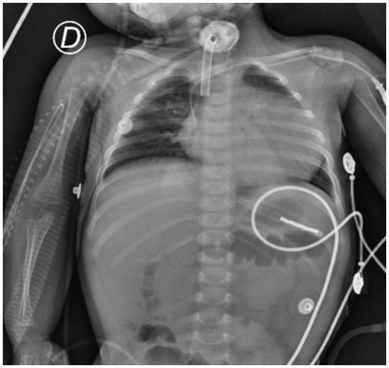
Chest X-ray of a neonate with IOPD showing severe cardiomegaly with globoid cardiac silhouette, endotracheal tube, and hepatomegaly.
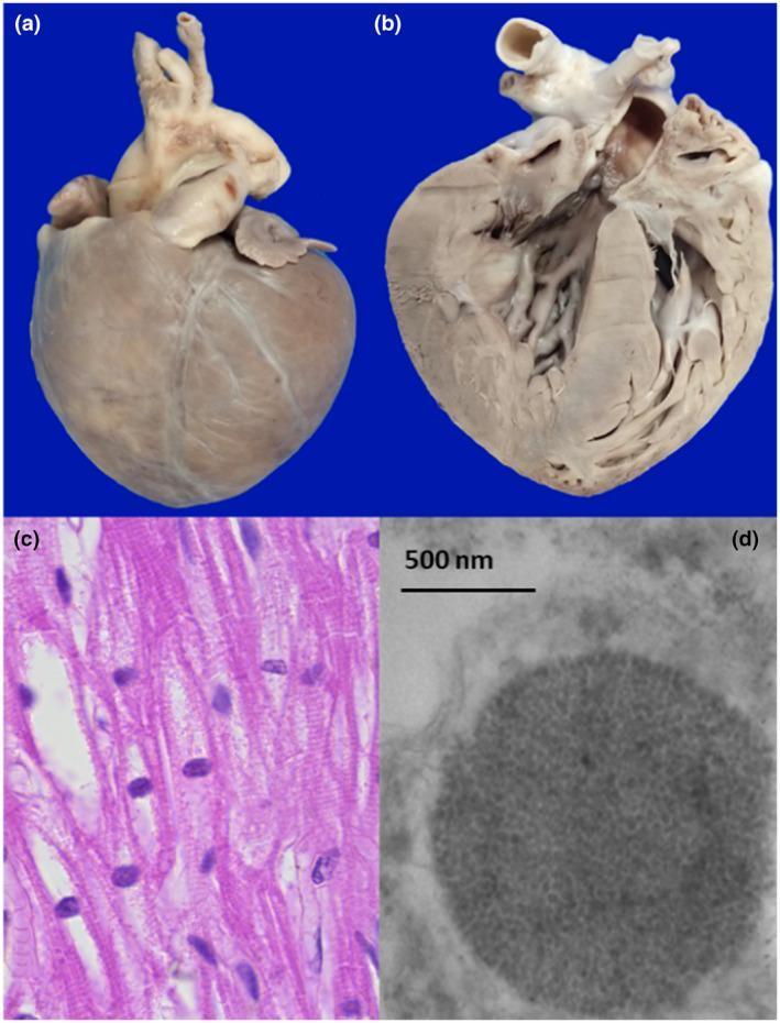
Gross specimen, H&E histology, and TEM of the heart in IOPD: globoid cardiomegaly, cytoplasmic vacuolation, and membrane-bound lysosomes packed with glycogen particles.
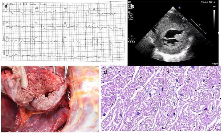
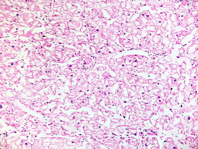
ECG showing high-voltage QRS (LVH), echocardiogram showing concentric hypertrophy, gross cardiac specimen, and PAS stain demonstrating massive intracellular glycogen accumulation in cardiomyocytes.
5. GENETICS
| Feature | Details |
|---|
| Gene | GAA (acid alpha-glucosidase) |
| Chromosomal locus | 17q25.3 |
| Inheritance | Autosomal recessive |
| OMIM | #232300 |
| Gene size | 28 kb, 20 exons |
| Number of known variants | >750 (ClinVar); >300 pathogenic/likely pathogenic |
Mutational Spectrum
- Most common mutation in LOPD (Caucasians): c.-32-13T>G (IVS1-13T>G) - a leaky splice site mutation that allows some residual GAA protein production (~1-10%), resulting in later onset and milder disease
- Most common in IOPD (non-Caucasians): Missense and nonsense mutations causing complete loss of enzyme activity (e.g., p.Arg854X, p.Trp746X)
- African Americans: Relatively high frequency of c.2560C>T (p.Arg854Cys) and deletion variants
- Dutch founder mutation: c.-32-13T>G present at high frequency in the Netherlands
Genotype-Phenotype Correlation
- Two null alleles (both alleles causing complete loss of function) -> Classic IOPD with cardiomyopathy, <1% GAA activity
- One null + one leaky allele (e.g., IVS1) -> Often IOPD without cardiomyopathy (atypical IOPD) or LOPD
- Two leaky alleles (e.g., IVS1/IVS1) -> LOPD, slowly progressive
- However, genotype does not fully predict phenotype - additional modifying factors (epigenetic, environmental, diet, exercise) influence clinical expression
Pseudodeficiency Alleles
The common pseudodeficiency haplotype (c.[1726A; 2065A]) produces low GAA enzyme activity on assays but NO clinical disease. This is more prevalent than true Pompe disease and creates false-positive NBS results. Molecular confirmation is essential.
CRIM Status (Cross-Reactive Immunological Material)
-
CRIM-positive: Patient produces some GAA protein (even if non-functional) - less likely to mount antibody response to ERT
-
CRIM-negative: No GAA protein produced (null/null genotype) - high risk of developing high-sustained antibody titres (HSAT) to ERT, drastically reducing therapeutic efficacy
-
Immunomodulation (rituximab + methotrexate before first ERT infusion) is now standard in CRIM-negative patients to prevent HSAT formation - Harrison's 22e
-
GeneReviews: Pompe Disease, NBK1261
-
Adams and Victor's Neurology, 12th ed.
6. CLINICAL MANIFESTATIONS
Pompe disease is traditionally divided into Infantile-Onset Pompe Disease (IOPD) and Late-Onset Pompe Disease (LOPD), based on age of onset, cardiomyopathy, and rate of progression. A "childhood onset" intermediate form also exists.
6a. SYMPTOMS
Infantile-Onset Pompe Disease (IOPD) - Classic Form
Age of onset: Birth to 6 months (typically 2-6 months)
| Symptom | Description |
|---|
| Profound hypotonia | "Floppy infant" - generalised severe muscle weakness from birth |
| Poor feeding | Sucking and swallowing difficulties due to oropharyngeal muscle weakness |
| Failure to thrive | Poor weight gain, feeding difficulties |
| Respiratory distress | Dyspnoea, tachypnoea, cyanosis due to diaphragm and intercostal weakness |
| Recurrent respiratory infections | Aspiration pneumonia, poor cough |
| Exercise intolerance | Inability to sustain any physical activity |
| Cardiomegaly/cardiac symptoms | Dyspnoea, cyanosis, pallor due to hypertrophic cardiomyopathy |
Late-Onset Pompe Disease (LOPD)
Age of onset: 1 year to late adulthood (childhood, adolescent, adult - all possible)
| Symptom | Description |
|---|
| Progressive proximal muscle weakness | Lower limb > upper limb; difficulty climbing stairs, rising from chairs, walking |
| Exertional dyspnoea | Early respiratory muscle involvement; orthopnoea in advanced cases |
| Sleep-disordered breathing | Nocturnal hypoventilation, morning headache, daytime somnolence |
| Recurrent respiratory infections | Reduced cough strength |
| Diaphragmatic weakness | Can be the presenting or dominant feature - may resemble ALS, nemaline myopathy, or myasthenia gravis |
| Exercise intolerance | Fatigue with exertion |
| Late LOPD | Dysphagia, dysarthria, bowel/bladder dysfunction |
| Scapular winging | Observed in some patients |
6b. SIGNS
IOPD
| Sign | Feature |
|---|
| Generalised hypotonia | Decreased tone throughout; frog-leg posture |
| Macroglossia | Enlarged tongue giving "cretinoid appearance" |
| Cardiomegaly | Markedly enlarged globoid heart on CXR |
| Hepatomegaly | Present but often not pronounced |
| Absent deep tendon reflexes | Due to muscle weakness |
| Respiratory failure signs | Tachypnoea, use of accessory muscles, paradoxical breathing |
| "Bulging" abdomen | Hepatomegaly + flaccid abdominal muscles |
| Fasciculations | Present in some - mimics Werdnig-Hoffmann disease (SMA type I) |
| Pseudo-hypertrophy | Paradoxically increased muscle bulk despite weakness (due to glycogen storage) |
LOPD
| Sign | Feature |
|---|
| Proximal muscle weakness | Limb-girdle pattern; waddling gait, Gowers' sign |
| Lumbar lordosis | Compensation for hip flexor and paraspinal weakness |
| Toe-walking | Weak ankle dorsiflexors |
| Calf pseudo-hypertrophy | Resembles Duchenne muscular dystrophy |
| Foot drop / foot-slapping gait | In advanced disease |
| Reduced FVC | Spirometry shows restrictive pattern |
| Orthopnoea | Diaphragmatic weakness - worse in supine position |
| Absent or reduced reflexes | Distal reflexes may be preserved early |
| Scapular winging | Periscapular muscle weakness |
6c. COMPLICATIONS
| System | Complication |
|---|
| Respiratory | Respiratory failure (leading cause of death), recurrent pneumonia, ventilatory failure (especially REM-related nocturnal hypoventilation), tracheostomy dependence |
| Cardiac (IOPD) | Hypertrophic cardiomyopathy with outflow tract obstruction, cardiac arrhythmias, heart failure, sudden cardiac death |
| Musculoskeletal | Contractures, scoliosis, osteoporosis (disuse), loss of ambulation |
| Neurological | Sensorineural hearing loss, autonomic dysfunction, white matter hyperintensities (on MRI brain), small fibre neuropathy, foot-slapping gait, bulbar weakness with dysarthria and oropharyngeal dysphagia |
| Vascular (LOPD) | Intracranial arterial abnormalities in ~60% of LOPD - vertebrobasilar dolichoectasia, unruptured intracranial aneurysms (risk of stroke, compression, haemorrhage) |
| Gastrointestinal | Dysphagia, aspiration, bowel dysfunction in late disease |
| Urological | Bladder dysfunction (urinary incontinence/retention) in late LOPD |
| Growth | Growth retardation in childhood-onset |
| ERT-related | Hypersensitivity/anaphylaxis to enzyme infusion, high-sustained antibody titres (HSAT) in CRIM-negative patients reducing ERT efficacy |
6d. DIFFERENTIAL DIAGNOSIS
For IOPD:
| Condition | Differentiating Feature |
|---|
| Spinal Muscular Atrophy type I (Werdnig-Hoffmann) | No cardiomegaly; SMN1 deletion; fasciculations prominent; GAA activity normal |
| Glycogen Storage Disease IIIa (Debrancher deficiency) | More hepatomegaly; cardiomegaly; hypoglycemia; normal GAA |
| GSD IV (Branching enzyme deficiency) | Hepatomegaly, hypotonia, cardiomegaly; GBE1 mutation |
| Idiopathic hypertrophic cardiomyopathy | No hypotonia; no skeletal myopathy; normal GAA |
| Endocardial fibroelastosis | No skeletal muscle disease; ECG pattern different |
| Myocarditis | Acute onset; viral history; troponin elevation; no skeletal myopathy |
| Other metabolic myopathies | Mitochondrial myopathy (ragged-red fibres); Danon disease (LAMP2 mutation, X-linked) |
| Neonatal/Infantile botulism | Descending paralysis; clostridial toxin; no cardiomegaly |
For LOPD:
| Condition | Differentiating Feature |
|---|
| Limb-girdle muscular dystrophy (LGMD) | No diaphragmatic predominance early; specific gene panel differs |
| Duchenne/Becker muscular dystrophy | X-linked; DMD deletion; very high CK; no respiratory onset |
| Polymyositis / Dermatomyositis | Inflammatory markers; skin features; biopsy shows inflammation |
| Endocrine myopathies | Thyroid/cortisol levels; reversible with hormone treatment |
| Myasthenia gravis | Fatigable weakness; acetylcholine receptor antibodies; positive Tensilon test |
| Motor neuron disease / ALS | UMN + LMN signs; EMG shows denervation; no GAA deficiency |
| Glycogen storage disease V (McArdle) | Exercise-induced cramps; myoglobinuria; no resting weakness; ischaemic forearm test |
| Nemaline myopathy | Nemaline rods on biopsy |
| GSD VI (Hers) / GSD IX | Predominantly hepatic; no significant myopathy |
- GeneReviews, NBK1261 - Differential Diagnosis Table
- Adams and Victor's Neurology, 12th ed.
- Goldman-Cecil Medicine
7. INVESTIGATIONS OF DISEASE
Biochemical / Enzymatic Investigations
| Test | Finding |
|---|
| Serum CK (Creatine Kinase) | Elevated (muscle-derived CK); may be normal in LOPD adults; aldolase also elevated |
| Serum aminotransferases (AST, ALT) | Mildly elevated (muscle origin, not hepatic) |
| Urinary Hex4 (glucotetrasaccharide, Glc4) | Elevated - biomarker of glycogen accumulation; useful for monitoring treatment response |
| Serum LDH | Often elevated |
Enzyme Activity Assays (Gold Standard Biochemical Test)
| Method | Details |
|---|
| Dried Blood Spot (DBS) GAA assay | First-line screening test; detects GAA enzyme activity from a blood spot; used for newborn screening; cannot reliably differentiate IOPD vs LOPD |
| Lymphocyte/Leukocyte GAA assay | More specific; low false-positive rate; diagnostic cutoff <1% of normal |
| Fibroblast GAA assay | Gold standard for confirmation; most reliable |
| Muscle tissue GAA assay | Definitive but invasive |
⚠️ Pseudodeficiency pitfall: Homozygous c.[1726A; 2065A] haplotype gives low enzyme activity on assays but NO clinical disease. Always confirm low DBS GAA with molecular genetic testing.
Molecular Genetic Testing
| Method | Details |
|---|
| GAA gene sequencing | Identifies biallelic pathogenic variants; required for definitive diagnosis |
| Multigene panel | Useful when clinical features overlap with other myopathies |
| Exome/Genome sequencing | When targeted testing is inconclusive |
Diagnostic criteria (GeneReviews): Pompe disease is confirmed by:
- Deficient GAA activity in leukocytes OR fibroblasts, AND/OR
- Identification of biallelic pathogenic GAA variants on molecular genetic testing
Muscle Biopsy
| Finding | Details |
|---|
| PAS stain | PAS-positive, diastase-digestible vacuoles (glycogen-laden) |
| Acid phosphatase stain | Intensely positive vacuoles (lysosomal marker) |
| H&E | Vacuolated fibres with cytoplasmic clearing; myofibril disruption |
| Electron microscopy | Membrane-bound lysosomes packed with monoparticulate glycogen; free glycogen in cytoplasm; autophagic vacuoles |
| Fibre type | Glycogen accumulation more pronounced in type 1 fibres |
| Late LOPD | May show only non-specific changes; biopsy can be normal |
- Adams and Victor's Neurology 12th ed.
- Goldman-Cecil Medicine, Ch. 391
8. INVESTIGATIONS AT DIAGNOSIS
Cardiac Assessment
| Investigation | Purpose |
|---|
| ECG | High-voltage QRS (short PR interval + delta-like waves), LVH pattern, arrhythmias - classic in IOPD |
| Echocardiography | Hypertrophic cardiomyopathy, outflow tract obstruction, wall thickness, EF - mandatory in IOPD; less common in LOPD |
| 24h Holter monitor | Arrhythmia surveillance |
Respiratory Assessment
| Investigation | Purpose |
|---|
| Spirometry (FVC, FEV1) | Restrictive pattern; FVC in upright and supine (>20% drop in supine = diaphragm weakness) |
| Arterial blood gas / Pulse oximetry | Detect hypercapnia, hypoxia |
| Sleep study (polysomnography) | Nocturnal hypoventilation - may be the first respiratory manifestation |
| Sniff nasal inspiratory pressure / MIP / MEP | Respiratory muscle strength |
| Peak cough flow | Cough efficacy |
Neuromuscular Assessment
| Investigation | Purpose |
|---|
| EMG / Nerve conduction study | Myopathic changes (short-duration, small-amplitude MUPs); fibrillation potentials, bizarre high-frequency discharges, pseudomyotonic discharges, positive sharp waves |
| Muscle MRI | Distribution of muscle involvement; L-spine paraspinals, posterior thigh muscles typically affected early |
| 6-Minute Walk Test (6MWT) | Functional assessment and treatment monitoring baseline |
| Timed Up and Go test (TUG) | Functional mobility baseline |
Hearing Assessment
- Audiometry - sensorineural hearing loss documented in IOPD
Brain/Spinal Cord Imaging
- MRI Brain - white matter hyperintensities reported in ERT-treated IOPD survivors; intracranial aneurysm screening in LOPD (MRA/CTA)
Baseline Laboratory Work-up
- CK, ALT, AST, LDH, urinary Hex4
- CRIM status determination (Western blot or genotype-based)
- Pre-ERT antibody baseline
Newborn Screening (NBS)
- DBS GAA enzyme activity -> if low: confirmatory GAA sequencing + CK + echocardiography
- A single abnormal NBS is NOT sufficient for diagnosis - requires confirmation
9. TREATMENT
Treatment of Pompe disease requires a multidisciplinary team including metabolic specialists, cardiologists (IOPD), pulmonologists, neurologists, physiotherapists, speech therapists, occupational therapists, dietitians, and genetic counsellors.
9a. Enzyme Replacement Therapy (ERT) - Disease-Modifying
ERT is the only approved disease-modifying therapy and the standard of care.
First-Generation ERT
| Drug | Alglucosidase alfa (Myozyme®/Lumizyme®) |
|---|
| Mechanism | Recombinant human acid alpha-glucosidase; taken up by CI-MPR pathway into lysosomes |
| Dose | 20 mg/kg IV every 2 weeks |
| Indication | All ages, all forms of Pompe disease |
| Efficacy - IOPD | Prolongs survival; cardiac function improves consistently; motor function improvement variable; respiratory improvement especially if started before 6 months |
| Efficacy - LOPD | FVC stabilisation; improved 6MWT; modest benefit overall; very advanced disease has significant irreversible components |
| Limitation | Antibody formation (HSAT) can reduce efficacy; insufficient M6P tags limit cellular uptake; limited penetration into fibrotic muscle and CNS |
Second-Generation ERT (Improved Uptake)
| Drug | Avalglucosidase alfa (Nexviazyme®) | Cipaglucosidase alfa + Miglustat (Pombiliti® + Opfolda®) |
|---|
| Mechanism | Higher mannose-6-phosphate content -> enhanced lysosomal targeting | Cipaglucosidase alfa: enhanced M6P tags; Miglustat: pharmacological chaperone stabilising the enzyme in circulation |
| Approval | FDA-approved (2021) | FDA-approved (2023) |
| Evidence | COMET trial (phase 3) vs. alglucosidase: significant improvement in 6MWT and FVC in naive LOPD patients | PROPEL trial: non-inferior to alglucosidase; improved in some endpoints |
| Dose | 20 mg/kg IV every 2 weeks | Cipaglucosidase 20 mg/kg IV q2w + miglustat 260 mg oral 1h before infusion |
- Goldman-Cecil Medicine: "Avalglucosidase alfa, a second-generation recombinant acid α-glucosidase replacement therapy with a significantly higher number of mannose-6-phosphate tags, can provide significant benefit over alglucosidase alfa."
- Harrison's 22e
CRIM-Negative Patients - Immunomodulation Protocol
- Pre-ERT immunomodulation: Rituximab + Methotrexate given before first ERT infusion
- This prevents development of high-sustained antibody titres (HSAT) which otherwise abolish ERT efficacy
- Has greatly improved therapeutic response and long-term survival in CRIM-negative IOPD
9b. Respiratory Support
- NIV (non-invasive ventilation): BiPAP or CPAP for nocturnal hypoventilation - often the first intervention needed
- Invasive ventilation: Tracheostomy in advanced respiratory failure
- Cough-assist devices / mechanical in-exsufflation (MI-E): To improve cough efficacy and prevent atelectasis
- Chest physiotherapy and airway clearance techniques
- Avoidance of sedatives and respiratory depressants
9c. Physical and Rehabilitative Therapy
- Regular physiotherapy to maintain strength, flexibility, and prevent contractures
- Aquatic therapy is well tolerated
- High-protein diet may be beneficial (preserves muscle mass; Umpleby et al. data)
- Low-carbohydrate, high-protein diet has been used, particularly in adults
- Occupational therapy: Energy conservation, assistive devices
- Speech therapy: For dysphagia, dysarthria - swallowing assessment and exercises
9d. Cardiac Management (IOPD)
- Medications with caution: Beta-blockers and digoxin may worsen outflow tract obstruction in HCM - use with extreme caution
- Anti-arrhythmic therapy as indicated
- ERT reverses cardiomyopathy in most IOPD patients who start early
9e. Surgical / Orthopaedic Interventions
- Scoliosis surgery if progressive
- Gastrostomy (PEG) for feeding in IOPD with feeding failure
- Hearing aids for sensorineural hearing loss
9f. Emerging Therapies
| Approach | Status |
|---|
| Gene therapy (AAV-based): Liver-directed or muscle-directed delivery of GAA transgene | Multiple clinical trials ongoing (as of 2025); liver-directed approach most advanced; aims to provide one-time treatment |
| Haematopoietic stem cell gene therapy (HSCGT): Ex vivo correction of HSCs and reinfusion | Early trials |
| Substrate reduction therapy | Theoretical; less explored in Pompe |
| Pharmacological chaperones | Miglustat used as co-therapy (see Pombiliti+Opfolda above) |
- George KA et al., "Pompe disease: Unmet needs and emerging therapies," Mol Genet Metab 2024 (PMID 39418752)
- Colella P, "Advances in Pompe Disease Treatment: From Enzyme Replacement to Gene Therapy," Mol Diagn Ther 2024 (PMID 39134822)
- Corsini A, "Improving the treatment of Pompe disease with enzyme replacement therapy," Expert Opin Pharmacother 2025 (PMID 40237692)
10. COUNSELLING
Genetic Counselling
Inheritance pattern: Autosomal recessive. Parents of an affected child are obligate heterozygous carriers.
Recurrence risk:
- Each subsequent sibling: 25% risk of being affected, 50% carrier, 25% unaffected non-carrier
- Affected individual's children: All will be obligate carriers (unless partner is also a carrier)
- If partner is a carrier: Each child has 50% affected, 50% carrier risk
Key counselling points:
- Cascade screening: Offer molecular genetic testing (GAA variant analysis) to all first-degree relatives once pathogenic variants identified in proband
- Prenatal testing: Chorionic villus sampling (CVS) at 10-12 weeks or amniocentesis at 15-18 weeks - enzyme activity and/or molecular testing of GAA
- Preimplantation Genetic Testing (PGT-M): Available for couples who are both carriers and wish to avoid affected pregnancy
- CRIM status counselling: Explain implications for ERT antibody response and need for pre-ERT immunomodulation in CRIM-negative patients
Disease Counselling
-
Explain the spectrum of disease - IOPD is life-threatening within the first year untreated; LOPD is slowly progressive but causes significant disability
-
Address prognosis with ERT: Cardiac disease reverses in IOPD; muscle and respiratory involvement may stabilise or improve but may not fully reverse, especially in advanced disease
-
Discuss disease monitoring needs (lifelong follow-up)
-
Address psychological burden - grief, caregiver stress, impact on schooling/employment
-
Connect with patient advocacy groups (e.g., Acid Maltase Deficiency Association - AMDA; International Pompe Association - IPA)
-
Address newborn screening for subsequent pregnancies
-
Discuss career and lifestyle modifications - activity planning, avoiding activities with fall risk, respiratory precautions during anaesthesia (avoid succinylcholine, careful use of sedatives)
-
GeneReviews Pompe Disease, NBK1261
11. ROUTINE FOLLOW-UP
Follow-up should be at a metabolic/neuromuscular disease specialist centre with a multidisciplinary team. Frequency: every 3-6 months in infants on ERT; every 6-12 months for stable LOPD patients.
Monitoring Schedule (Based on MetabERN 2024 CPR, GeneReviews, and Practice Guidelines)
| Domain | Assessment | Frequency |
|---|
| Enzyme Replacement Therapy | Infusion reactions, antibody titres (GAA IgG), HSAT monitoring | Each infusion; titres every 3-6 months |
| Biomarkers | Serum CK, LDH, AST/ALT; urine Hex4 (Glc4) | Every 6 months |
| Respiratory | Spirometry (FVC upright + supine), MIP/MEP, peak cough flow | Every 6-12 months; more frequently if declining |
| Sleep | Polysomnography or overnight oximetry | Annually or if symptoms |
| Cardiac (IOPD) | ECG, echocardiography | Every 6-12 months; more frequently if abnormal |
| Neuromuscular | 6-Minute Walk Test, timed tests (TUG, 10m walk), manual muscle testing | Every 6-12 months |
| Swallowing | Speech therapy assessment, videofluoroscopy if swallowing issues | As needed; annually in advanced disease |
| Hearing | Audiometry | Annually in IOPD |
| Neurological | Neurological exam; MRI brain if white matter changes; MRA/CTA for intracranial aneurysm in LOPD | Every 1-2 years; lower threshold if symptomatic |
| Musculoskeletal | Scoliosis surveillance, contractures, bone density (DXA) | Annually |
| Quality of life / Mental health | Standardised QoL questionnaires; psychological assessment | Annually |
| Nutrition | Dietitian review, body composition | Annually |
| Genetics | Family cascade testing; reproductive counselling | As needed |
Treatment Targets
-
FVC maintained above 50% predicted (ventilatory support threshold)
-
Motor function stabilisation or improvement
-
Urinary Hex4 within or near normal range
-
Antibody titres below HSAT threshold
-
Cardiac dimensions normalising on echo (IOPD)
-
Parenti G et al., MetabERN Clinical Pathway Recommendations for Pompe disease, Orphanet J Rare Dis 2024 (PMID 39482698)
-
GeneReviews: Pompe Disease, NBK1261 (Updated August 21, 2025)
12. APPROACH TO DISEASE - DIAGNOSTIC AND MANAGEMENT FLOWCHART
                          CLINICAL SUSPICION FOR POMPE DISEASE
                                         |
           _____________________________|______________________________
           |                                                          |
    INFANT (< 12 months)                              CHILD/ADULT (≥ 1 year)
    - Floppy infant (hypotonia)                       - Proximal limb weakness
    - Cardiomegaly (on CXR/Echo)                      - Respiratory insufficiency
    - Poor feeding, macroglossia                       - Elevated CK, diaphragm weakness
    - Elevated CK                                      - Sleep-disordered breathing
           |                                                          |
           v                                                          v
    ┌─────────────────────────────────────────────────────────────────────────┐
    │                  STEP 1: INITIAL SCREENING                              │
    │  Dried Blood Spot (DBS) GAA Enzyme Activity Assay                       │
    │  + Serum CK, LDH, urine Hex4                                            │
    │  + ECG + Echocardiogram (IOPD)                                          │
    └─────────────────────────────────────────────────────────────────────────┘
                         |
          _______________|________________
          |                              |
    GAA ACTIVITY LOW               GAA ACTIVITY NORMAL
    (or out-of-range NBS)          → Consider alternative diagnoses
          |
          v
    ┌─────────────────────────────────────────────────────────────┐
    │         STEP 2: CONFIRMATORY TESTING                        │
    │  1. GAA molecular genetic testing (biallelic GAA variants)  │
    │  2. Leukocyte / fibroblast GAA enzyme assay                 │
    │  3. Muscle biopsy if needed (PAS, acid phosphatase, EM)     │
    │  [Rule out pseudodeficiency allele c.[1726A;2065A]]         │
    └─────────────────────────────────────────────────────────────┘
                         |
          _______________|________________
          |                              |
   CONFIRMED POMPE DISEASE         PSEUDODEFICIENCY / CARRIER
                 |                  → No treatment; family counselling
                 v
    ┌─────────────────────────────────────────────────────────────┐
    │              STEP 3: FULL BASELINE EVALUATION               │
    │  - CRIM status (Western blot / genotype)                    │
    │  - Echo, ECG, Holter                                        │
    │  - Spirometry (upright + supine), sleep study               │
    │  - 6MWT, muscle MRI, neurological exam                      │
    │  - Audiometry (IOPD), baseline antibody titres              │
    │  - Genetic counselling; family cascade testing              │
    └─────────────────────────────────────────────────────────────┘
                         |
                         v
    ┌─────────────────────────────────────────────────────────────┐
    │                   STEP 4: TREATMENT                         │
    │                                                             │
    │  CRIM-NEGATIVE IOPD:                                        │
    │   → Rituximab + Methotrexate FIRST (immunomodulation)       │
    │   → Then: Avalglucosidase alfa 20 mg/kg IV q2w              │
    │                                                             │
    │  CRIM-POSITIVE IOPD / LOPD:                                 │
    │   → Avalglucosidase alfa 20 mg/kg IV q2w (1st choice)       │
    │     OR Alglucosidase alfa 20 mg/kg IV q2w                   │
    │     OR Cipaglucosidase + Miglustat (oral chaperone combo)   │
    │                                                             │
    │  SUPPORTIVE:                                                │
    │   → NIV / invasive ventilation as needed                    │
    │   → Physiotherapy, speech therapy, occupational therapy     │
    │   → High-protein diet; cough assist devices                 │
    │   → Cardiac management, hearing aids                        │
    └─────────────────────────────────────────────────────────────┘
                         |
                         v
    ┌─────────────────────────────────────────────────────────────┐
    │              STEP 5: ROUTINE FOLLOW-UP                      │
    │  Every 3-6 months (IOPD) / 6-12 months (LOPD)              │
    │  Monitor: CK, Hex4, spirometry, 6MWT, echo, ERT titres     │
    │  Adjust ERT; escalate respiratory support as needed         │
    │  Genetic counselling; reproductive planning support         │
    └─────────────────────────────────────────────────────────────┘
13. REFERENCES
Textbooks
-
Goldman L, Schafer AI (eds). Goldman-Cecil Medicine, 27th ed. Philadelphia: Elsevier, 2024. Chapter 391 (Glycogen Storage Diseases) and Chapter 192 (Lysosomal Storage Diseases).
-
Loscalzo J, Fauci A, Kasper D, et al. (eds). Harrison's Principles of Internal Medicine, 22nd ed. New York: McGraw Hill, 2025. Chapter on Lysosomal Storage Diseases and Glycogen Storage Disorders.
-
Ropper AH, Samuels MA, Klein JP, Prasad S. Adams and Victor's Principles of Neurology, 12th ed. New York: McGraw-Hill, 2023. Chapter 45 (Acid Maltase Deficiency / GSD II).
-
Fishman A, Elias J, Fishman J, et al. (eds). Fishman's Pulmonary Diseases and Disorders, 5th ed. New York: McGraw-Hill. Section on Metabolic Myopathies (Acid Maltase Deficiency/Pompe Disease).
-
Emery's Elements of Medical Genetics and Genomics, 16th ed. Edinburgh: Elsevier, 2022. Chapter on Pompe Disease / GSD II.
Online Databases
-
OMIM #232300. Pompe Disease; Glycogen Storage Disease II. Johns Hopkins University. Available at:
https://www.omim.org/entry/232300
-
Sperry E, Leslie N, Berry L, Pena L. Pompe Disease. In: Adam MP, et al. (eds).
GeneReviews®. Seattle: University of Washington; Initial posting August 31, 2007;
Last updated August 21, 2025. Available at:
https://www.ncbi.nlm.nih.gov/books/NBK1261
Journal Articles
-
Labella B, Cotti Piccinelli S, Risi B, et al. A Comprehensive Update on Late-Onset Pompe Disease. Biomolecules. 2023 Aug 22. [PMID: 37759679]
-
Do H, Meena NK, Raben N. Failure of Autophagy in Pompe Disease. Biomolecules. 2024 May 13. [PMID: 38785980]
-
George KA, Anding AL, van der Flier A, et al. Pompe disease: Unmet needs and emerging therapies. Mol Genet Metab. 2024 Nov. [PMID: 39418752]
-
Colella P. Advances in Pompe Disease Treatment: From Enzyme Replacement to Gene Therapy. Mol Diagn Ther. 2024 Nov. [PMID: 39134822]
-
Parenti G, Fecarotta S, Alagia M, et al. The European reference network for metabolic diseases (MetabERN) clinical pathway recommendations for Pompe disease. Orphanet J Rare Dis. 2024 Nov 1. [PMID: 39482698]
-
Corsini A. Improving the treatment of Pompe disease with enzyme replacement therapy: current strategies and clinical evidence. Expert Opin Pharmacother. 2025 May. [PMID: 40237692]
-
Sperry E, Leslie N, et al. Clinical features of Pompe disease. Acta Myologica. 2013. [PMC3866902]
-
Diaz-Manera J, Kishnani PS, Kushlaf H, et al. Safety and efficacy of avalglucosidase alfa versus alglucosidase alfa in patients with late-onset Pompe disease (COMET): a phase 3, randomised, multicentre trial. Lancet Neurol. 2021;20:1012-1026.
-
Schoser B, Roberts M, Byrne BJ, et al. Safety and efficacy of cipaglucosidase alfa plus miglustat versus alglucosidase alfa plus placebo in late-onset Pompe disease (PROPEL). Lancet Neurol. 2021;20:1027-1037.
Key take-home points:
- Pompe disease = GAA deficiency -> lysosomal glycogen accumulation in muscle/heart
- IOPD: fatal in <1 year untreated (cardiomyopathy + hypotonia); LOPD: slow proximal myopathy + diaphragm weakness
- Diagnosis by DBS GAA activity (screen) -> confirmed by leukocyte/fibroblast enzyme assay + GAA biallelic variants
- Beware pseudodeficiency alleles - always confirm with molecular testing
- ERT (avalglucosidase alfa preferred) is standard of care; pre-ERT immunomodulation essential in CRIM-negative patients
- Multidisciplinary lifelong follow-up: respiratory, cardiac, motor, swallowing, hearing, genetic counselling