
Reddish blisters on lower limb with pedal edema with pain . What would be the diagnosis
Reading File
The clinical picture of reddish/hemorrhagic blisters on the lower limb with pedal edema and pain is highly suggestive of acute lower limb cellulitis with bullae, possibly erysipelas, or necrotizing fasciitis in severe cases. Let me search the medical library for a thorough answer.
Searching sources...
Finding Sources
Finding Sources
Searching the Web
reddish blisters lower limb pedal edema pain diagnosis differential
Finding Sources
Finding Sources
Good results. Let me now read the relevant sections from Andrews' Diseases of the Skin and Tintinalli's for detailed clinical information.
Reading File
Reading File
Excellent - I now have very detailed and authoritative information. Let me also fetch the erysipelas image and check for necrotizing fasciitis content.
Loading Image
Finding Sources
Reading File
Reading File
I now have comprehensive information. Let me compile the full answer.
Diagnosis: Bullous Erysipelas (Most Likely) / Bullous Cellulitis
The clinical triad of reddish/hemorrhagic blisters on the lower limb + pedal edema + pain is the classic presentation of bullous erysipelas or bullous cellulitis, both acute bacterial skin infections of the lower extremity.
Most Likely Diagnosis: Erysipelas with Bullae
Erysipelas is an acute infection of the superficial dermal lymphatics, most commonly caused by beta-hemolytic Group A Streptococcus (Streptococcus pyogenes). On the lower limbs, edema and bullous lesions are prominent features in many patients - exactly matching this presentation.
Andrews' Diseases of the Skin, p. 308
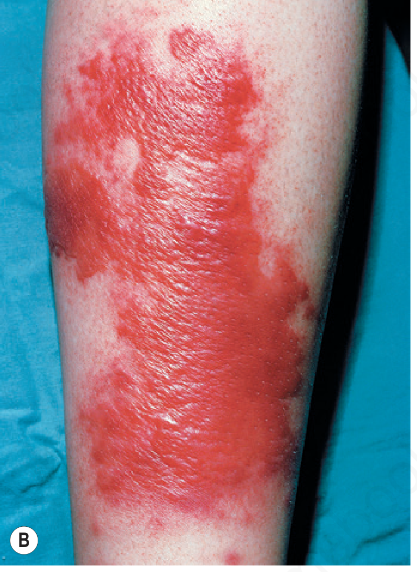
Key clinical features of erysipelas:
| Feature | Description |
|---|---|
| Onset | Abrupt, with prodrome of fever, chills, malaise, headache |
| Skin lesion | Bright red, hot, swollen, raised/indurated sharply demarcated border |
| Blisters | Vesicles or bullae containing seropurulent fluid (more common on legs) |
| Edema | Prominent, especially on legs |
| Pain | Severe, burning sensation |
| Systemic signs | High fever, leukocytosis (WBC >20,000), malaise |
Bullous erysipelas represents a more severe form - it can reflect synergy between beta-hemolytic streptococci and MRSA and is treated with broader coverage. (Tintinalli's Emergency Medicine, p. 1048)
Differential Diagnosis
| Condition | Distinguishing Features |
|---|---|
| Bullous erysipelas (most likely) | Sharply raised demarcated border, intense redness, bullae filled with seropurulent fluid, severe systemic symptoms |
| Bullous cellulitis | Deeper infection (dermis/subcutaneous), less distinct border, "peau d'orange" skin, more gradual onset |
| Necrotizing fasciitis | Pain out of proportion to findings, blisters become serosanguineous then purple/gangrenous by day 4-5, skin anesthesia - surgical emergency |
| DVT + secondary changes | Unilateral swelling, no fever, positive Doppler; blistering can occur with severe venous congestion |
| Acute lipodermatosclerosis | Chronic venous disease, painful red-brown induration, no fever |
| Contact dermatitis | No fever, prominent pruritus not pain, no systemic signs |
Red Flag - Rule Out Necrotizing Fasciitis
If there is:
- Pain out of proportion to external findings
- Skin anesthesia (numbness over affected area)
- Rapid spread despite antibiotics
- Serosanguineous (blood-tinged) blister fluid turning purple/dusky
- Systemic sepsis signs
Consider necrotizing fasciitis urgently - this requires emergency surgical debridement; mortality can reach 20% even with optimal care. MRI is the most definitive non-surgical confirmatory test. (Andrews' Diseases of the Skin, p. 309)
Predisposing Factors to Ask About
- Tinea pedis (toe web fissure - common portal of entry)
- Lymphedema, chronic leg ulcers
- Venous insufficiency
- Obesity, diabetes, immunosuppression
- Prior episodes of cellulitis/erysipelas
Management Outline
Erysipelas/Non-purulent cellulitis:
- First-line: Systemic penicillin (IV benzylpenicillin for severe; oral phenoxymethylpenicillin for mild-moderate)
- If MRSA suspected (purulent, bullous): trimethoprim-sulfamethoxazole, clindamycin, or doxycycline (oral); vancomycin IV for severe cases
- Limb elevation to reduce edema
- Mark the border with an indelible pen to monitor spread
Investigations:
- CBC (leukocytosis expected), CRP, blood cultures
- Wound/blister swab culture
- LFTs, RFTs, blood glucose (assess comorbidities)
- Consider Doppler USS to exclude DVT if response is slow
- MRI if necrotizing fasciitis is suspected
The IDSA guidelines on skin and soft tissue infections provide the most current treatment algorithms.
This is a shared conversation. Sign in to Orris to start your own chat.