
Functional areas of cerberal cortex
Reading File
The cerebral cortex is divided into functional areas including the primary motor cortex (precentral gyrus), primary somatosensory cortex (postcentral gyrus), visual cortex (occipital lobe), auditory cortex (temporal lobe), and association areas for higher cognitive functions.
Searching sources...
Finding Sources
Finding Sources
Good results. Let me read the dedicated "Review of Main Functional Areas" section from Neuroanatomy through Clinical Cases, plus the Guyton physiology content and the Neuroanatomy motor/sensory section in parallel.
Reading File
Reading File
Reading File
Now let me read the dedicated "Review of Main Functional Areas" section from Neuroanatomy through Clinical Cases, and also get the sensory/association cortex content from Guyton.
Reading File
Reading File
Now let me get the sensory cortex, visual cortex, auditory cortex, and association cortex content from Guyton, and also fetch the key diagram image.
Finding Sources
Loading Image
Loading Image
Excellent! I have very rich content and great images. Now let me get the somatosensory cortex, association areas, prefrontal, and language areas content from Guyton.
Reading File
Finding Sources
Now let me read the association areas, prefrontal, Wernicke, and auditory cortex content in parallel.
Reading File
Reading File
I now have comprehensive content from the textbooks. Let me also fetch the functional areas diagram from Guyton (Fig 58.3 and 58.4).
Loading Image
Reading File
I now have everything I need for a comprehensive answer. Let me compile it.
Functional Areas of the Cerebral Cortex
The cerebral cortex is only 2-5 mm thick but contains over 80 billion neurons organized into six layers. Its surface area is approximately 0.25 m². Functional areas are classically mapped using Brodmann areas (52 distinct cytoarchitectonic regions) and are broadly grouped into primary motor, premotor, primary sensory, and association cortices.
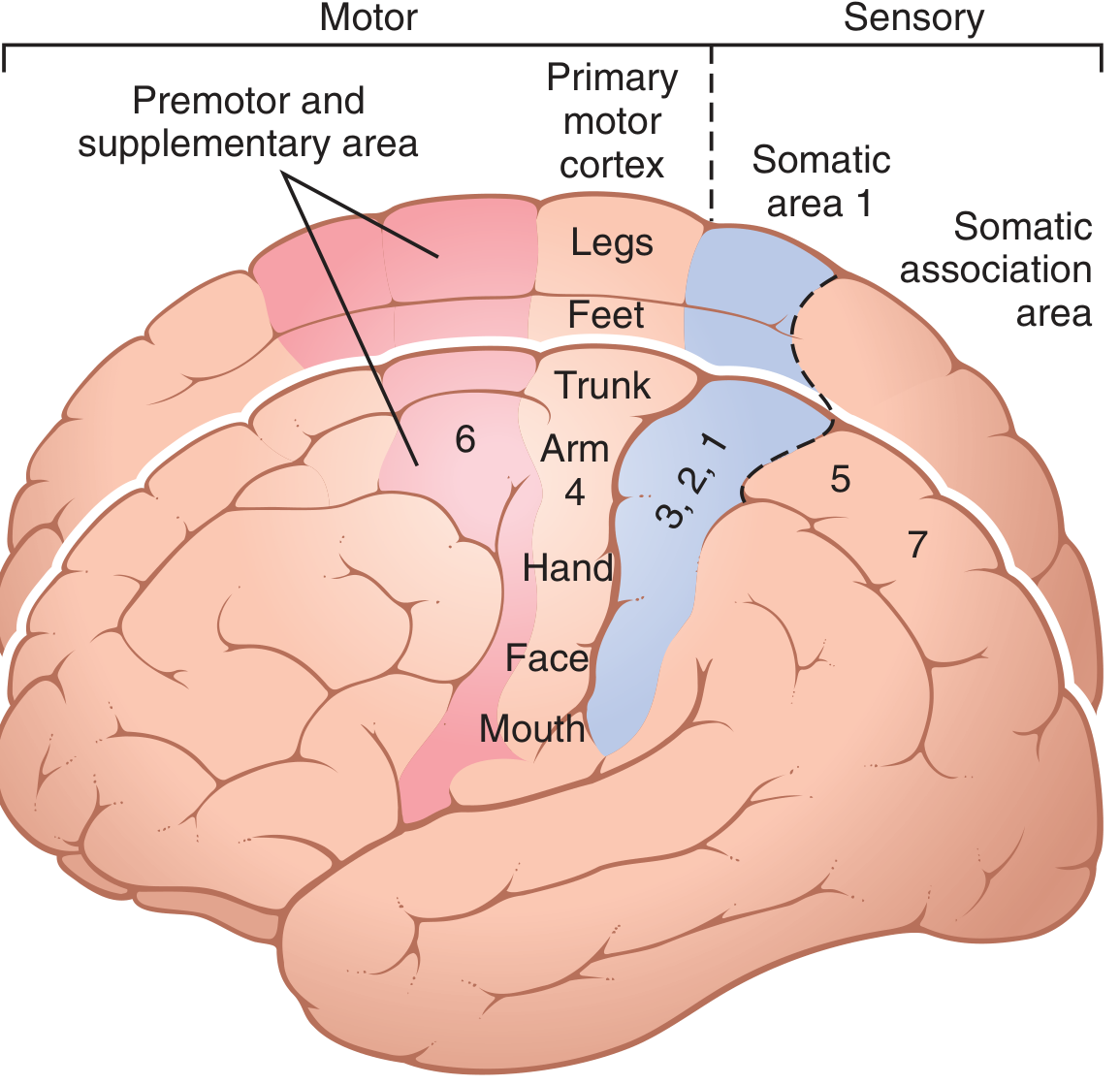
Diagram: Motor and Sensory Functional Areas
(Guyton & Hall, Fig. 56.1 - Motor and somatosensory areas with Brodmann numbers)
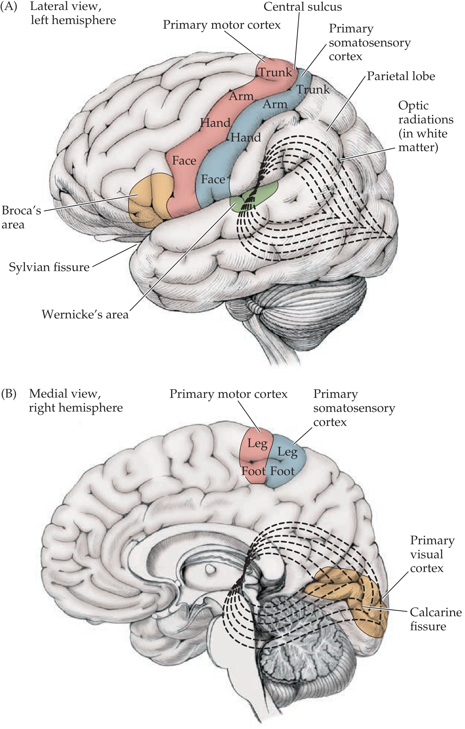
(Neuroanatomy through Clinical Cases Fig. 10.1 - Lateral and medial views showing key functional areas)
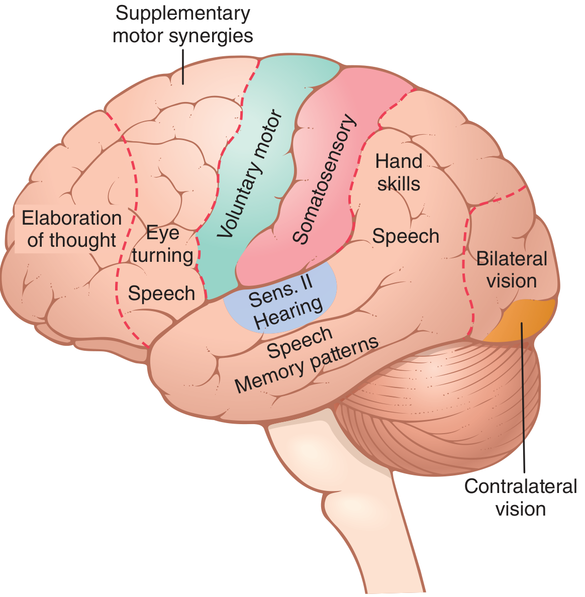
(Guyton & Hall, Fig. 58.3 - Functional areas as mapped by electrical stimulation)
I. Motor Areas (Anterior to the Central Sulcus)
1. Primary Motor Cortex (Brodmann Area 4)
- Located on the precentral gyrus, immediately anterior to the central sulcus
- Contains the motor homunculus - a somatotopic map of the body where face and hand areas are on the lateral convexities and the leg area dips into the interhemispheric fissure
- More than half the primary motor cortex is devoted to controlling the hands and muscles of speech
- Stimulation of a single neuron usually causes contraction of a group of muscles ("a specific movement rather than one specific muscle")
- Output travels via the corticospinal (pyramidal) tract to the spinal cord
2. Premotor Area (Brodmann Area 6)
- Lies 1-3 cm anterior to the primary motor cortex
- Generates complex "patterns" of movement - e.g., positioning the shoulder and arm before hand use
- Contains mirror neurons (active both when performing a task and when observing someone else perform it)
- The anterior part creates a "motor image" of the total movement; the posterior part executes it sequentially, sending signals either directly to the primary motor cortex or via the basal ganglia-thalamic circuit
3. Supplementary Motor Area (also Brodmann Area 6)
- Located on the medial surface, just above the premotor area, extending into the longitudinal fissure
- Works with the premotor area to plan bimanual and complex sequential movements
- Important for initiating voluntary movement (lesions cause akinesia)
4. Frontal Eye Field (Brodmann Area 8)
- Located anterior to the premotor area in the prefrontal cortex
- Controls voluntary conjugate eye movements (turning the eyes)
- Stimulation causes eyes to deviate to the contralateral side
II. Sensory Areas (Posterior to the Central Sulcus)
5. Primary Somatosensory Cortex - Somatosensory Area I (Brodmann Areas 3, 1, 2)
- Located on the postcentral gyrus, immediately posterior to the central sulcus
- Receives cutaneous, proprioceptive, and kinesthetic signals relayed via the ventrobasal thalamus
- Has high-degree somatotopic organization (sensory homunculus) - face and hand have disproportionately large representation
- Sensory input arrives at layer IV; output leaves via layers V and VI
- Area 3a - deep muscle/joint sensation; Area 3b - cutaneous touch; Areas 1 and 2 - texture and size discrimination
6. Somatosensory Area II (Brodmann Area 40, lateral parietal)
- Smaller secondary somatosensory area located in the upper wall of the Sylvian fissure
- Also has body representation but less precise; important for bilateral sensory integration
7. Somatosensory Association Area (Brodmann Areas 5, 7)
- Located in the posterior parietal lobe, posterior to the primary somatosensory cortex
- Interprets the meaning of somatosensory signals - stereognosis (recognizing objects by touch), shape, texture
- The nondominant (usually right) parietal association cortex is critical for attention to the contralateral body and space - its damage causes hemispatial neglect
8. Primary Visual Cortex (Brodmann Area 17)
- Located along the calcarine fissure of the occipital lobe (medial surface)
- Receives visual signals from the contralateral visual hemifield via the optic radiations (Meyer's loop passes under the temporal lobe; superior fibers pass under the parietal lobe)
- Highly organized retinotopic map; responds to contralateral visual field
- Damage causes contralateral homonymous hemianopia
9. Visual Association Cortex (Brodmann Areas 18, 19)
- Surrounds the primary visual cortex
- Processes color, form, movement, and spatial relationships
- Two main pathways:
- Dorsal stream ("where/how"): to posterior parietal cortex for spatial processing
- Ventral stream ("what"): to inferior temporal lobe for object and face recognition
10. Primary Auditory Cortex (Brodmann Areas 41, 42)
- Located in the superior temporal gyrus (Heschl's gyri), within the lateral/Sylvian fissure
- Receives input from the medial geniculate body of the thalamus
- Has at least 6 tonotopic maps - low frequencies anteriorly, high frequencies posteriorly
- Determines pitch, loudness, and sound localization
11. Auditory Association Cortex (Brodmann Area 22 and surroundings)
- Located adjacent and posterior to the primary auditory cortex
- Processes patterns of sound, sequences of tones, and auditory memory
- Extends into the parieto-occipital junction where it can associate auditory with somatosensory information
- Destruction causes inability to distinguish different sound patterns despite intact hearing
III. Association Areas
12. Parieto-Occipito-Temporal Association Area
- Located at the junction of the parietal, occipital, and temporal lobes (Brodmann areas 5, 7, 39, 40)
- The major "integrative" area of the brain - combines visual, auditory, and somatosensory inputs
- Functions include: analysis of spatial coordinates, language, reading, and complex object recognition
- Provides preanalyzed sensory input to the prefrontal cortex for planning
13. Wernicke's Area (Brodmann Area 22 posterior / Areas 39, 40)
- Located in the posterior superior temporal gyrus of the dominant (usually left) hemisphere, adjacent to the primary auditory cortex
- The "language comprehension" center - interprets the meaning of spoken and written words
- Receives input from visual and auditory cortices; integrates language comprehension
- Damage causes Wernicke's (receptive) aphasia: fluent but meaningless speech; poor comprehension
14. Prefrontal Association Area (Brodmann Areas 9, 10, 11, 12, 46)
- Located in the anterior frontal lobe, anterior to the premotor area
- Functions:
- "Elaboration of thought" - storing working memory for combining new thoughts
- Planning complex sequences of actions
- Executive functions: judgment, impulse control, social behavior, abstract reasoning
- Receives massive input from the parieto-occipito-temporal association area via a subcortical fiber bundle
- Output to motor system passes through the caudate-thalamic circuit
15. Broca's Area (Brodmann Areas 44, 45)
- Located in the inferior frontal gyrus of the dominant hemisphere, just anterior to the primary motor cortex face area
- Contains the neural circuits for word formation and speech production - planning the articulatory motor program
- Works closely with Wernicke's area via the arcuate fasciculus
- Damage causes Broca's (expressive/non-fluent) aphasia: comprehension intact, speech is labored and reduced
16. Limbic Association Area
- Located in the anterior temporal pole, ventral frontal lobe, and cingulate gyrus (deep in the longitudinal fissure)
- Concerned primarily with behavior, emotions, and motivation
- Part of the broader limbic system that provides emotional drive and motivational impetus for learning
17. Facial Recognition Area
- Located on the inferior medial surface of the occipital and temporal lobes
- Specialized for recognition of faces
- Damage causes prosopagnosia (inability to recognize familiar faces)
Summary Table
| Functional Area | Brodmann Area(s) | Location | Key Function |
|---|---|---|---|
| Primary Motor | 4 | Precentral gyrus | Voluntary movement (motor homunculus) |
| Premotor | 6 | Anterior to area 4 | Complex movement patterns, mirror neurons |
| Supplementary Motor | 6 (medial) | Medial frontal | Bimanual and sequential movement initiation |
| Frontal Eye Field | 8 | Anterior frontal | Voluntary eye movements |
| Primary Somatosensory | 3, 1, 2 | Postcentral gyrus | Touch, pain, proprioception (sensory homunculus) |
| Somatosensory Association | 5, 7 | Posterior parietal | Stereognosis, spatial awareness, neglect |
| Primary Visual | 17 | Calcarine fissure (medial occipital) | Contralateral visual field processing |
| Visual Association | 18, 19 | Surrounding occipital | Object recognition ("what") and spatial ("where") |
| Primary Auditory | 41, 42 | Superior temporal gyrus (Heschl's) | Pitch, loudness, tonotopic mapping |
| Auditory Association | 22 | Superior temporal | Sound pattern recognition |
| Wernicke's Area | 22 post / 39, 40 | Posterior superior temporal | Language comprehension |
| Broca's Area | 44, 45 | Inferior frontal gyrus | Speech production |
| Prefrontal Association | 9, 10, 11, 46 | Anterior frontal lobe | Working memory, executive function, thought |
| Limbic Association | 24, 28, 38 | Cingulate + temporal pole | Emotions, behavior, motivation |
| Facial Recognition | Medial temporal-occipital | Inferior temporal lobe | Face recognition |
Key Concepts to Remember
- The central sulcus is the key landmark - motor anterior, sensory posterior
- Dominance: Language areas (Broca's + Wernicke's) are in the dominant (left) hemisphere in ~95% of right-handed people; spatial/attentional areas are in the nondominant (right) hemisphere
- Thalamocortical unity: Each cortical area works in concert with a specific thalamic nucleus - cutting thalamic connections nearly abolishes cortical function in that area
- Layer specificity: Sensory signals arrive at layer IV; motor output leaves from layers V-VI
- Lesion consequences: Understanding which area is damaged predicts the neurological deficit (aphasia type, neglect, visual field defect, apraxia, etc.)
Sources: Guyton and Hall Textbook of Medical Physiology (Chapters 48, 56, 58); Neuroanatomy through Clinical Cases, 3rd Edition (Chapter 10)
This is a shared conversation. Sign in to Orris to start your own chat.