
Kindly explain this question for dnb answer taking reference from Williams Obs and recent guidelines uski g simple flow charts and diagrams
Please paste or describe the DNB question you want explained.

Kindly explain this question for dnb answer taking reference from Williams Obs and recent guidelines uski g simple flow charts and diagrams
WHO labour care guide partogram labor monitoring
WHO Labour Care Guide 2020 components advantages over partogram
PMID: 36381845
https://www.figo.org/news/who-labour-care-guide-new-global-s…
https://www.europeanjournalofmidwifery.eu/WHO-Labor-Care-Gui…
A clean medical educational flowchart/diagram titled "WHO Labour Care Guide (LCG) 2020 - 7 Sections". Show 7 color-coded boxes arranged vertically or as a structured layout, each numbered and labeled: Section 1 - Identifying Information and Labor Characteristics at Admission (patient details, gestational age, membrane status, mode of onset), Section 2 - Supportive Care (companionship, mobility, hydration, emotional support, pain relief), Section 3 - Care of the Baby (fetal heart rate every 30 min in 1st stage/every 5 min in 2nd stage, liquor color, moulding, position), Section 4 - Care of the Woman (pulse, BP, temperature, urine output, contractions), Section 5 - Labor Progress (cervical dilation starting at 5cm active phase, descent, time-based alert system), Section 6 - Medications (oxytocin, antibiotics, IV fluids, analgesia), Section 7 - Shared Decision-Making (informed consent, communication, birth preferences). Use medical blue and white color scheme with clear text.
A clean medical comparison table diagram for exam study titled "WHO Labour Care Guide vs Conventional Partogram - Key Differences". Create a two-column comparison table with these rows: Feature | Conventional Partogram | WHO Labour Care Guide; Year introduced | 1972 (Philpott) / 1994 WHO modified | 2020 WHO; Active phase start | 3-4 cm cervical dilation | 5 cm cervical dilation (based on new evidence); Alert/Action lines | Yes (action line 4h after alert) | No action line - uses time-based alert column; 2nd stage monitoring | Not included | Included; Fetal heart rate 1st stage | Every 30-60 min | Every 30 min (documented numerically); Fetal heart rate 2nd stage | Not specified | Every 5 minutes; Supportive care | Not documented | Dedicated section - companionship, hydration, mobility; Shared decision making | Absent | Dedicated section; Latent phase | Monitored on partogram | Not monitored on LCG; Format | Graphical plot with lines | Tabular numerical documentation with alert column; Use clean medical colors, easy to read for exam preparation.
Anesthetic mngment of 25 yr old women collapsed after delievery
postpartum collapse maternal cardiac arrest management
anesthetic management postpartum collapse causes treatment guidelines 2023 2024
PMID: 36594762
A clean medical educational flowchart titled "Causes of Postpartum Collapse - HAEMORRHAGE Mnemonic + Structured Approach". Show a structured diagram with the main heading "25-year-old Woman Collapsed After Delivery". Below it, show two parallel columns: LEFT COLUMN titled "Common Causes (ABCDE Framework)" with boxes: A - AFE (Amniotic Fluid Embolism), B - Bleeding / PPH (most common cause), C - Cardiac causes (peripartum cardiomyopathy, MI, arrhythmia), D - DVT / Pulmonary Embolism, E - Eclampsia / HELLP, plus extras: Sepsis/septic shock, Local anaesthetic toxicity (LAST), Vasovagal/spinal hypotension. RIGHT COLUMN titled "Key Features" listing brief clinical signs for each. Use medical blue, red for emergencies, clean white background. Suitable for exam study.
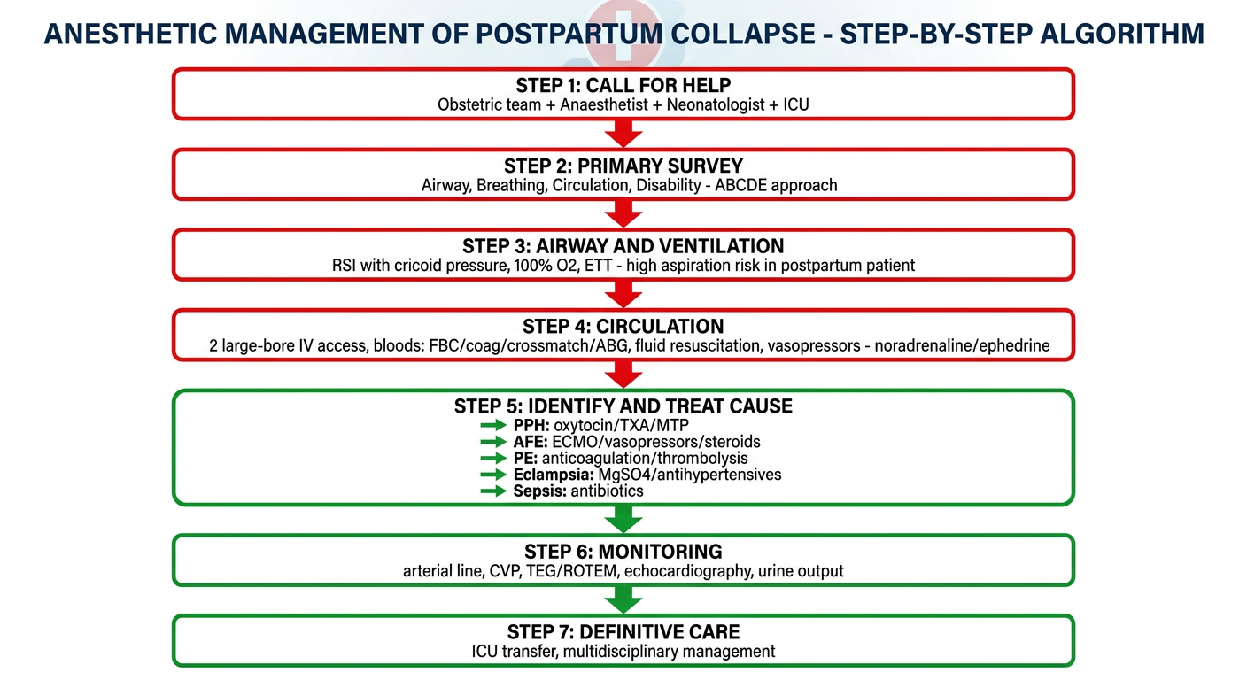
A detailed medical flowchart titled "Anesthetic Management of Postpartum Collapse - Step-by-Step Algorithm". Show a top-to-bottom emergency protocol flowchart with these sequential steps connected by arrows: Step 1 - CALL FOR HELP (Obstetric team + Anaesthetist + Neonatologist + ICU); Step 2 - PRIMARY SURVEY (Airway, Breathing, Circulation, Disability - ABCDE approach); Step 3 - AIRWAY AND VENTILATION (RSI with cricoid pressure, 100% O2, ETT - high aspiration risk in postpartum patient); Step 4 - CIRCULATION (2 large-bore IV access, bloods: FBC/coag/crossmatch/ABG, fluid resuscitation, vasopressors - noradrenaline/ephedrine); Step 5 - IDENTIFY AND TREAT CAUSE (PPH: oxytocin/TXA/MTP; AFE: ECMO/vasopressors/steroids; PE: anticoagulation/thrombolysis; Eclampsia: MgSO4/antihypertensives; Sepsis: antibiotics); Step 6 - MONITORING (arterial line, CVP, TEG/ROTEM, echocardiography, urine output); Step 7 - DEFINITIVE CARE (ICU transfer, multidisciplinary). Use red for critical steps, green for treatments, clean medical style.
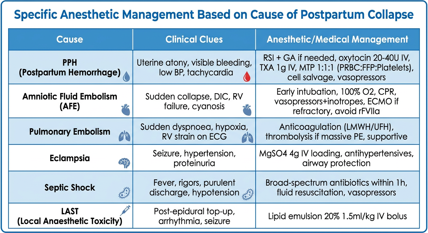
A clean medical comparison table/flowchart titled "Specific Anesthetic Management Based on Cause of Postpartum Collapse". Show a three-column table with columns: Cause | Clinical Clues | Anesthetic/Medical Management. Include these rows: 1. PPH (Postpartum Hemorrhage) | Uterine atony, visible bleeding, low BP, tachycardia | RSI + GA if needed, oxytocin 20-40U IV, TXA 1g IV, MTP 1:1:1 (PRBC:FFP:Platelets), cell salvage, vasopressors; 2. Amniotic Fluid Embolism (AFE) | Sudden collapse, DIC, RV failure, cyanosis | Early intubation, 100% O2, CPR, vasopressors+inotropes, ECMO if refractory, avoid rFVIIa; 3. Pulmonary Embolism | Sudden dyspnoea, hypoxia, RV strain on ECG | Anticoagulation (LMWH/UFH), thrombolysis if massive PE, supportive; 4. Eclampsia | Seizure, hypertension, proteinuria | MgSO4 4g IV loading, antihypertensives, airway protection; 5. Septic Shock | Fever, rigors, purulent discharge, hypotension | Broad-spectrum antibiotics within 1h, fluid resuscitation, vasopressors; 6. LAST (Local Anaesthetic Toxicity) | Post-epidural top-up, arrhythmia, seizure | Lipid emulsion 20% 1.5ml/kg IV bolus. Use clean medical blue/white color scheme, easy to read.
Anesthetic Management of Postpartum Collapse
References: Miller's Anesthesia 10e, Creasy & Resnik's Maternal-Fetal Medicine, Knapp & Bhatia (Br J Hosp Med 2022, PMID 36594762), ASA Practice Guidelines for Obstetric Anesthesia, SOAP Consensus Statement on Cardiac Arrest in Pregnancy
STEP 1 - DEFINITION AND SCOPE
DIAGRAM 1 - Causes of Postpartum Collapse
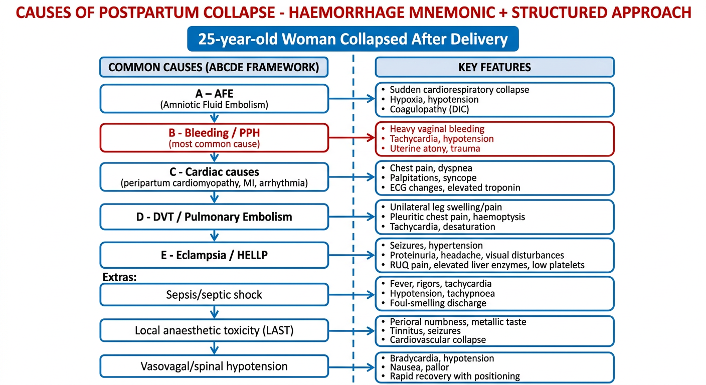
Causes - Mnemonic "HAEMORRHAGE" or ABCDE Framework:
| System | Cause |
|---|---|
| A | Amniotic Fluid Embolism (AFE) |
| B | Bleeding - PPH (most common) |
| C | Cardiac - Peripartum cardiomyopathy, arrhythmia, MI |
| D | DVT/Pulmonary Embolism |
| E | Eclampsia / HELLP syndrome |
| + | Septic shock |
| + | Local Anaesthetic Systemic Toxicity (LAST) |
| + | Vasovagal / Spinal hypotension (if recent neuraxial block) |
| + | Uterine inversion |
| + | Anaphylaxis |
STEP 2 - IMMEDIATE RESPONSE (First 5 Minutes)
Call for Help - Assemble the Team:
- Obstetric anaesthetist (lead airway + resuscitation)
- Obstetrician + Senior midwife
- Neonatology team
- ICU/HDU team
- Blood bank - alert early
DIAGRAM 2 - Anesthetic Management Algorithm
STEP 3 - PRIMARY SURVEY (ABCDE)
A - Airway
- Assess patency; protect if reduced GCS
- High aspiration risk - postpartum patients have full stomach, delayed gastric emptying, raised intragastric pressure
- If unconscious or GCS <8: Rapid Sequence Induction (RSI)
- Preoxygenate 3 min (or 8 vital capacity breaths) with 100% O₂
- Induction: Ketamine 1-2 mg/kg (preserves haemodynamics) OR Etomidate 0.2-0.3 mg/kg if haemodynamically unstable
- Avoid propofol if hypotensive
- Suxamethonium 1.5 mg/kg + cricoid pressure
- Cuffed ETT; confirm with capnography and bilateral air entry
B - Breathing
- 100% FiO₂ post-intubation
- Maintain SpO₂ >95%, ETCO₂ 35-40 mmHg
- Ventilate with tidal volume 6-8 mL/kg ideal body weight
- PEEP 5-8 cm H₂O for AFE/pulmonary oedema
C - Circulation
- 2 large-bore IV cannulae (14-16G) in antecubital fossae
- Bloods: FBC, coagulation screen, fibrinogen, U&E, LFTs, ABG, crossmatch 6 units, TEG/ROTEM
- IV fluids: Crystalloids (Hartmann's/Normal Saline) for initial resuscitation
- Vasopressors: Noradrenaline (first choice in septic/AFE collapse) OR Ephedrine/Phenylephrine (for spinal hypotension)
- Invasive monitoring: Arterial line (radial - continuous BP + ABG sampling), CVP via internal jugular or subclavian
- Urine output: Foley catheter - target >0.5 mL/kg/hr
D - Disability
- GCS, pupils, blood glucose (hypoglycaemia can cause collapse)
- Check for seizures (eclampsia)
- Neurological status: posture, reflexes
E - Exposure + Environment
- Full exposure, keep warm (warm IV fluids, warming blanket)
- Examine for: uterine atony, retained placenta, perineal tears, uterine inversion
STEP 4 - INVESTIGATE TO IDENTIFY CAUSE
| Investigation | Purpose |
|---|---|
| ABG | Hypoxia, acidosis, lactate |
| 12-lead ECG | PE (S1Q3T3), arrhythmia, MI |
| POCUS / Echo (bedside) | RV dysfunction (AFE/PE), LV failure, pericardial effusion |
| Chest X-ray | Pulmonary oedema, pneumothorax |
| FBC + Coagulation + Fibrinogen | DIC (AFE, PPH, abruption) |
| TEG/ROTEM | Directed blood product therapy |
| Blood cultures | Sepsis |
| Serum lactate | Tissue hypoperfusion |
DIAGRAM 3 - Cause-Specific Anesthetic Management
STEP 5 - CAUSE-SPECIFIC MANAGEMENT IN DETAIL
5A. POSTPARTUM HAEMORRHAGE (PPH) - Most Common Cause
- Secure airway early if haemodynamic instability (RSI + ETT)
- Uterotonic agents:
- Oxytocin 20-40 units in 500 mL NS (slow infusion - avoid bolus as causes hypotension)
- Ergometrine 0.2 mg IM (avoid in hypertension)
- Carboprost 250 mcg IM every 15 min (max 8 doses; avoid in asthma)
- Misoprostol 800-1000 mcg SL/PR
- Tranexamic Acid (TXA) 1g IV within 3 hours (WHO recommended; repeat if bleeding continues at 30 min)
- Massive Transfusion Protocol (MTP):
- PRBC : FFP : Platelets = 1:1:1 ratio
- Target fibrinogen >2g/L - give cryoprecipitate if <1.5g/L
- Use TEG/ROTEM to guide ratios
- Warm all blood products (hotline warmer)
- Cell salvage - acceptable in PPH; use leucocyte depletion filter
- Invasive options if pharmacological fails: Balloon tamponade, compression sutures, uterine artery ligation, interventional radiology (uterine artery embolisation), hysterectomy as last resort
5B. AMNIOTIC FLUID EMBOLISM (AFE)
- Sudden cardiorespiratory compromise
- DIC (documented)
- Onset during labour or within 30 min of placental delivery
- No other obvious diagnosis
- Immediate CPR if cardiac arrest
- Early intubation + 100% O₂ + PEEP
- Vasopressors (noradrenaline) + Inotropes (dobutamine) for RV failure
- Transoesophageal echocardiography post-intubation to assess RV function
- Treat DIC: 1:1:1 blood products + cryoprecipitate; TEG/ROTEM guided
- ECMO for persistent haemodynamic instability unresponsive to medical management
- Avoid recombinant FVIIa - associated with major organ thrombosis and worse outcomes (Creasy & Resnik)
5C. MATERNAL CARDIAC ARREST
- No left uterine displacement needed postpartum (uterus involuting)
- Standard CPR position (supine, firm surface)
- Earlier and more aggressive airway management (high aspiration risk)
- Chest compressions at same position as non-pregnant adults after delivery
- Perimortem Caesarean Delivery - not needed postpartum (fetus already delivered)
- CPR quality is compromised by transfer to OT - perform resuscitation at the bedside
- PPH > Heart failure > AFE > Sepsis (Creasy & Resnik, PMID 36594762)
5D. PULMONARY EMBOLISM (PE)
- Anticoagulate with unfractionated heparin IV (80 units/kg bolus then infusion)
- If massive PE with haemodynamic collapse: IV thrombolysis (alteplase 100 mg over 2 hours)
- Supportive: O₂, vasopressors, PEEP-cautious ventilation (avoid worsening RV afterload)
- POCUS shows RV dilation, D-sign, McConnell's sign
5E. ECLAMPSIA
- Secure airway (seizure risk → aspiration)
- MgSO₄: 4-6g IV over 15-20 min loading dose, then 1-2g/hour infusion
- Antihypertensives: Labetalol 20 mg IV or Hydralazine 5 mg IV if BP >160/110
- Watch for: MgSO₄ toxicity (loss of patellar reflexes, respiratory arrest) - antidote is Calcium gluconate 10 mL 10% IV
5F. SEPTIC SHOCK
- Sepsis-3 criteria: Suspected infection + organ dysfunction (SOFA ≥2)
- 1-hour bundle: Blood cultures, lactate, broad-spectrum antibiotics (within 1 hour), 30 mL/kg crystalloid if lactate >4, vasopressors (noradrenaline)
- Source control: drain collections, remove infected products of conception
5G. LOCAL ANAESTHETIC SYSTEMIC TOXICITY (LAST)
- Stop LA infusion immediately
- Seizure: benzodiazepines; cardiac arrest: CPR
- 20% Lipid emulsion (Intralipid): 1.5 mL/kg IV bolus over 1 min, then 0.25 mL/kg/min infusion
- Avoid propofol as lipid substitute
- May need prolonged CPR
STEP 6 - MONITORING
| Monitor | Target |
|---|---|
| Arterial line (radial) | Continuous BP, ABG sampling |
| SpO₂ | >95% |
| ETCO₂ | 35-40 mmHg |
| Urine output | >0.5 mL/kg/hr |
| CVP | 8-12 mmHg (fluid status) |
| TEG/ROTEM | Targeted blood product therapy |
| Bedside POCUS/Echo | Cardiac function, RV strain |
| Temperature | Normothermia |
STEP 7 - DEFINITIVE CARE
- Transfer to ICU/HDU once stabilised
- Multidisciplinary review: Obstetrician, Anaesthetist, Haematologist, Intensivist, Cardiologist (as needed)
- Debrief the team and document clearly
- Thromboprophylaxis as soon as haemostasis achieved
- Family communication and psychological support
KEY EXAM POINTS (DNB Recall)
| Point | Fact |
|---|---|
| Most common cause of postpartum collapse | PPH (haemorrhage) |
| Airway risk in postpartum patient | High - full stomach, RSI mandatory |
| TXA dose in PPH | 1g IV, repeat if needed at 30 min |
| Blood product ratio in MTP | 1:1:1 (PRBC:FFP:Platelets) |
| AFE diagnostic triad | Cardiorespiratory collapse + DIC + intrapartum/30 min post-delivery onset |
| ECMO indication | AFE or refractory cardiogenic shock |
| Drug to AVOID in AFE-DIC | rFVIIa (causes organ thrombosis) |
| LAST treatment | 20% Intralipid 1.5 mL/kg bolus |
| MgSO₄ toxicity antidote | Calcium gluconate 10 mL 10% IV |
| Perimortem CS threshold | ≥20 weeks gestation (not applicable post-delivery) |