
Cardiac cycle for 10 mark gk apl
I'll look up the cardiac cycle from the medical library for a comprehensive 10-mark answer.
Searching sources...
Reading File
Finding Sources
Finding Sources
Now let me read the key physiology sources in parallel - Guyton & Hall, Costanzo, and Ganong's for the most comprehensive cardiac cycle content:
Reading File
Reading File
Reading File
Reading File
Reading File
I now have comprehensive content from multiple physiology textbooks. Let me fetch the key cardiac cycle diagram:
Loading Image
I now have everything needed for a thorough 10-mark answer. Here is the complete answer:
Cardiac Cycle - 10 Marks
Definition
The cardiac cycle refers to the sequence of mechanical and electrical events that repeats with every heartbeat - from the beginning of one heartbeat to the beginning of the next. At a normal heart rate of 72 beats/min, each cycle lasts approximately 0.833 seconds (reciprocal of heart rate).
- Guyton and Hall Textbook of Medical Physiology
- Costanzo Physiology 7th Edition, p. 160
Initiation
Each cardiac cycle is initiated by spontaneous action potential generation at the sinoatrial (SA) node (located in the superior lateral wall of the right atrium near the opening of the superior vena cava). The impulse travels through both atria, then through the AV node/bundle into the ventricles. The AV node delay (~0.1 second) allows the atria to contract first, acting as "primer pumps" before the powerful ventricular contraction begins.
Phases of the Cardiac Cycle
The cycle is divided into 7 phases (A-G) based on Costanzo's classification:
1. ATRIAL SYSTOLE (Phase A) - Duration: ~0.1 s
- ECG: P wave (atrial depolarization), PR interval
- Events: Atria contract, ejecting the remaining ~30% of blood into ventricles (70% fills passively before this)
- Valves: Mitral and tricuspid (AV) valves open; aortic and pulmonary valves closed
- Pressures: Left atrial pressure rises; small "a wave" seen on venous pulse tracing
- Heart Sound: S4 (not normally audible; heard in ventricular hypertrophy)
- LV Volume: Reaches maximum (End-Diastolic Volume, EDV ~130 mL)
2. ISOVOLUMETRIC VENTRICULAR CONTRACTION (Phase B) - Duration: ~0.05 s
- ECG: QRS complex (ventricular depolarization)
- Events: Ventricles begin contracting; intraventricular pressure rises sharply; all valves are closed - so ventricular volume remains constant (no ejection yet)
- Valve event: Mitral valve closes → generates S1 (first heart sound - "lub")
- Pressure: LV pressure rises rapidly from ~5 mmHg toward 80 mmHg (aortic diastolic pressure)
- AV valves bulge into atria, causing a small rise in atrial pressure (c wave on venous pulse)
3. RAPID VENTRICULAR EJECTION (Phase C)
- ECG: ST segment
- Events: LV pressure exceeds aortic pressure (80 mmHg) → aortic valve opens; blood ejected rapidly into aorta
- Valve event: Aortic and pulmonary valves open
- Pressure: LV pressure peaks at ~120 mmHg; aortic pressure also rises to ~120 mmHg
- Volume: LV volume falls rapidly
4. REDUCED VENTRICULAR EJECTION (Phase D)
- ECG: T wave (ventricular repolarization begins)
- Events: Ejection continues at a slower rate; ventricular pressure starts to fall; aortic pressure begins to fall as blood runs off into arteries
- Volume: LV volume reaches its minimum = End-Systolic Volume (ESV) ~50 mL
- Stroke Volume (SV) = EDV - ESV = 130 - 50 = ~80 mL
- Ejection Fraction (EF) = SV/EDV = 80/130 ≈ 65%
5. ISOVOLUMETRIC VENTRICULAR RELAXATION (Phase E) - Duration: ~0.08 s
- ECG: After T wave (isoelectric)
- Events: Ventricular muscle relaxes; LV pressure falls rapidly; all valves closed - volume remains constant
- Valve event: Aortic valve closes → generates S2 (second heart sound - "dub"); causes the dicrotic notch on aortic pressure tracing
- Protodiastole precedes this (~0.04 s when momentum still keeps valves open)
6. RAPID VENTRICULAR FILLING (Phase F)
- ECG: Isoelectric (after T wave)
- Events: LV pressure falls below left atrial pressure → mitral valve opens; blood rushes rapidly from atria into ventricles
- Heart Sound: S3 (third heart sound, not normally audible in adults; heard in heart failure / dilated cardiomyopathy) - due to vibration when blood hits ventricular walls
- Volume: LV volume increases quickly
7. REDUCED VENTRICULAR FILLING / DIASTASIS (Phase G)
- Events: Ventricular filling slows as the pressure gradient between atrium and ventricle equalizes; this is the longest phase
- Merges into the next atrial systole
Cardiac Cycle Diagram
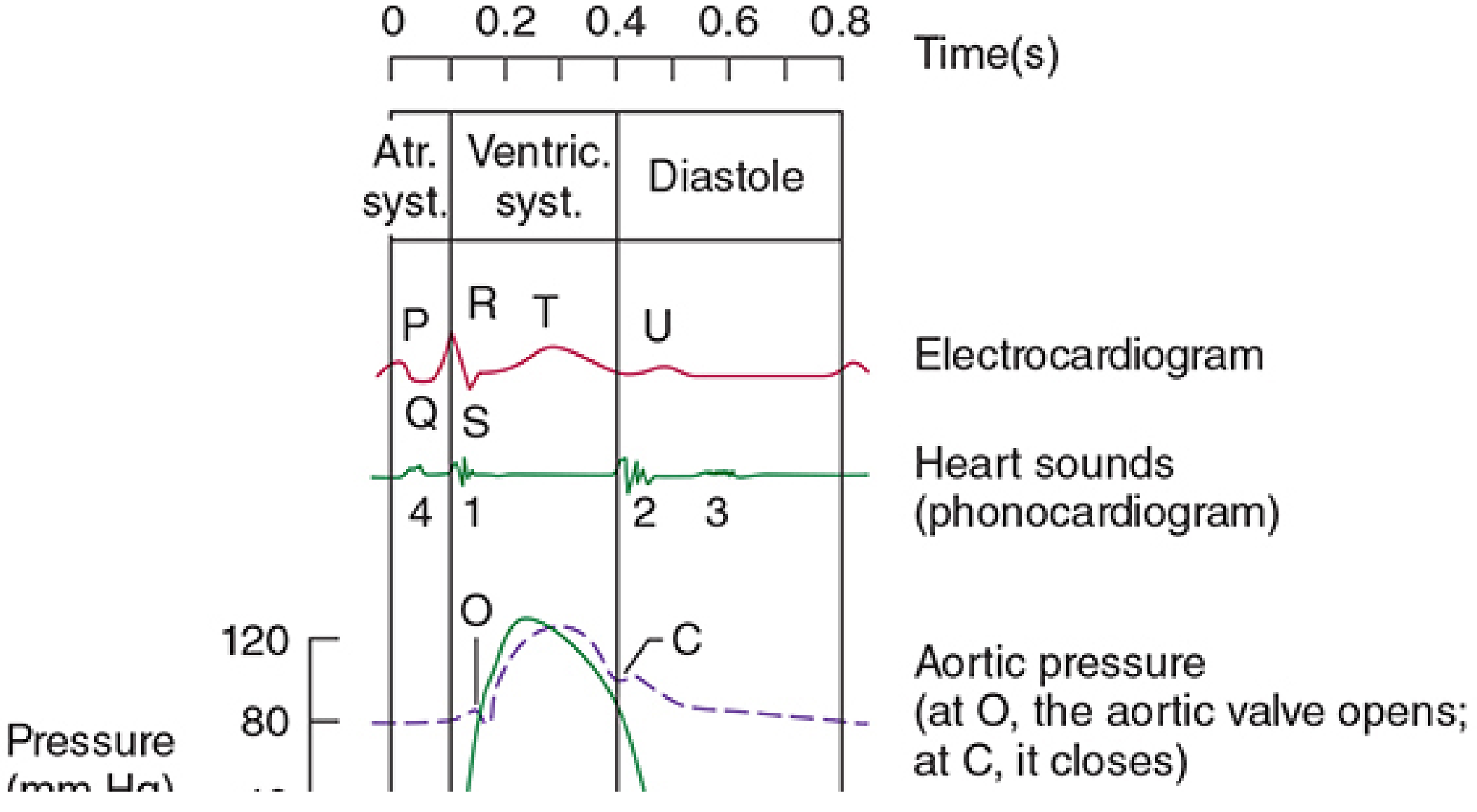
Summary Table
| Phase | Duration | Valves | ECG | Heart Sound |
|---|---|---|---|---|
| Atrial Systole | ~0.1 s | AV open, SL closed | P wave | S4 (abnormal) |
| Isovolumetric Contraction | ~0.05 s | All closed | QRS | S1 ("lub") |
| Rapid Ejection | - | SL open, AV closed | ST segment | - |
| Reduced Ejection | - | SL open, AV closed | T wave | - |
| Isovolumetric Relaxation | ~0.08 s | All closed | Post-T | S2 ("dub") |
| Rapid Filling | - | AV open, SL closed | - | S3 (abnormal) |
| Diastasis | - | AV open, SL closed | - | - |
SL = semilunar (aortic + pulmonary); AV = atrioventricular (mitral + tricuspid)
Key Pressure Values (Left Heart)
| Parameter | Value |
|---|---|
| LV end-diastolic pressure | ~5 mmHg |
| LV peak systolic pressure | ~120 mmHg |
| Aortic systolic pressure | ~120 mmHg |
| Aortic diastolic pressure | ~80 mmHg |
| Left atrial pressure | ~5-10 mmHg |
| EDV | ~130 mL |
| ESV | ~50 mL |
| Stroke Volume | ~80 mL |
| Ejection Fraction | ~65% |
Effect of Heart Rate on Cardiac Cycle
When heart rate increases, the total cycle duration decreases. Diastole shortens more than systole. At very high heart rates, incomplete ventricular filling can compromise cardiac output. At 72 bpm, systole occupies ~0.4 of the cycle; at 3x normal heart rate, systole occupies ~0.65 of the cycle.
Venous Pulse Waves (JVP) Correlated with Cardiac Cycle
| Wave | Cause |
|---|---|
| a wave | Atrial contraction (atrial systole) |
| c wave | AV valve bulging into atria during isovolumetric contraction |
| v wave | Venous filling of atria while AV valves are closed (during ventricular systole) |
| x descent | Atrial relaxation + AV valve pulled downward during ejection |
| y descent | AV valve opens → blood rushes into ventricle |
Sources: Costanzo Physiology 7th Edition | Ganong's Review of Medical Physiology 26th Edition | Guyton and Hall Textbook of Medical Physiology
This is a shared conversation. Sign in to Orris to start your own chat.