
"lichen planus"[MeSH Terms] AND "treatment"
- Several round to oval violaceous (purple-grey) papules and small plaques, about 0.5 to 1.5 cm.
- Some have a fine, adherent white-grey scale and a lacy surface pattern that may be Wickham striae. Dermoscopy would show these more clearly.
- A few lesions have joined into a short linear band near the wrist crease. This suggests a Koebner phenomenon, where lesions appear along sites of rubbing or scratching.
- Several smaller, paler pink-grey spots sit around the larger lesions.
- The classic lesion is a flat-topped, polygonal, violaceous papule with a shiny, dry surface and scant adherent scale, crossed by gray or white Wickham striae.
- The flexor wrists are a typical site, and Koebnerization is recognized in LP (Andrews' Diseases of the Skin, Lichen Planus chapter).
- In darker skin, the colour tends to look grey-violet, and resolving lesions turn hyperpigmented. That matches this image.
- LP is usually very itchy, often with rubbing rather than scratching.
- Hypertrophic LP, if lesions are thicker and warty.
- Lichenoid drug eruption. Ask about recent medications such as thiazides, beta-blockers, ACE inhibitors, antimalarials, NSAIDs, anti-TB drugs and antiepileptics.
- Psoriasis, which has thicker silvery scale and no violaceous hue.
- Discoid lupus, which shows follicular plugging and atrophy.
- Bowen's disease, if lesions are solitary and persistent.
- Secondary syphilis, which can look similar and calls for serology if there is any risk.
- Lichen simplex chronicus, or a lichenoid or sarcoid reaction.
- Examine the mouth, genitals, nails and scalp. Oral involvement is common and changes the follow-up.
- Take a drug history and ask about itch and duration.
- Do a 4 mm punch biopsy from an intact lesion. It should show interface dermatitis with a band-like lymphocytic infiltrate and a saw-tooth rete pattern.
- Check hepatitis C and B serology, especially in an endemic region. Add fasting glucose and lipids, since LP is associated with diabetes and hyperlipidemia. Consider VDRL/RPR if syphilis is a differential.
- First line is a potent or super-potent topical steroid (clobetasol 0.05%) once or twice daily for 2 to 4 weeks, then taper. Occlusion or intralesional triamcinolone can be used for thick lesions.
- Add an oral antihistamine for itch.
- For widespread or resistant disease, options include a short oral prednisolone course, acitretin, methotrexate, narrowband UVB or PUVA, and other systemic agents. I haven't checked doses or regimens against a source, so confirm them with a dermatology reference.
- The disease is variable. About two-thirds of cutaneous cases clear within a year. Hypertrophic and mucosal forms are more chronic, and recurrences are common (Andrews').
Mangment
"lichen planus"[MeSH Terms] AND "cutaneous"
- Stop any suspected culprit drug if this could be a lichenoid drug eruption.
- Reduce rubbing and trauma, because lichen planus koebnerizes.
- Use emollients and an oral antihistamine (e.g., cetirizine, or a sedating antihistamine at night) for itch.
- Screen for hepatitis C and B, diabetes and lipids. Examine the mouth, genitals and nails.
- First line: a high-potency topical corticosteroid, applied twice daily. The book notes the evidence is limited, but this is the accepted first-line treatment.
- Use occlusion for thick or hypertrophic plaques.
- If there is no response after 2 to 4 weeks, move to a higher potency steroid or use intralesional injection.
- Intralesional triamcinolone: 5 to 10 mg/mL monthly for resistant or hypertrophic lesions.
- Avoid excess trauma, because it can koebnerize.
- Combine it with topical therapy or another steroid-sparing agent, because relapse is common.
- Topical tacrolimus 0.1%: a second-line option and a steroid-sparing add-on. It is mainly supported by oral lichen planus data, where it was as effective as clobetasol 0.05%.
- It lets you taper the steroid and reduce the risk of atrophy.
- Calcipotriene: the only randomized trial in cutaneous lichen planus found it equal to betamethasone valerate over 12 weeks.
- Topical retinoids: of limited value.
- Narrowband UVB (311 nm): three times weekly until remission, then taper over 3 to 6 weeks.
- One series reported a 70% remission rate.
- It has largely replaced PUVA.
- Oral corticosteroids: give a 4 to 6 week taper for rapid control.
- Relapse is high after stopping, so always pair them with topical therapy or a steroid-sparing agent.
- Do not use them as long-term monotherapy.
- Steroid-sparing second-line agents:
| Agent | Dose and evidence | Best suited to |
|---|---|---|
| Methotrexate | 15 to 20 mg weekly for 4 to 24 weeks. Complete response in 58% to 91%. | Preferred and cost-effective. Good for hypertrophic or recalcitrant disease. |
| Acitretin | 30 mg/day for 8 weeks. Marked improvement in 64%. | Hypertrophic disease. Watch lipids and mucocutaneous dryness. |
| Sulfasalazine | Start at 1 g/day, increase by 0.5 g every 3 days to a maximum of 2.5 g/day. Highest level of evidence, with 83% skin improvement. | Generalized disease. Monitor blood count and liver tests. |
| Metronidazole | 250 mg three times daily for 12 weeks. 74% response. | Generalized disease. Watch for neuropathy. |
| Hydroxychloroquine | 200 to 400 mg/day for 6 to 12 months. | Second line, and first line for actinic lichen planus. |
| Mycophenolate or azathioprine | Mycophenolate 2 to 3 g/day, with delayed response. | Refractory or ulcerative disease. |
| Cyclosporine | Third-line option. | Reserve for resistant cases. |
- Start clobetasol 0.05% ointment twice daily for 2 to 4 weeks, with occlusion for the thicker plaques.
- Add an antihistamine at night.
- Review at 4 weeks.
- If improving, taper to a lower-potency steroid or tacrolimus.
- If thick lesions persist, give intralesional triamcinolone.
- If the disease spreads or does not respond, refer to dermatology for narrowband UVB or a systemic agent (methotrexate is a reasonable first choice).
- Counsel on the course. Most skin disease clears within 1 to 2 years, but hypertrophic and mucosal forms are more chronic, and recurrence is common (Andrews' Diseases of the Skin).
- Expect post-inflammatory hyperpigmentation in darker skin. It fades slowly and is not a treatment failure.
How to confirm this lichen planus
lichen planus histopathology band-like infiltrate sawtooth rete ridges hypergranulosis
Wickham striae dermoscopy lichen planus
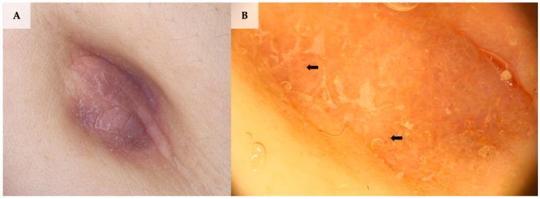
This composite image illustrates the clinical and dermoscopic presentation of umbilical lichen planus. Panel A shows a macro clinical photograph of a well-demarcated, violaceous-to-pink, oval-shaped plaque within the umbilical fold. The surface exhibits characteristic fine white reticular lines (Wickham striae) and a subtle texture suggestive of surface scale. Panel B provides a dermoscopic view (20x magnification) highlighting diagnostic features: a brown structureless background interspersed with prominent white intersecting lines (Wickham striae, indicated by black arrows), white surface scale, and fine coiled (glomerular) vessels. These findings are hallmarks of lichen planus and aid in differentiating this inflammatory dermatosis from other umbilical pathologies like melanoma or seborrheic keratosis. The visual serves as an educational tool for dermatological diagnosis and dermoscopy training, focusing on the identification of specific vascular and structural patterns in inflammatory skin disease.
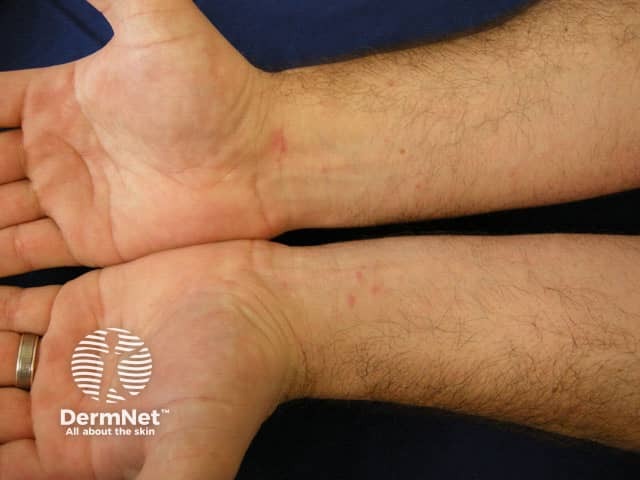
Clinical photography of bilateral wrists demonstrating classic cutaneous lichen planus. In vivo skin image documenting the palmar/flexor surfaces of the wrists with low to moderate magnification. Primary subject: violaceous, flat-topped papules arranged in a symmetric, polygonal pattern along the flexor aspects. The lesions exhibit a glossy, slightly scaly surface and subtle Wickham’s striae in some areas; pruritus is often present but not uniformly required for diagnosis. The distribution is predominantly on the wrists with potential mild edema or erythema at lesion margins. The overall appearance is briskly ink-purple to brown-purple, with well-defined borders and a tendency to coalesce into plaques over time. There is no obvious mucosal involvement in this image. This photograph supports the clinical diagnosis of lichen planus by morphology and anatomical distribution, aiding differentiation from psoriasis, eczema, pityriasis rosea, and tinea infections. Relevant features for search include lichen planus, violaceous papules, flat-topped papules, Wickham lines, wrists, flexor surfaces, bilateral distribution, dermatology photography, clinical dermoscopy correlation not shown. This image is primarily for educational and diagnostic reference, enabling clinicians and trainees to recognize typical cutaneous LP patterns, document treatment response, and correlate with histopathology in uncertain cases. Thanks.
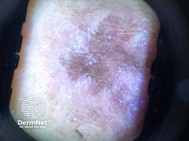
This in vivo dermoscopic image (Dermoscopy) captures a recent-onset annular lichen planus lesion on the forearm. Examined with polarized dermoscopy at standard magnification, the lesion shows an annular plaque with a pale, slightly erythematous center and a thin, scaly periphery. Under dermoscopy, characteristic Wickham striae manifest as interlacing white lines across the surface, most evident around the inner border, aligned with the lesion’s contours. The pale center displays subtle translucency with mild pigment network disruption. There may be faint perifollicular hyperpigmentation and minimal vascularity. The border remains well defined, with a subtle reddish halo suggesting active dermatitis. Combined findings—white reticulation, peripheral scaling, and central clearing—are typical for LP and help distinguish from tinea and psoriasis. The image provides clues for clinical diagnosis, such as recent onset, annular morphology, and lack of pustular features. Clinically, LP often presents pruritic, violaceous, polygonal papules; annular variants may exhibit central clearing and active borders. Dermoscopy enhances diagnostic confidence by highlighting Wickham striae and interface changes; these features correlate with histopathology showing interface lichenoid inflammation, sawtooth rete ridges, Civatte bodies, and basal vacuolization. Relevant clinical scenarios include evaluating suspicious annular lesions on extremities, differentiating LP from pityriasis rosea, psoriasis, or fungal infections; guiding biopsy decisions and treatment planning.
- Purple, polygonal, pruritic, planar (flat-topped) papules and plaques. The lesions in your photo already fit the purple and plaque parts.
- Look for Wickham striae (fine white lines on the surface). Wiping with oil or water helps you see them.
- Look for Koebner phenomenon, flexor wrist and forearm sites, and symmetry.
- Check for mucosal, nail and scalp involvement. Oral reticular white lacy lesions on the buccal mucosa support the diagnosis strongly (Andrews' Diseases of the Skin, Lichen Planus chapter).
- Look for white interlacing Wickham striae over a violaceous-brown background. In darker skin, expect a grey-brown background and peripheral dots.
- Coiled (glomerular) vessels and fine peripheral scale may also be present.
- This helps separate lichen planus from psoriasis, Bowen's disease and discoid lupus.
- Technique:
- Take a 4 mm punch from a fully developed, intact, untreated papule or plaque.
- Avoid excoriated lesions and avoid lesions treated with topical steroids in the past 1 to 2 weeks.
- Send it in formalin.
- Take a second sample for direct immunofluorescence (DIF) only if lupus or an immunobullous disease is a concern.
- Histology:
- A dense, band-like lymphocytic infiltrate that obscures the dermo-epidermal junction.
- Liquefactive (hydropic) degeneration of the basal layer.
- Hypergranulosis (wedge-shaped), with orthokeratosis and no parakeratosis.
- Saw-tooth rete ridges, Civatte (colloid) bodies, and sometimes Max-Joseph clefts.
- Pigment incontinence in darker skin.
- The Yamada's textbook confirms the core criteria: a distinct band-like lymphocytic infiltrate, liquefactive basal cell degeneration, and overlying hypergranulosis (Yamada's Textbook of Gastroenterology, Lichen Planus).
- Features that point away from lichen planus:
- Parakeratosis, eosinophils, plasma cells, or a deep or perivascular infiltrate suggest a lichenoid drug eruption.
- Follicular plugging, basement membrane thickening and mucin suggest discoid lupus.
- Atypical keratinocytes suggest Bowen's disease.
- Epidermal psoriasiform change suggests psoriasis.
- Drug history: the main way to separate lichenoid drug eruption from true lichen planus. Look for recent thiazides, beta-blockers, ACE inhibitors, antimalarials, NSAIDs, anti-TB drugs, antiepileptics, and gold.
- Serology: VDRL/RPR for secondary syphilis, which can mimic lichen planus.
- Hepatitis: HCV and HBV serology.
- Metabolic screen: fasting glucose and lipid profile. I'm adding the metabolic screen from clinical knowledge, not from the retrieved text.
- Skin scraping: KOH if there is any annular scale that could be tinea.
- Anti-nuclear antibodies: only if lupus is suspected.
- the picture is atypical, hypertrophic or solitary;
- lesions are non-healing;
- they are not responding to treatment;
- you are suspicious of a drug cause, Bowen's disease or lupus.
Make a presentation on lichen planus diagnosis and management
lichen planus histology band-like lymphocytic infiltrate saw-tooth rete ridges
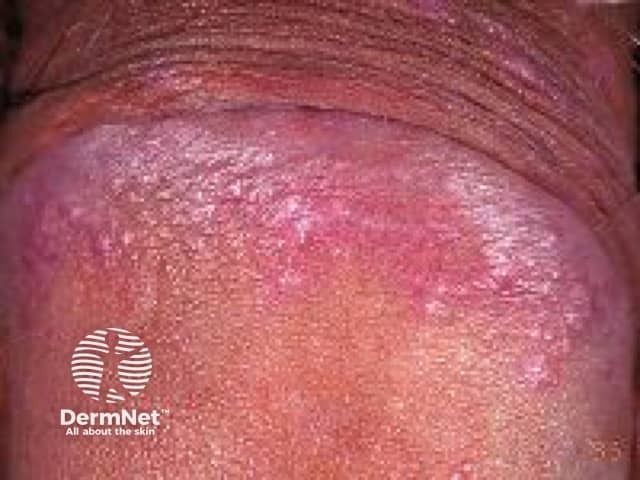
Penile lichen planus is a mucocutaneous inflammatory condition affecting external genitalia. This clinical photograph shows erythematous to violaceous plaques on the penile shaft and glans, with a shiny, indented surface and possible Wickham striae along the lesion margins. The distribution is confined to the genital skin, often involving the foreskin or balanus region, and may be accompanied by superficial erosions or fissures. Lesions are typically pruritic or painful, and chronicity can lead to scarring or stenosis in severe cases. The appearance described here—flat-topped, polygonal papules with well-defined borders and a violaceous hue—fits the classic dermatologic phenotype of lichen planus affecting mucocutaneous sites. Clinically, differential diagnoses include lichen sclerosus, genital psoriasis, candidal balanitis, fixed drug eruption, and other inflammatory dermatoses. Correlation with patient history and examination of additional mucocutaneous sites (oral, genital across other areas) is helpful. If biopsy is obtained, histology typically reveals a lichenoid interface dermatitis with saw-tooth rete ridges, a dense band-like lymphocytic infiltrate, and Civatte bodies; treatment usually starts with potent topical corticosteroids or calcineurin inhibitors, with close follow-up for symptom control and prevention of scarring. This image is relevant for dermatology, urology, medical education, and mucosal disease research.
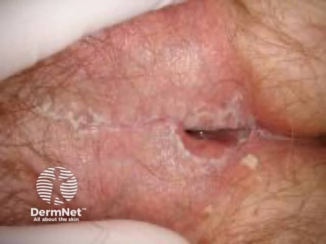
This clinical photograph depicts vulvar skin with features typical of vulvar lichen planus. The close-up view shows erythematous to violaceous, shiny plaques with polygonal contours and mild mucosal erosion at the vestibule. Surface markings such as Wickham striae may be subtle or absent on vulvar mucosa. The lesions are localized to the vulvar mucocutaneous region, involving the labia majora/minora and vestibular lips. From a dermatologic perspective, the appearance suggests a lichenoid inflammatory dermatosis characterized by flat-topped papules and well-demarcated plaques. Although histology is not available in this image, biopsy would be expected to reveal lichenoid interface dermatitis with a band-like lymphocytic infiltrate, basal cell degeneration, hyperkeratosis, and saw-tooth rete ridges. Clinically, vulvar LP presents with pruritus, burning, dyspareunia, and mucosal fragility, and requires exclusion of other vulvar conditions such as lichen sclerosus, erosive LP, lichen simplex chronicus, candidiasis, and psoriasis. This image is valuable for educational, diagnostic, and differential-diagnostic training, and supports clinical-pathology correlation, patient counseling, and dermatology illustration repositories. Potential clinical use cases include dermatology consultation, gynecologic evaluation, medical education, and AI-driven vulvar disease datasets. It thereby enhances recognition by clinicians, improves triage decisions, and supports research into pathogenetic mechanisms, treatment response, and patient education resources for patient care.
oral lichen planus reticular Wickham striae buccal mucosa

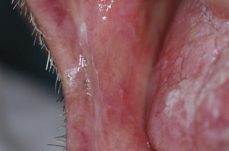
A composite of three clinical photographs showing intraoral manifestations of Oral Lichen Planus. The top-left image displays a well-demarcated, irregularly shaped white plaque (plaque-type lichen planus) on the right dorsum of the tongue, which clinically resembles leukoplakia or hyperplastic candidiasis. The top-right and bottom images show the bilateral buccal mucosa, highlighting classic reticular lichen planus characterized by a network of fine, white, interconnected lacy lines known as Wickham striae. These striae appear against a background of erythematous (pinkish-red) mucosa. The presence of these reticular lesions elsewhere in the oral cavity assists in the differential diagnosis of the isolated lingual plaque. This educational visual demonstrates common diagnostic challenges in oral medicine, specifically the distinction between plaque-type lichen planus, leukoplakia, and lichenoid reactions.
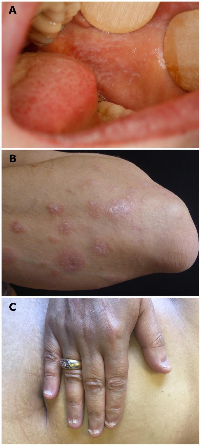
This composite clinical photograph illustrates characteristic clinical manifestations of lichen planus (LP) across mucosal, cutaneous, and ungual sites. Panel A displays a reticular pattern of lacy, white interconnected lines known as Wickham striae on the buccal mucosa, typical of oral lichen planus. Panel B shows the cutaneous presentation on a forearm, featuring multiple well-demarcated, violaceous, flat-topped polygonal papules; close inspection reveals the presence of surface Wickham striae. Panel C demonstrates lichen planus onychodystrophy, specifically showing pterygium formation where the proximal nail fold has fused with the nail bed, leading to splitting and destruction of the nail plate. These images serve as a teaching reference for the diagnostic 'P's' of lichen planus (purple, polygonal, planar, pruritic, papules, and plaques) and the multisystem involvement of the condition in dermatology and oral medicine.
This clinical photograph shows an intraoral view of the right buccal mucosa, demonstrating classic signs of reticular oral lichen planus. The primary visual finding is a network of interlacing white, keratotic lines known as Wickham striae. These thin, lace-like striae form a reticulated pattern over an erythematous and slightly irregular mucosal background. The texture appears somewhat roughened in the areas of keratosis. The distribution of these white markings is localized to the posterior and middle regions of the buccal mucosa. Clinically, this visual presentation is highly characteristic of lichen planus, a chronic inflammatory condition. The image serves as a high-quality educational example for dermatology and oral medicine, illustrating the classic reticular morphology used to distinguish this condition from other oral white lesions such as leukoplakia or candidiasis.
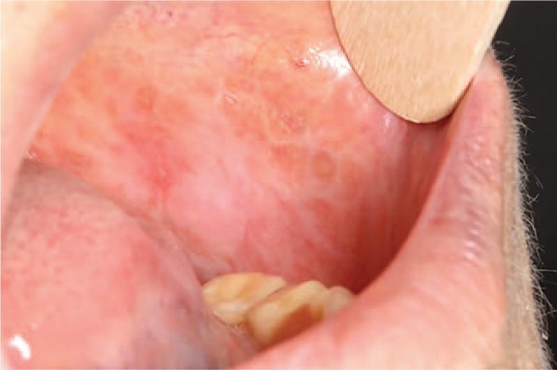
This clinical photograph displays an intraoral view of the left buccal mucosa, highlighting manifestations of oral lichen planus. The mucosa exhibits a characteristic mixed appearance featuring both reticular and atrophic elements. Predominant white, interlacing striae (Wickham striae) form a fine, web-like reticular pattern across a background of erythematous, thinned (atrophic) tissue. The affected area shows signs of chronic inflammation without overt ulceration or erosions. A wooden tongue depressor is visible in the upper right quadrant of the frame, retracting the lip for better visualization of the oral cavity. This image serves as a representative example of oral lichen planus, illustrating the key morphological features used to differentiate subtypes and monitor disease progression in oral medicine and dermatology.
hypertrophic lichen planus shins
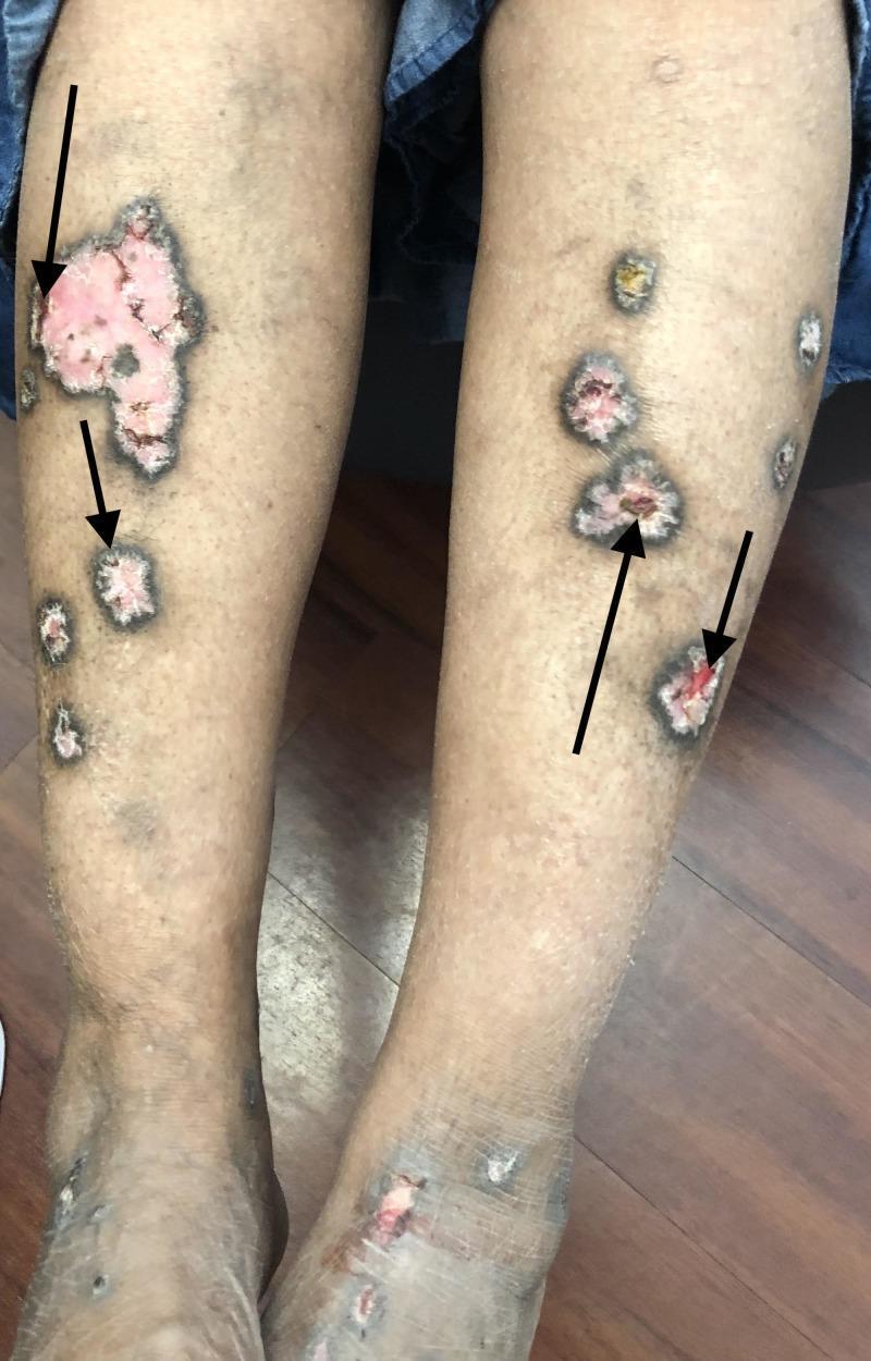
A clinical photograph of the lower extremities of a 63-year-old patient demonstrating hypertrophic lichen planus. The image shows multiple, well-circumscribed, pink-to-violaceous hypertrophic plaques distributed over the distal shins and dorsal feet. Key morphological features include a prominent border of peripheral hyperpigmentation and central areas of ulceration, as indicated by black arrows. The ulcerated regions reveal raw, erythematous tissue with focal loss of the epidermal barrier. The non-ulcerated portions of the plaques appear thickened with a rough, verrucous texture. These findings are characteristic of the hypertrophic variant of lichen planus, emphasizing the chronic, inflammatory nature of the disease and its potential for ulceration. The presentation serves as an educational example for dermatological diagnosis, highlighting the hallmark features of plaque morphology, pigmentary changes, and tissue erosion in a typical anatomical distribution.
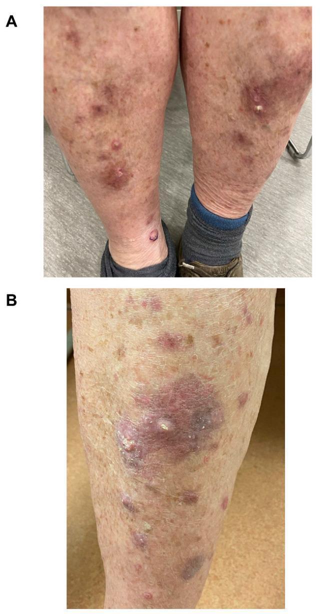
Clinical photograph of the bilateral lower limbs (Figure A) and a close-up detail (Figure B) demonstrating a cutaneous eruption characterized by violaceous papules and hypertrophic nodules. The lesions are distributed primarily over the shins and anterior surfaces of the lower legs. Individual lesions range from small, well-demarcated erythematous-to-violaceous papules to larger, indurated nodules with a rough, hyperkeratotic surface texture. Figure B shows a prominent violaceous plaque with central crusting and focal white scaling, suggestive of pseudoepitheliomatous hyperplasia or lichenoid dermatitis. The surrounding skin exhibits signs of chronological aging with mild diffuse erythema and mottled hyperpigmentation. This presentation is clinically representative of hypertrophic lichen planus, specifically appearing as a cutaneous immune-related adverse event (irAE) following immune checkpoint inhibitor therapy.
ls -la /tmp/workspace/; ls /tmp/skills/shared/scripts/
mkdir -p /tmp/workspace/lp-ppt/assets /tmp/workspace/lp-ppt/output && cd /tmp/workspace/lp-ppt && python /tmp/skills/shared/scripts/fetch_images.py "https://cdn.orris.care/cdss_images/pmc_clinical_VQA_446b1ed4dff9724e6e6bb3c32957463e5dd8bf93a7650e70ba0543b9189220d9.jpg" "https://cdn.orris.care/cdss_images/pmc_clinical_VQA_039e9a327efd138ce88d66ca630de0c28e27f7a60c99ee30c73fd0c03e1b4dfa.jpg" "https://cdn.orris.care/cdss_images/pmc_clinical_VQA_60440e12212a8200b901fb720f1680fb59f957574f05746d8913c4693bfd8f15.jpg" "https://cdn.orris.care/cdss_images/DermNetNZ_1760035570110_16dad32f-bb5a-4d8a-a54f-86d3e627dacd.jpg" "https://cdn.orris.care/cdss_images/pmc_clinical_VQA_5e87415cd2f81071c5928974a7afc7eb7d4e574eda2acd92e85e2182bcf1f51b.jpg" "https://cdn.orris.care/cdss_images/DermNetNZ_1760029487713_3587cfec-61aa-43d6-8a3e-e85010edf0fc.jpg" > assets/imgs.json; python - <<'E' import json,base64 d=json.load(open('/tmp/workspace/lp-ppt/assets/imgs.json')) names=['composite','oral','umbilical_derm','wrists','hypertrophic','annular_derm'] from PIL import Image for n,i in zip(names,d): if i['base64']: b=i['base64'].split('base64,')[1] ext='jpg' open(f'/tmp/workspace/lp-ppt/assets/{n}.jpg','wb').write(base64.b64decode(b)) im=Image.open(f'/tmp/workspace/lp-ppt/assets/{n}.jpg');print(n,im.size) else: print(n,i['error']) E cp /tmp/workspace/attachments/*/IMG_3724.jpg /tmp/workspace/lp-ppt/assets/case.jpg; python -c " from PIL import Image;im=Image.open('/tmp/workspace/lp-ppt/assets/case.jpg');print(im.size)"
~/lp-ppt/build.py
from pptx import Presentation
from pptx.util import Inches, Pt
from pptx.dml.color import RGBColor
from pptx.enum.text import PP_ALIGN, MSO_ANCHOR
from pptx.enum.shapes import MSO_SHAPE
from PIL import Image

A = "/tmp/workspace/lp-ppt/assets/"
PLUM = RGBColor(0x4A, 0x1F, 0x5E)
VIOLET = RGBColor(0x7B, 0x4B, 0x94)
LILAC = RGBColor(0xEE, 0xE6, 0xF3)
GOLD = RGBColor(0xE0, 0xA4, 0x2B)
INK = RGBColor(0x2B, 0x2B, 0x33)
GREY = RGBColor(0x6B, 0x6B, 0x75)
WHITE = RGBColor(0xFF, 0xFF, 0xFF)
FONT = "Calibri"

prs = Presentation()
prs.slide_width = Inches(13.333)
prs.slide_height = Inches(7.5)
BLANK = prs.slide_layouts[6]
W, H = 13.333, 7.5


def rect(s, x, y, w, h, fill, line=None, shape=MSO_SHAPE.RECTANGLE):
    r = s.shapes.add_shape(shape, Inches(x), Inches(y), Inches(w), Inches(h))
    r.fill.solid()
    r.fill.fore_color.rgb = fill
    if line is None:
        r.line.fill.background()
    else:
        r.line.color.rgb = line
    r.shadow.inherit = False
    return r


def text(s, x, y, w, h, content, size=16, bold=False, color=INK, align=PP_ALIGN.LEFT,
         anchor=MSO_ANCHOR.TOP, bullets=False, spacing=6):
    tb = s.shapes.add_textbox(Inches(x), Inches(y), Inches(w), Inches(h))
    tf = tb.text_frame
    tf.word_wrap = True
    tf.vertical_anchor = anchor
    tf.margin_left = tf.margin_right = Inches(0.05)
    tf.margin_top = tf.margin_bottom = Inches(0.03)
    items = content if isinstance(content, list) else [content]
    for i, it in enumerate(items):
        p = tf.paragraphs[0] if i == 0 else tf.add_paragraph()
        p.alignment = align
        p.space_after = Pt(spacing)
        label = None
        if isinstance(it, tuple):
            label, it = it
        pre = "\u2022  " if bullets else ""
        if label:
            r0 = p.add_run()
            r0.text = pre + label + " "
            r0.font.bold = True
            r0.font.size = Pt(size)
            r0.font.name = FONT
            r0.font.color.rgb = VIOLET if color == INK else color
            pre = ""
        r = p.add_run()
        r.text = pre + it
        r.font.size = Pt(size)
        r.font.bold = bold
        r.font.name = FONT
        r.font.color.rgb = color
    return tb


def pic(s, path, x, y, bw, bh, caption=None, align="center"):
    im = Image.open(path)
    ar = im.size[0] / im.size[1]
    w, h = bw, bw / ar
    if h > bh:
        h, w = bh, bh * ar
    px = x + (bw - w) / 2 if align == "center" else x
    py = y + (bh - h) / 2 if align == "center" else y
    s.shapes.add_picture(path, Inches(px), Inches(py), Inches(w), Inches(h))
    if caption:
        text(s, x, y + bh + 0.03, bw, 0.4, caption, size=11, color=GREY, align=PP_ALIGN.CENTER)
    return px, py, w, h


def header(s, title, sub=None, n=None):
    rect(s, 0, 0, W, 1.15, PLUM)
    rect(s, 0, 1.15, W, 0.06, GOLD)
    text(s, 0.6, 0.12, 11.5, 0.7, title, size=30, bold=True, color=WHITE, anchor=MSO_ANCHOR.MIDDLE)
    if sub:
        text(s, 0.6, 0.72, 11.5, 0.4, sub, size=14, color=RGBColor(0xD9, 0xC8, 0xE6))
    if n:
        text(s, W - 1.1, 7.05, 0.8, 0.3, str(n), size=11, color=GREY, align=PP_ALIGN.RIGHT)


def footer(s, src):
    text(s, 0.6, 7.05, 10.5, 0.3, src, size=10, color=GREY)


def table(s, x, y, colw, rows, size=13, rowh=0.5, head_fill=VIOLET):
    nrows, ncols = len(rows), len(colw)
    gs = s.shapes.add_table(nrows, ncols, Inches(x), Inches(y), Inches(sum(colw)), Inches(rowh * nrows))
    t = gs.table
    for i, w in enumerate(colw):
        t.columns[i].width = Inches(w)
    for r in range(nrows):
        t.rows[r].height = Inches(rowh)
        for c in range(ncols):
            cell = t.cell(r, c)
            cell.margin_left = cell.margin_right = Inches(0.08)
            cell.margin_top = cell.margin_bottom = Inches(0.04)
            cell.vertical_anchor = MSO_ANCHOR.MIDDLE
            cell.fill.solid()
            if r == 0:
                cell.fill.fore_color.rgb = head_fill
            else:
                cell.fill.fore_color.rgb = LILAC if r % 2 == 1 else WHITE
            tf = cell.text_frame
            tf.word_wrap = True
            p = tf.paragraphs[0]
            run = p.add_run()
            run.text = rows[r][c]
            run.font.name = FONT
            run.font.size = Pt(size)
            run.font.bold = (r == 0) or (c == 0)
            run.font.color.rgb = WHITE if r == 0 else (PLUM if c == 0 else INK)
    return t


def notes(s, t):
    s.notes_slide.notes_text_frame.text = t


n = 0
# ---------- 1. Title ----------
s = prs.slides.add_slide(BLANK)
rect(s, 0, 0, W, H, PLUM)
rect(s, 0.6, 3.55, 2.2, 0.07, GOLD)
text(s, 0.6, 1.7, 8.2, 1.8, "Lichen Planus", size=54, bold=True, color=WHITE, anchor=MSO_ANCHOR.BOTTOM)
text(s, 0.6, 3.8, 7.8, 1.2, "Diagnosis and management of cutaneous disease", size=24, color=RGBColor(0xE6, 0xD9, 0xEE))
text(s, 0.6, 6.3, 8, 0.5, "Based on Fitzpatrick's Dermatology and Andrews' Diseases of the Skin", size=14, color=RGBColor(0xC9, 0xB3, 0xD8))
pic(s, A + "wrists.jpg", 8.9, 1.3, 3.9, 2.9)
pic(s, A + "annular_derm.jpg", 8.9, 4.3, 3.9, 2.9)
notes(s, "Overview: how to recognise, confirm and treat cutaneous lichen planus.")

# ---------- 2. Overview ----------
n = 2
s = prs.slides.add_slide(BLANK)
header(s, "Lichen planus at a glance", "A T-cell mediated inflammatory disease of skin, mucosa, nails and hair follicles", n)
cards = [
    ("Who", "Mostly adults aged 40 to 70. Childhood LP is 5% or less overall, but 10 to 20% in the Indian subcontinent, Arab countries and Mexico."),
    ("How common", "Cutaneous LP affects about 0.3% of men and 0.1% of women. Oral LP is more common: 1.5% of men, 2.3% of women."),
    ("Mechanism", "T-cell disorder with raised Th1 cytokines and T-cell activity at the basement membrane zone. HLA and TNF-alpha variants reported."),
    ("Course", "About two thirds of skin cases clear within a year. Hypertrophic and mucosal disease is more chronic. Recurrence is common."),
]
for i, (t, b) in enumerate(cards):
    x = 0.6 + (i % 2) * 6.15
    y = 1.6 + (i // 2) * 2.55
    rect(s, x, y, 5.95, 2.3, LILAC)
    rect(s, x, y, 0.09, 2.3, VIOLET)
    text(s, x + 0.3, y + 0.15, 5.4, 0.5, t, size=22, bold=True, color=PLUM)
    text(s, x + 0.3, y + 0.75, 5.4, 1.5, b, size=16)
footer(s, "Source: Andrews' Diseases of the Skin, Lichen Planus chapter")

# ---------- 3. Clinical features ----------
n = 3
s = prs.slides.add_slide(BLANK)
header(s, "Clinical features: the classic lesion", "Small, flat-topped, polygonal papules that expand to 0.5 to 1.5 cm plaques", n)
text(s, 0.6, 1.5, 7.6, 5.4, [
    ("Purple:", "erythematous early, violaceous when developed, hyperpigmented as it resolves (especially in darker skin)."),
    ("Polygonal, planar:", "flat-topped, shiny, dry surface with scant adherent scale."),
    ("Wickham striae:", "white-grey lines or puncta across the surface, best seen with dermoscopy."),
    ("Pruritic:", "often marked, may precede the rash. Patients rub rather than scratch."),
    ("Koebner phenomenon:", "new lesions along sites of trauma, giving linear arrangements."),
    ("Favoured sites:", "flexor wrists, trunk, medial thighs, shins, dorsal hands, glans penis. Usually bilateral and symmetric."),
    ("Nails:", "involved in 5 to 10%: longitudinal ridging, splitting, pterygium."),
], size=16, bullets=True, spacing=9)
pic(s, A + "composite.jpg", 8.7, 1.45, 4.0, 5.45, "Oral, forearm and nail lichen planus")
footer(s, "Source: Andrews' Diseases of the Skin, Lichen Planus chapter")

# ---------- 4. Variants ----------
n = 4
s = prs.slides.add_slide(BLANK)
header(s, "Sites and variants", "Recognising the variant guides the choice of treatment", n)
table(s, 0.6, 1.55, [2.6, 5.4, 4.1], [
    ["Variant", "Typical features", "Practical point"],
    ["Classic", "Violaceous flat-topped papules, flexor wrists, trunk, legs", "Topical steroid first"],
    ["Hypertrophic", "Thick warty plaques, anterior shins, intense itch", "Occlusion, intralesional steroid, acitretin"],
    ["Oral", "Reticular white striae, or painful erosions of buccal mucosa", "Check for genital disease; long-term follow-up"],
    ["Genital / vulvovaginal", "Papules or annular lesions; erosive disease causes pain, scarring", "Distinguish from lichen sclerosus"],
    ["Nail", "Ridging, thinning, pterygium, anonychia", "Early treatment prevents scarring"],
    ["Scalp (planopilaris)", "Perifollicular scale and scarring alopecia", "Needs urgent treatment"],
    ["Actinic", "Photo-distributed, often annular", "Hydroxychloroquine first line"],
], size=14, rowh=0.62)
footer(s, "Sources: Andrews' Diseases of the Skin; Fitzpatrick's Dermatology")

# ---------- 5. Case ----------
n = 5
s = prs.slides.add_slide(BLANK)
header(s, "Case image: forearm lesions", "Likely lichen planus; confirm before long-term treatment", n)
pic(s, A + "case.jpg", 0.8, 1.45, 3.3, 5.5)
text(s, 4.6, 1.5, 8.2, 0.5, "What the image shows", size=22, bold=True, color=PLUM)
text(s, 4.6, 2.1, 8.2, 2.3, [
    "Several round to oval grey-violet papules and small plaques on the flexor forearm and wrist.",
    "Fine adherent white-grey scale with a lacy surface pattern, possibly Wickham striae.",
    "Some lesions merge into a short linear band, suggesting Koebnerization.",
], size=16, bullets=True, spacing=7)
text(s, 4.6, 4.45, 8.2, 0.5, "What is still needed", size=22, bold=True, color=PLUM)
text(s, 4.6, 5.0, 8.2, 2.0, [
    "Itch, duration, drug history.",
    "Mouth, genitals, nails and scalp examination.",
    "Dermoscopy, then a 4 mm punch biopsy.",
], size=16, bullets=True, spacing=7)
footer(s, "Atypical or non-classic features justify a biopsy")

# ---------- 6. Diagnosis pathway ----------
n = 6
s = prs.slides.add_slide(BLANK)
header(s, "Diagnosis: step by step", "Clinical pattern first, then dermoscopy, then histology when in doubt", n)
steps = [
    ("1", "Clinical", "6 P's: purple, polygonal, planar, pruritic papules and plaques. Check Wickham striae, Koebner sign, mucosa, nails."),
    ("2", "Dermoscopy", "White interlacing Wickham striae on a violaceous-brown background; coiled vessels and fine scale."),
    ("3", "Biopsy", "4 mm punch from an intact, untreated lesion. DIF only if lupus or immunobullous disease is suspected."),
    ("4", "Exclude mimics", "Drug history, syphilis serology, hepatitis B and C, fasting glucose and lipids."),
]
for i, (num, t, b) in enumerate(steps):
    x = 0.6 + i * 3.1
    rect(s, x, 1.7, 2.95, 3.6, LILAC)
    c = rect(s, x + 0.2, 1.9, 0.6, 0.6, VIOLET, shape=MSO_SHAPE.OVAL)
    text(s, x + 0.2, 1.9, 0.6, 0.6, num, size=22, bold=True, color=WHITE, align=PP_ALIGN.CENTER, anchor=MSO_ANCHOR.MIDDLE)
    text(s, x + 0.2, 2.65, 2.6, 0.5, t, size=20, bold=True, color=PLUM)
    text(s, x + 0.2, 3.2, 2.6, 2.1, b, size=14)
rect(s, 0.6, 5.65, 12.1, 1.15, PLUM)
text(s, 0.85, 5.7, 11.6, 1.05,
     "A clinical diagnosis is acceptable when the picture is classic. Biopsy when atypical, hypertrophic, solitary, non-healing, drug-related or not responding to treatment.",
     size=16, color=WHITE, anchor=MSO_ANCHOR.MIDDLE)
footer(s, "Source: Andrews' Diseases of the Skin; Yamada's Textbook of Gastroenterology")

# ---------- 7. Dermoscopy and clinical images ----------
n = 7
s = prs.slides.add_slide(BLANK)
header(s, "Seeing the diagnosis", "Wickham striae on clinical and dermoscopic views", n)
pic(s, A + "umbilical_derm.jpg", 0.6, 1.5, 6.0, 2.3, "Umbilical LP (left) with dermoscopy (right): white interlacing striae")
pic(s, A + "annular_derm.jpg", 7.0, 1.5, 5.7, 4.3, "Dermoscopy, annular LP on the forearm")
pic(s, A + "oral.jpg", 0.6, 4.35, 6.0, 2.4, "Oral reticular LP: lacy white striae on the buccal mucosa")
text(s, 7.0, 6.3, 5.7, 0.7, "Dermoscopy helps separate LP from psoriasis, Bowen's disease and discoid lupus.", size=14, color=PLUM, bold=True)
footer(s, "Images: Orris medical image library (DermNet and PMC clinical figures)")

# ---------- 8. Histology ----------
n = 8
s = prs.slides.add_slide(BLANK)
header(s, "Biopsy and histology", "The confirmatory test", n)
text(s, 0.6, 1.5, 6.2, 0.5, "Technique", size=22, bold=True, color=PLUM)
text(s, 0.6, 2.05, 6.2, 2.6, [
    "4 mm punch from a fully developed, intact papule or plaque.",
    "Avoid excoriated lesions and areas treated with topical steroid in the past 1 to 2 weeks.",
    "Formalin for routine histology; separate sample for DIF only if needed.",
], size=16, bullets=True, spacing=8)
text(s, 0.6, 4.45, 6.2, 0.5, "If the biopsy is not typical", size=22, bold=True, color=PLUM)
text(s, 0.6, 5.0, 6.2, 2.0, [
    "Parakeratosis, eosinophils or plasma cells suggest a lichenoid drug eruption.",
    "Follicular plugging and a thick basement membrane suggest discoid lupus.",
], size=16, bullets=True, spacing=8)
rect(s, 7.2, 1.5, 5.5, 5.35, LILAC)
text(s, 7.5, 1.6, 5.0, 0.5, "Typical findings", size=22, bold=True, color=PLUM)
text(s, 7.5, 2.2, 5.0, 4.6, [
    "Dense band-like lymphocytic infiltrate hugging the dermo-epidermal junction",
    "Liquefactive degeneration of the basal layer",
    "Wedge-shaped hypergranulosis",
    "Orthokeratosis, without parakeratosis",
    "Saw-tooth rete ridges",
    "Civatte (colloid) bodies; pigment incontinence",
], size=16, bullets=True, spacing=9)
footer(s, "Source: Yamada's Textbook of Gastroenterology (Lichen planus); Dermatology 5e")

# ---------- 9. Differential ----------
n = 9
s = prs.slides.add_slide(BLANK)
header(s, "Differential diagnosis", "What else looks like lichen planus on the forearm", n)
table(s, 0.6, 1.55, [3.2, 4.3, 4.6], [
    ["Condition", "Clue for it", "Clue against LP"],
    ["Lichenoid drug eruption", "Recent new drug; wider, eczematous spread", "Parakeratosis, eosinophils on biopsy"],
    ["Psoriasis", "Thick silvery scale, extensor sites", "No violaceous hue or Wickham striae"],
    ["Discoid lupus", "Follicular plugging, atrophy, scarring", "Basement membrane thickening, mucin"],
    ["Bowen's disease", "Solitary, persistent scaly plaque", "Atypical keratinocytes"],
    ["Secondary syphilis", "Systemic features, palm and sole lesions", "Positive serology"],
    ["Lichen simplex chronicus", "Lichenified, rubbed area", "Compact hyperkeratosis, no interface change"],
    ["Hypertrophic LP vs. prurigo", "Thick plaques on the shins", "Biopsy helps; exclude SCC"],
], size=14, rowh=0.62)
footer(s, "Compiled from Andrews' Diseases of the Skin and Dermatology 5e")

# ---------- 10. Work-up ----------
n = 10
s = prs.slides.add_slide(BLANK)
header(s, "Work-up and associations", "Look beyond the skin", n)
cols = [
    ("Examine", ["Mouth: lacy white striae, erosions", "Genitals and anus", "Nails and scalp", "Other Koebner sites"]),
    ("Ask", ["Itch severity and duration", "New drugs: thiazides, beta-blockers, ACE inhibitors, antimalarials, NSAIDs, anti-TB, antiepileptics", "Pain with eating or sex"]),
    ("Test", ["Biopsy if atypical", "HCV and HBV serology", "Fasting glucose and lipids", "VDRL/RPR if risk", "KOH if annular scale"]),
]
for i, (t, items) in enumerate(cols):
    x = 0.6 + i * 4.1
    rect(s, x, 1.6, 3.9, 5.2, LILAC)
    rect(s, x, 1.6, 3.9, 0.7, VIOLET)
    text(s, x, 1.6, 3.9, 0.7, t, size=22, bold=True, color=WHITE, align=PP_ALIGN.CENTER, anchor=MSO_ANCHOR.MIDDLE)
    text(s, x + 0.2, 2.5, 3.5, 4.2, items, size=15, bullets=True, spacing=8)
footer(s, "Hepatitis C and metabolic screening are clinical-practice additions, not drawn from the retrieved text")

# ---------- 11. Management algorithm ----------
n = 11
s = prs.slides.add_slide(BLANK)
header(s, "Management overview", "Stepwise approach for cutaneous lichen planus", n)
boxes = [
    ("General", "Stop culprit drug. Avoid rubbing and trauma. Emollient and antihistamine for itch.", LILAC, INK),
    ("Limited disease", "High-potency topical steroid twice daily for 2 to 4 weeks. Occlusion for thick plaques.", VIOLET, WHITE),
    ("Not responding or hypertrophic", "Higher potency steroid, intralesional triamcinolone, add tacrolimus.", VIOLET, WHITE),
    ("Widespread or refractory", "Narrowband UVB, or systemic: oral steroid taper plus methotrexate, acitretin, sulfasalazine and others.", PLUM, WHITE),
]
for i, (t, b, f, tc) in enumerate(boxes):
    y = 1.55 + i * 1.3
    rect(s, 0.6, y, 3.4, 1.15, f if f != LILAC else LILAC)
    text(s, 0.7, y, 3.2, 1.15, t, size=18, bold=True, color=PLUM if f == LILAC else WHITE, anchor=MSO_ANCHOR.MIDDLE)
    rect(s, 4.1, y, 8.6, 1.15, WHITE, line=VIOLET)
    text(s, 4.3, y, 8.3, 1.15, b, size=16, anchor=MSO_ANCHOR.MIDDLE)
text(s, 0.6, 6.75, 12, 0.3, "Review at 4 weeks and step up if there is no response.", size=14, bold=True, color=PLUM)
footer(s, "Source: Fitzpatrick's Dermatology, Skin-Directed and Systemic Therapies")

# ---------- 12. Topical ----------
n = 12
s = prs.slides.add_slide(BLANK)
header(s, "Skin-directed therapy", "First line for limited disease", n)
table(s, 0.6, 1.55, [3.0, 5.0, 4.1], [
    ["Treatment", "Use", "Notes"],
    ["High-potency topical steroid", "Twice daily; first line for limited disease", "Move to higher potency or intralesional if no response in 2 to 4 weeks"],
    ["Occlusion", "Thick or hypertrophic plaques", "Improves penetration"],
    ["Intralesional steroid", "5 to 10 mg/mL monthly for resistant or hypertrophic lesions", "Avoid excess trauma; combine with topical or sparing agent"],
    ["Topical tacrolimus 0.1%", "Second line, steroid-sparing", "As effective as clobetasol 0.05% in oral LP"],
    ["Calcipotriene", "Alternative topical", "Equal to betamethasone valerate over 12 weeks in one RCT"],
    ["Narrowband UVB (311 nm)", "Three times weekly until remission, then taper over 3 to 6 weeks", "About 70% remission in one series"],
], size=14, rowh=0.72)
footer(s, "Source: Fitzpatrick's Dermatology, p. 573")

# ---------- 13. Systemic ----------
n = 13
s = prs.slides.add_slide(BLANK)
header(s, "Systemic therapy", "For severe, widespread or protracted disease", n)
table(s, 0.6, 1.5, [2.7, 5.2, 4.2], [
    ["Agent", "Dose and evidence", "Best for / caution"],
    ["Oral prednisolone", "4 to 6 week taper; rapid control", "High relapse. Never as long-term monotherapy"],
    ["Methotrexate", "15 to 20 mg weekly for 4 to 24 weeks; complete response 58 to 91%", "Preferred, cost-effective; hypertrophic, recalcitrant"],
    ["Acitretin", "30 mg/day for 8 weeks; marked improvement in 64%", "Hypertrophic; watch lipids, dryness"],
    ["Sulfasalazine", "1 g/day, up by 0.5 g every 3 days to 2.5 g/day; 83% improved", "Generalized; monitor blood count, LFTs"],
    ["Metronidazole", "250 mg three times daily for 12 weeks; 74% response", "Generalized; watch neuropathy"],
    ["Hydroxychloroquine", "200 to 400 mg/day for 6 to 12 months", "Second line; first line in actinic LP"],
    ["MMF, azathioprine, cyclosporine", "MMF 2 to 3 g/day; delayed response", "Refractory or ulcerative disease"],
], size=13, rowh=0.64)
footer(s, "Source: Fitzpatrick's Dermatology, Systemic Therapies, p. 573-574. Verify doses locally before prescribing.")

# ---------- 14. Hypertrophic ----------
n = 14
s = prs.slides.add_slide(BLANK)
header(s, "Hypertrophic and special-site disease", "Harder to treat and more likely to scar or persist", n)
pic(s, A + "hypertrophic.jpg", 0.6, 1.45, 3.7, 5.35, "Hypertrophic LP, distal shins")
text(s, 4.8, 1.5, 7.9, 5.4, [
    ("Hypertrophic:", "thick, warty plaques with intense itch. Use occlusion, intralesional steroid, then acitretin or methotrexate. Biopsy if ulcerated to exclude SCC."),
    ("Oral:", "potent topical steroid or tacrolimus in an adhesive base; antifungal cover for candidiasis; long-term mucosal follow-up."),
    ("Genital:", "potent or moderately potent steroid ointment controls most classic disease; hypertrophic or erosive disease needs specialist care."),
    ("Nail and scalp:", "treat early to prevent permanent scarring; methotrexate and hydroxychloroquine are used."),
    ("Actinic:", "hydroxychloroquine is first line, with sun protection."),
], size=16, bullets=True, spacing=11)
footer(s, "Sources: Fitzpatrick's Dermatology; Dermatology 5e (genital LP)")

# ---------- 15. Counselling ----------
n = 15
s = prs.slides.add_slide(BLANK)
header(s, "Counselling and follow-up", "Set expectations early", n)
items = [
    ("Natural history", "Most skin disease clears within 1 to 2 years. Mucosal and hypertrophic forms last longer."),
    ("Pigmentation", "Post-inflammatory hyperpigmentation is common in darker skin and fades slowly."),
    ("Relapse", "Expect recurrence. Taper treatment rather than stopping abruptly."),
    ("Review", "4 weeks after starting, then every 3 to 6 months for oral, genital or chronic disease."),
    ("Red flags", "Ulceration, a rapidly growing plaque, or erosive mucosal disease needs biopsy and specialist review."),
    ("Safety", "Steroid atrophy with prolonged potent use. Monitor bloods on systemic agents."),
]
for i, (t, b) in enumerate(items):
    x = 0.6 + (i % 2) * 6.15
    y = 1.55 + (i // 2) * 1.75
    rect(s, x, y, 5.95, 1.6, LILAC)
    rect(s, x, y, 0.09, 1.6, GOLD)
    text(s, x + 0.3, y + 0.1, 5.5, 0.45, t, size=19, bold=True, color=PLUM)
    text(s, x + 0.3, y + 0.6, 5.5, 1.0, b, size=14)
footer(s, "Source: Andrews' Diseases of the Skin; Fitzpatrick's Dermatology")

# ---------- 16. Take-home ----------
n = 16
s = prs.slides.add_slide(BLANK)
rect(s, 0, 0, W, H, PLUM)
text(s, 0.8, 0.6, 11, 0.9, "Key points", size=40, bold=True, color=WHITE)
rect(s, 0.8, 1.55, 1.6, 0.07, GOLD)
text(s, 0.8, 1.9, 11.7, 4.2, [
    "Think lichen planus for pruritic violaceous flat-topped papules on flexor wrists and forearms.",
    "Confirm with dermoscopy and a punch biopsy when the picture is atypical.",
    "Exclude drug-induced lichenoid eruption, and screen for hepatitis C and metabolic disease.",
    "Start with a high-potency topical steroid; add intralesional steroid, tacrolimus or NB-UVB if needed.",
    "Use methotrexate, acitretin or sulfasalazine for widespread or refractory disease.",
    "Always examine the mouth and genitals, and follow up chronic disease.",
], size=20, color=WHITE, bullets=True, spacing=14)
text(s, 0.8, 6.3, 11.7, 0.8,
     "References: Fitzpatrick's Dermatology (Lichen planus, therapy); Andrews' Diseases of the Skin (Lichen planus); Yamada's Textbook of Gastroenterology; Dermatology 5e. Recent review: PMID 42296289.",
     size=12, color=RGBColor(0xC9, 0xB3, 0xD8))

prs.save("/tmp/workspace/lp-ppt/output/Lichen_Planus_Diagnosis_and_Management.pptx")
print("saved")
Why hcq