
Peripartum cardiomyopathy management
"peripartum cardiomyopathy" AND management
peripartum cardiomyopathy echocardiogram dilated cardiomyopathy
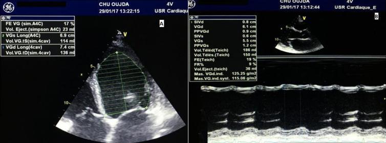
Transthoracic echocardiogram (TTE) in a patient with dilated cardiomyopathy, presented in two panels. Panel A displays a 2D apical 4-chamber view during systole. The Simpson's biplane method is applied to the dilated left ventricle, showing severe global hypokinesia with a measured ejection fraction (LVEF) of 17% and an end-systolic volume (ESV) of 114 ml. Panel B shows a parasternal long-axis view with M-mode tracing across the left ventricle. Quantitative analysis indicates a dilated left ventricular end-diastolic diameter (LVEDD) of 6.1 cm (61 mm) and significantly reduced wall motion, with a Teichholz-calculated ejection fraction of 19% and a fractional shortening of 9%. The interventricular septum and posterior wall thicknesses are relatively thin (0.8 cm and 0.9 cm respectively), consistent with eccentric remodeling. These findings demonstrate severe systolic dysfunction and ventricular dilatation, characteristic of peripartum cardiomyopathy as described in the clinical context.
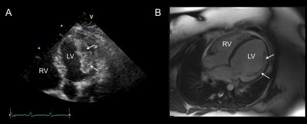
This dual-panel diagnostic image compares a transthoracic echocardiogram (TTE) and a cardiac magnetic resonance (CMR) image of the heart in a patient with peripartum cardiomyopathy. Image A (left) is an apical five-chamber view on TTE, showing a dilated left ventricle (LV) and right ventricle (RV). White arrows point to a large, heterogeneous, layered echodense mass adherent to the lateral wall of the LV, characteristic of an intra-cavitary thrombus. Image B (right) is a four-chamber view from a balanced steady-state free precession (SSFP) CMR sequence taken four days after initiation of anticoagulation. The follow-up image demonstrates near-complete resolution of the previously seen echodense mass. White arrows in Image B highlight the now-visible underlying muscular trabeculations on the endocardial surface of the lateral wall, which were previously obscured by the thrombus. This comparison illustrates the rapid resolution of left ventricular thrombi following medical management in the context of severe biventricular systolic dysfunction.
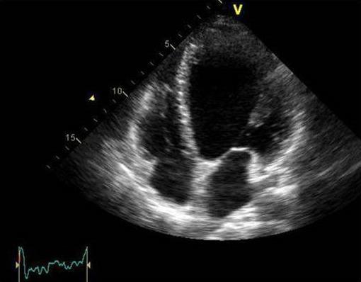
This diagnostic ultrasound image shows a grayscale transthoracic echocardiogram (TTE) in the apical four-chamber (A4C) view. The image demonstrates a significantly dilated and globular left ventricle (LV), which has lost its normal elliptical shape, indicative of dilated cardiomyopathy. All four cardiac chambers are visible: the left ventricle, right ventricle, left atrium, and right atrium. The left ventricle appears disproportionately large compared to the right-sided chambers, and there is evidence of global hypokinesia suggested by the myocardial wall appearance. The interventricular septum and mitral valve apparatus are visible, though the valve leaflets appear stretched due to ventricular remodeling. This clinical finding is characteristic of severe systolic dysfunction, such as peripartum cardiomyopathy, consistent with a reduced ejection fraction. A synchronized ECG tracing is visible in the lower-left corner for timing the cardiac cycle during the imaging study.
Peripartum Cardiomyopathy (PPCM): Management
Definition & Diagnostic Criteria
- LVEF < 45%
- M-mode fractional shortening < 30%
- LV end-diastolic dimension > 2.7 cm/m²
Epidemiology & Risk Factors
- Incidence: 1:1000-4000 deliveries in the US; up to 1:100 in Africa and Haiti
- Incidence in the US has been rising (1:4350 in early 1990s to 1:2230 currently)
- Risk factors: older maternal age, multiparity, twin/multifetal pregnancy, African/Black race (5-15x higher risk), preeclampsia (~25% of PPCM cases), malnutrition, tocolytic therapy, obesity, fertility treatments
- Most diagnoses made within 4 months postpartum; prepartum cases usually in the last month
Pathobiology
- Genetic susceptibility - TTN (titin) truncating variants in ~15% of cases, similar frequency to dilated cardiomyopathy (DCM); also TTNC1, STAT3 mutations
- Prolactin cleavage - The 16-kDa prolactin fragment (vasoinhibin) from late-pregnancy oxidative stress causes myocardial damage and impaired endothelial function
- Antiangiogenic signaling - Placental soluble fms-like tyrosine kinase-1 (sFLT-1) inhibits VEGF, predicting worse outcomes
- Autoimmune/inflammatory mechanisms in some
Management
Immediate Stabilization
| Measure | Notes |
|---|---|
| Loop diuretics | Restore euvolemia; use cautiously in pregnancy (avoid fetal volume depletion) |
| Supplemental oxygen | For hypoxia or pulmonary edema |
| Fluid restriction | Standard heart failure approach |
| Hemodynamic monitoring | If hemodynamically unstable; plan for emergency delivery if needed |
| Delivery planning | Prompt delivery after maternal stabilization if diagnosed antenatally |
Drug Therapy: ANTEPARTUM (fetal safety constraints)
| Drug | Status in Pregnancy | Notes |
|---|---|---|
| Loop diuretics | Safe (use cautiously) | Furosemide preferred |
| Beta-blockers | Safe | Metoprolol preferred over carvedilol |
| Digoxin | Safe | For arrhythmia rate control or adjunct inotropic support |
| Hydralazine + isosorbide dinitrate | Safe (vasodilators of choice) | Replace ACE inhibitors/ARBs during pregnancy |
| ACE inhibitors / ARBs | CONTRAINDICATED | Fetotoxic (renal agenesis, oligohydramnios, skull ossification defects) |
| Sacubitril/valsartan | CONTRAINDICATED | Same fetal risks as ARBs |
| Spironolactone | Use cautiously in late pregnancy | Antiandrogenic effects on fetus; eplerenone should be avoided |
| LMWH | Preferred anticoagulant | For LVEF < 35% or marked dilation; LMWH preferred over UFH; warfarin not recommended in pregnancy |
Drug Therapy: POSTPARTUM
| Drug | Notes |
|---|---|
| ACE inhibitors (e.g., enalapril) | First-line; compatible with breastfeeding |
| Beta-blockers (metoprolol tartrate) | Compatible with breastfeeding |
| Spironolactone | Compatible with breastfeeding in stable patients |
| Sacubitril/valsartan | Can be introduced postpartum (avoid if breastfeeding concerns) |
| Loop diuretics | Continue as needed |
| Anticoagulation | LMWH or warfarin for LVEF < 35% or LV thrombus; continue ~6 weeks postpartum once obstetric bleeding resolved |
Bromocriptine
- Small uncontrolled and non-placebo-controlled trials showed improved EF and clinical outcomes
- A randomized trial (IPAC-like studies) showed no convincing benefit with significant adverse effects
- Current status: Considered experimental/investigational; ongoing large randomized trial pending
- Mostly considered in non-breastfeeding patients with severe PPCM (LVEF < 35%)
Anticoagulation
- Indicated when LVEF < 35% or marked LV dilation (high risk for LV thrombus and systemic embolism)
- LMWH is preferred over UFH and warfarin during pregnancy
- Postpartum: LMWH or warfarin for ~6 weeks once obstetric bleeding has resolved
- Echocardiogram findings of intracavitary thrombus mandate anticoagulation
Arrhythmia Management
- Both atrial and ventricular arrhythmias can occur
- Beta-blockers (metoprolol) and digoxin are safe in pregnancy and postpartum
- Exclude pulmonary embolism and spontaneous coronary artery dissection (SCAD) as mimics
Advanced Heart Failure / Refractory Cases
- Mechanical circulatory support (e.g., IABP, Impella, ECMO) for cardiogenic shock
- Cardiac transplantation: PPCM is the 4th most common indication in women in the US; ~5% of female cardiac transplants; post-transplant outcomes are similar to other indications
Echocardiographic Findings
Prognosis & Recovery
- LVEF normalizes (≥50%) in 50-80% of cases, usually within 6 months of standard therapy
- Recovery is less likely with:
- LVEF < 30% at presentation
- Marked LV dilation
- African/Black race (both slower and less complete recovery)
- TTN truncating variants (predict lower EF at 1 year)
- Spontaneous improvement can continue over 5 years
Subsequent Pregnancy Counseling
| Prior Recovery Status | HF Recurrence Risk | Maternal Mortality |
|---|---|---|
| LVEF normalized | ~20% recurrence | Low (0% in one study) |
| Persistent LV dysfunction | ~40% recurrence | ~19% |
Genetic Evaluation
- Genetic testing (TTN, DCM gene panel) is recommended given ~15% carry truncating TTN variants
- Positive genetic testing predicts less recovery and has implications for first-degree relatives
- Clinical screening of first-degree relatives is recommended
- PPCM may represent DCM triggered by the physiologic stress of pregnancy
Key Management Summary
PPCM Diagnosed
    │
    ├─ ANTEPARTUM
    │   ├─ Diuretics (cautious), O₂, fluid restriction
    │   ├─ Hydralazine + nitrates (vasodilators)
    │   ├─ Metoprolol ± digoxin (arrhythmia)
    │   ├─ LMWH if LVEF <35%
    │   ├─ NO ACEi/ARB/sacubitril
    │   └─ Plan urgent delivery if hemodynamically unstable
    │
    └─ POSTPARTUM
        ├─ Full GDMT: ACEi + beta-blocker + spironolactone ± sacubitril/valsartan
        ├─ Anticoagulation x 6 weeks if LVEF <35%
        ├─ Bromocriptine (experimental, non-breastfeeding, severe cases)
        ├─ Genetic testing
        ├─ Echo at 6 months to assess recovery
        └─ Counsel on subsequent pregnancy risk