
Explain the topic of anti tubercular drug of kdt detailed
anti-tubercular drugs mechanism of action diagram
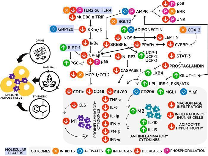
A complex pathophysiology diagram and signaling pathway illustrating the mechanism of action for anti-inflammatory agents (drugs, natural, and synthetic compounds) on inflamed adipose tissue. The visual maps a cascade of intracellular and extracellular molecular players within a hypothetical adipocyte and surrounding immune environment. Key signaling nodes include the inhibition of TLR2/4, MyD88, and TRIF, alongside the activation of AMPK, GRP120, and SIRT-1. The diagram details the downregulation of pro-inflammatory pathways, specifically the NF-kβ complex and its downstream chemokines like MCP-1/CCL2. It also highlights the modulation of metabolic regulators such as PPAR-γ, SREBP1c, and UCP-1/2/3. In the lower section, the diagram shows the transition from a pro-inflammatory M1 macrophage phenotype (characterized by TNF-α, IL-6, and IL-1β) toward an anti-inflammatory M2 phenotype (indicated by IL-10, IL-13, and Arg1). The overall clinical outcomes depicted include reduced macrophage infiltration, decreased adipocyte hypertrophy, and improved insulin sensitivity markers such as GLUT-4 and IRS-1. Symbol legends indicate activation, inhibition, and phosphorylation states for each molecular player.
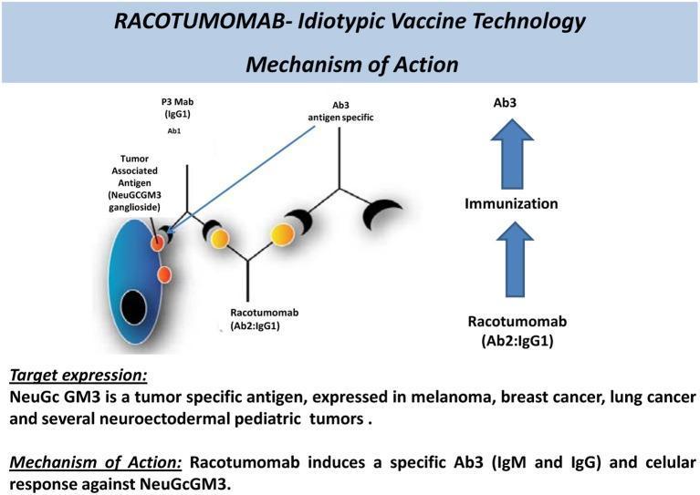
A pathophysiology diagram illustrating the mechanism of action of Racotumomab (Ab2:IgG1), an anti-idiotypic vaccine targeting the NeuGcGM3 ganglioside tumor-associated antigen. The visual shows a tumor cell (blue ellipse) expressing NeuGcGM3 (orange circles). An initial antibody (Ab1, P3 Mab) recognizes this antigen. Racotumomab is depicted as an Ab2 anti-idiotype antibody that mimics the original antigen's structure. The diagram uses a branched Y-shaped schematic to show how Racotumomab administration triggers an active immune response (labeled 'Immunization'), resulting in the production of antigen-specific Ab3 antibodies (IgM and IgG). These Ab3 antibodies are shown binding to the NeuGcGM3 antigen on the tumor cell surface, demonstrating the vaccine's ability to overcome the poor immunogenicity of gangliosides. The text highlights that NeuGcGM3 is expressed in melanoma, breast cancer, lung cancer, and neuroectodermal pediatric tumors, establishing the clinical relevance of this immunotherapy in oncology.
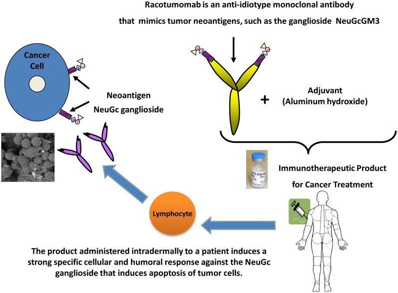
This pathophysiology diagram illustrates the mechanism of action of Racotumomab, an anti-idiotype monoclonal antibody used in cancer immunotherapy. The diagram shows a cancer cell expressing the neoantigen NeuGc ganglioside (specifically NeuGcGM3) on its surface. Racotumomab is depicted as a yellow Y-shaped molecule that mimics these tumor neoantigens. The immunotherapeutic product is formed by combining Racotumomab with an aluminum hydroxide adjuvant, shown alongside a pharmaceutical vial and a syringe icon indicating intradermal administration to a human patient. The flowchart pathways demonstrate that upon administration, the product stimulates lymphocytes to induce a specific cellular and humoral immune response. This response targets the NeuGc ganglioside on the tumor cells, leading to apoptosis. A grayscale inset image displays microscopic evidence of tumor cell destruction. The graphic effectively summarizes the therapeutic sequence from molecular mimicry to the systemic immune-mediated destruction of cancer cells.
Anti-Tubercular Drugs (KDT Style - Detailed)
Overview
Classification
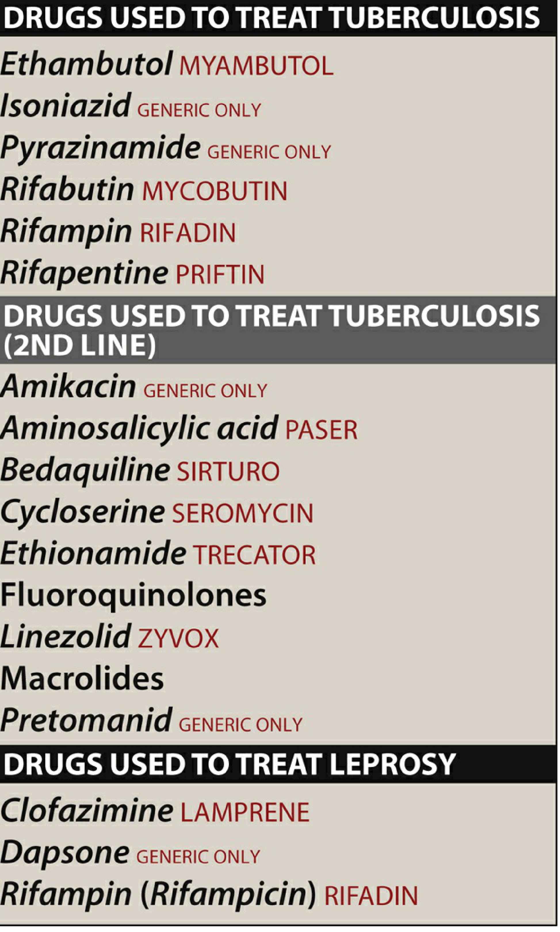
First-Line (Preferred) Drugs
| Drug | Abbreviation | Daily Adult Dose |
|---|---|---|
| Isoniazid | H (INH) | 300 mg/day (5 mg/kg) |
| Rifampicin | R | 600 mg/day (10 mg/kg) |
| Pyrazinamide | Z | 25 mg/kg/day |
| Ethambutol | E | 15-25 mg/kg/day |
| Streptomycin | S | 15 mg/kg/day (IM) |
Second-Line Drugs
Standard Treatment Regimen
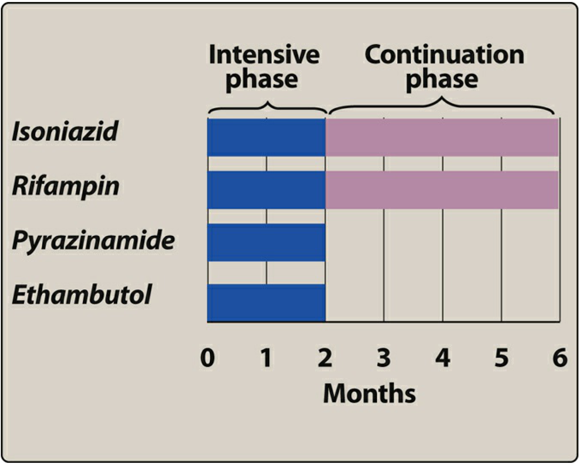
FIRST-LINE DRUGS (Detailed)
1. ISONIAZID (INH)
- INH is a prodrug activated by the mycobacterial catalase-peroxidase enzyme KatG
- The activated form covalently binds to acyl carrier protein reductase (InhA) and β-ketoacyl-ACP synthase (KasA)
- This inhibits mycolic acid synthesis, destroying the integrity of the mycobacterial cell wall
- Active against both intracellular (inside macrophages) and extracellular organisms
- MIC: inhibits most M. tuberculosis at ≤0.2 mcg/mL
- Well absorbed orally; peak plasma level 3-5 mcg/mL at 1-2 hours; absorption reduced by 50% with fatty meals
- Penetrates all body fluids including CSF (CSF level = 20-100% of serum level), caseous material, and macrophages
- Metabolized by N-acetyltransferase (NAT2) in the liver - genetically polymorphic
- Fast acetylators: half-life <1 hour (common in East Asians, Eskimos)
- Slow acetylators: half-life ~3 hours (common in Egyptians, Scandinavians)
- Slow acetylators accumulate more drug and parent compound but generally similar efficacy with daily dosing; risk of toxicity is higher
- Inhibits CYP450 enzymes - raises levels of phenytoin, carbamazepine, benzodiazepines
- Mutation/deletion of KatG gene → inability to activate prodrug (high-level resistance)
- Overexpression of inhA → low-level resistance (also cross-resistance with ethionamide)
- Mutations in KasA or ahpC
- Peripheral neuropathy (most common): due to pyridoxine (B6) deficiency caused by INH competing with pyridoxal phosphate. Prevented by co-administering pyridoxine 10-25 mg/day. Seen more in slow acetylators, malnourished, diabetics, alcoholics, pregnant women
- Hepatotoxicity: elevated transaminases in 10-20% of patients; clinical hepatitis in 1%; more common in >35 years and with alcohol use. Fatal hepatitis rare
- CNS effects: memory impairment, psychosis, seizures (especially in overdose)
- Lupus-like syndrome (antinuclear antibodies)
- Drug interactions: inhibits metabolism of phenytoin (toxicity), carbamazepine, warfarin
2. RIFAMPICIN (Rifampin)
- Binds to the beta subunit of DNA-dependent RNA polymerase (rpoB gene product)
- Blocks RNA transcription → inhibits mRNA synthesis
- Bactericidal; active against M. tuberculosis, MAC, M. leprae, as well as many gram-positive and gram-negative bacteria
- Active against both intracellular and extracellular organisms, including dormant/persister bacilli
- Good oral absorption; taken on an empty stomach (food reduces absorption)
- Widely distributed; penetrates CSF, cells, and caseous material
- Half-life 1.5-5 hours; undergoes enterohepatic recirculation
- Metabolized to desacetylrifampin (still active); excreted primarily in bile/feces
- Colours body secretions red-orange (urine, tears, saliva, sweat, sputum) - warn patients; can stain soft contact lenses
- Mutations in rpoB gene (beta subunit of RNA polymerase) - >96% of resistant strains
- Resistance develops rapidly with monotherapy (never give alone for active TB)
- Cross-resistance between rifampicin and rifabutin is virtually complete
- Hepatotoxicity: liver enzyme elevation; dose-related
- GI upset: nausea, vomiting, abdominal pain
- "Flu-like" syndrome: fever, chills, myalgias - especially with intermittent therapy (hypersensitivity)
- Thrombocytopenia, hemolytic anemia (rare)
- Drug interactions (most important): Rifampicin is a potent inducer of CYP3A4 and other CYP enzymes and P-glycoprotein. It reduces levels of: oral contraceptives (use alternative contraception!), warfarin, antiretrovirals (PIs, NNRTIs), methadone, corticosteroids, azole antifungals, digoxin, phenytoin, sulfonylureas
3. PYRAZINAMIDE (PZA)
- Pyrazinamide is a prodrug hydrolyzed by pyrazinamidase (encoded by pncA gene) to the active form pyrazinoic acid
- Exact mechanism not fully established; likely acidifies intracellular environment of mycobacteria
- Only active in acidic pH (inside macrophage phagolysosomes and necrotic/caseous lesions - pH 5-5.5)
- Active against intracellular organisms and slowly metabolizing "persister" bacilli
- Bactericidal at acidic pH; bacteriostatic at neutral pH
- This unique activity in acidic environments allows the treatment duration to be shortened from 9 months to 6 months
- Good oral absorption; widely distributed; penetrates CSF
- Metabolized by liver; excreted by kidneys
- Half-life ~9 hours
- Loss of pyrazinamidase activity (mutations in pncA)
- Resistance is NOT cross-resistant with other first-line drugs (important advantage)
- Hyperuricemia (most common): inhibits renal tubular secretion of uric acid; asymptomatic in most; rarely precipitates gout
- Hepatotoxicity: most significant toxicity; dose-related elevation of liver enzymes; serious hepatitis possible
- Arthralgia/joint pains (common, related to hyperuricemia)
- Nausea, rash
- Used only for the first 2 months in the standard 6-month regimen (most benefit early in treatment)
4. ETHAMBUTOL (EMB)
- Inhibits arabinosyl transferase (encoded by embCAB operon)
- Arabinosyl transferase is essential for synthesis of arabinogalactan, a major component of the mycobacterial cell wall
- Bacteriostatic (not bactericidal); specific for mycobacteria
- Prevents incorporation of arabinose into the cell wall
- Good oral absorption; penetrates most tissues well
- CNS penetration is variable (adequate in meningeal inflammation, questionably adequate in tuberculous meningitis)
- Excreted mainly in urine (parent drug + metabolites); dose reduction needed in renal failure
- Mutations in embB gene (arabinosyl transferase)
- Optic neuritis (most important and distinctive toxicity): dose-dependent
- Reduced visual acuity, loss of red-green color discrimination, central scotoma
- More common at higher doses (25 mg/kg) and in renal impairment
- Baseline visual acuity and color vision testing is mandatory; repeat monthly
- Usually reversible if drug stopped early
- Hyperuricemia (decreased uric acid excretion)
- Peripheral neuropathy (rare)
5. STREPTOMYCIN
- Aminoglycoside that binds to 30S ribosomal subunit → inhibits protein synthesis
- Bactericidal; active against extracellular organisms
- Poor intracellular penetration (inactive against intracellular bacilli)
- Given intramuscularly (IM) or IV; not absorbed orally
- Does NOT penetrate CSF (poor in normal meningitis; better in inflamed meninges)
- Excreted unchanged by kidneys; accumulates in renal failure
- Mutations in 16S rRNA (rrs gene) or ribosomal protein S12 (rpsL)
- Ototoxicity: both vestibular (balance disturbance, nystagmus, vertigo) and auditory (irreversible hearing loss)
- Nephrotoxicity: dose-dependent; monitor renal function
- Neuromuscular blockade
SECOND-LINE DRUGS
Bedaquiline
- First novel anti-TB drug approved in 40 years (FDA 2012) - a diarylquinoline
- Mechanism: inhibits mycobacterial ATP synthase (novel target) → depletes energy
- Active against both replicating and non-replicating bacilli
- Used in MDR-TB and XDR-TB regimens; part of the BPaL regimen (Bedaquiline + Pretomanid + Linezolid)
- Adverse effects: QT prolongation (serious - ECG monitoring required), hepatotoxicity, nausea
- Dose: 400 mg/day for 2 weeks, then 200 mg 3x/week for 22 weeks
Fluoroquinolones (Levofloxacin, Moxifloxacin)
- Inhibit DNA gyrase (topoisomerase II) and topoisomerase IV
- Used in MDR-TB regimens
- Moxifloxacin is part of the newer 4-month regimen (RPMZ = Rifapentine + Moxifloxacin + Pyrazinamide + INH)
- Adverse effects: QT prolongation (moxifloxacin), tendinopathy, GI upset
Cycloserine
- Structural analogue of D-alanine
- Inhibits D-alanine racemase and D-Ala-D-Ala ligase → blocks cell wall peptidoglycan synthesis
- Used in MDR-TB
- Adverse effects: CNS toxicity (seizures, depression, psychosis) - pyridoxine supplementation required
- Dose: 500-1000 mg/day in divided doses
Ethionamide
- Prodrug structurally similar to INH; also inhibits InhA (mycolic acid synthesis)
- Cross-resistance with INH (inhA mutations)
- Used in MDR-TB
- Adverse effects: GI intolerance (major problem), hepatotoxicity, hypothyroidism (with prolonged use), teratogenic
Para-Aminosalicylic Acid (PAS)
- Inhibits folate synthesis in mycobacteria (similar to sulfonamides)
- Also interferes with iron metabolism and mycobactin synthesis
- Second-line; used in MDR-TB
- Adverse effects: severe GI intolerance, hepatotoxicity, hypothyroidism
Rifabutin
- Rifamycin derivative; similar mechanism to rifampicin (rpoB inhibition)
- Cross-resistance with rifampicin nearly complete
- Preferred over rifampicin in HIV patients on antiretroviral therapy (less potent CYP inducer)
- Adverse effects: hepatotoxicity, leukopenia, thrombocytopenia, optic neuritis; uveitis (dose-related)
Linezolid
- Oxazolidinone; inhibits 50S ribosomal subunit (23S rRNA binding site)
- Active against MDR-TB and XDR-TB; part of BPaL regimen
- Adverse effects: bone marrow suppression (anemia, thrombocytopenia), peripheral neuropathy, optic neuropathy (with prolonged use), serotonin syndrome (with serotonergic agents)
- Pyridoxine supplementation recommended
Capreomycin / Amikacin
- Capreomycin: cyclic polypeptide antibiotic; inhibits protein synthesis (30S ribosome)
- Amikacin: aminoglycoside; inhibits 30S ribosome
- Both given parenterally (IM/IV)
- Adverse effects: ototoxicity, nephrotoxicity
Pretomanid
- Nitroimidazole prodrug; activated in anaerobic conditions
- Inhibits cell wall synthesis and generates cytotoxic nitric oxide
- Used in combination (BPaL regimen) for MDR/XDR-TB
- FDA approved 2019
MDR-TB and XDR-TB Treatment
| Type | Definition | Treatment |
|---|---|---|
| MDR-TB | Resistant to INH + rifampicin | Fluoroquinolone + second-line injectables + other second-line drugs; ~18-24 months |
| XDR-TB | MDR + resistant to fluoroquinolones + second-line injectables | BPaL (Bedaquiline + Pretomanid + Linezolid); newer regimens 6-9 months |
Adverse Effects - High-Yield Summary
| Drug | Key Adverse Effect | Mechanism/Notes |
|---|---|---|
| Isoniazid | Peripheral neuropathy | Pyridoxine deficiency; give B6 supplement |
| Isoniazid | Hepatotoxicity | More in >35 yrs, alcoholics |
| Rifampicin | Red-orange secretions | Harmless; warn patient |
| Rifampicin | Drug interactions | CYP3A4 inducer; reduces OCP, warfarin, ARVs |
| Pyrazinamide | Hyperuricemia | Inhibits uric acid secretion |
| Pyrazinamide | Hepatotoxicity | Most hepatotoxic first-line drug |
| Ethambutol | Optic neuritis | Dose-dependent; test visual acuity monthly |
| Streptomycin | Ototoxicity + nephrotoxicity | Aminoglycoside |
Key Pharmacological Facts (KDT High-Yield Points)
- INH is the most active drug for drug-susceptible TB; the only one with confirmed bactericidal activity across all mycobacterial populations
- Rifampicin is the most important "sterilizing" drug - eradicates persister bacilli
- Pyrazinamide - allows shortening from 9 to 6 months due to activity in acidic environments
- Ethambutol - primarily bacteriostatic; used to protect against undetected resistance
- Fast vs. slow acetylators of INH: peripheral neuropathy more in slow acetylators; hepatotoxicity more in fast acetylators
- INH + rifampicin + B6 is the regime for latent TB infection (LTBI) prophylaxis (preventive therapy)
- Directly Observed Therapy (DOT) is the standard strategy to improve adherence
- Rifampicin increases metabolism of oral contraceptive pills - use alternative contraception
- All first-line drugs are active orally except streptomycin (parenteral only)
- Bedaquiline has the novel mechanism of ATP synthase inhibition - first new TB drug in 40 years
For antifungal topic
antifungal drugs mechanism of action ergosterol cell wall
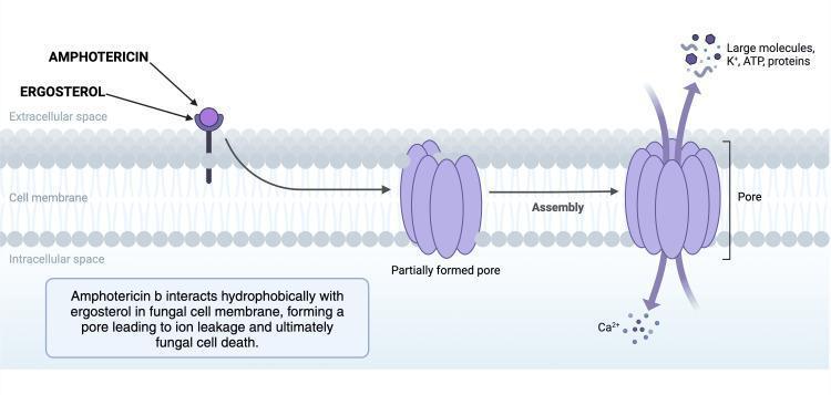
This pathophysiology diagram illustrates the mechanism of action of Amphotericin B, a polyene antifungal agent, on a fungal cell membrane. The diagram depicts the cell membrane as a phospholipid bilayer separating the extracellular space from the intracellular space. The process is shown in a three-step progression from left to right: (1) Initial interaction where Amphotericin B molecules bind hydrophobically to ergosterol, a primary sterol in the fungal membrane; (2) An intermediate state showing a 'partially formed pore' resulting from this interaction; and (3) The final 'Assembly' stage where a complete transmembrane pore is established. The diagram highlights the clinical consequence of pore formation: altered membrane permeability leading to the efflux of large molecules, potassium ions (K+), ATP, and proteins, and the influx of calcium ions (Ca2+). This disruption of cellular homeostasis results in fungal cell death. The visual is intended for medical education regarding antifungal pharmacology and is relevant to the treatment of invasive fungal infections like Cryptococcosis or Candidiasis.
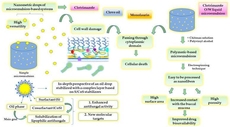
This medical pharmacology diagram illustrates the mechanism and processing of microemulsion-based drug delivery systems for antifungal therapy, specifically targeting Candida albicans. The flowchart is divided into three functional segments: mechanism of action, formulation chemistry, and advanced drug delivery processing. The top left depicts nanometric drops of microemulsion containing antifungal agents like clotrimazole, clove oil, or monolaurin. The mechanism of action shows these droplets causing cell wall damage and passing through the cytoplasmic domain of fungi, ultimately resulting in cellular death. The bottom left details the biochemical composition of the system, highlighting the use of an oil phase combined with surfactants (S) and cosurfactants (CoS) to achieve the solubilization of lipophilic antifungals, aiming for enhanced activity and new molecular targets. The right side of the diagram outlines the pharmaceutical processing where an oil-in-water (O/W) liquid microemulsion is modified with chitosan and polyvinyl alcohol to create polymeric-based microemulsions. These undergo electrospinning to form nanofibers. The educational focus is on how the resulting high surface area and porosity of these nanofibers increase contact with the buccal mucosa, thereby improving drug bioavailability for oral candidiasis treatment.
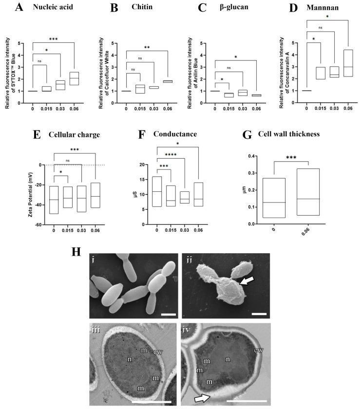
This composite educational image illustrates the effects of the antifungal agent olorofim on the pathogenic yeast Sporothrix brasiliensis. The upper panels (A–G) consist of box-and-whisker plots showing dose-dependent changes in cellular composition and physiological properties across olorofim concentrations (0 to 0.06 µM). Key findings include significant increases in nucleic acid accumulation (A), chitin (B), mannan (D), and cell wall thickness (G), alongside decreases in β-glucan (C), electronegativity (E), and conductance (F). Panel H provides visual confirmation via electron microscopy. Scanning electron microscopy (SEM) compares healthy, elongated untreated yeasts (Hi) with treated cells exhibiting granular surface alterations and compromised wall integrity (Hii, arrow). Transmission electron microscopy (TEM) displays untreated cells (Hiii) with distinct organelles—nucleus (n) and mitochondria (m)—and a thin cell wall (cw). Treated cells (Hiv) exhibit an amorphous shape and significant cell wall remodeling, characterized by increased thickness and an electron-dense modified layer (arrow). This figure serves as a diagnostic reference for understanding antifungal mechanisms of action and fungal cell wall remodeling in medical mycology.
Antifungal Drugs (KDT Style - Detailed)
Why Fungal Infections Are Difficult to Treat
Classification of Antifungal Drugs
By Site of Action:
| Drug Class | Target |
|---|---|
| Polyenes (Amphotericin B, Nystatin) | Cell membrane - bind ergosterol, form pores |
| Azoles (Imidazoles + Triazoles) | Ergosterol synthesis - inhibit CYP51 (lanosterol demethylase) |
| Allylamines (Terbinafine) | Ergosterol synthesis - inhibit squalene epoxidase |
| Echinocandins (Caspofungin, Micafungin) | Cell wall - inhibit β-1,3-glucan synthase |
| Pyrimidine analogs (Flucytosine) | DNA/RNA synthesis |
| Griseofulvin | Mitotic spindle - inhibits microtubule assembly |
Sites of action diagram:
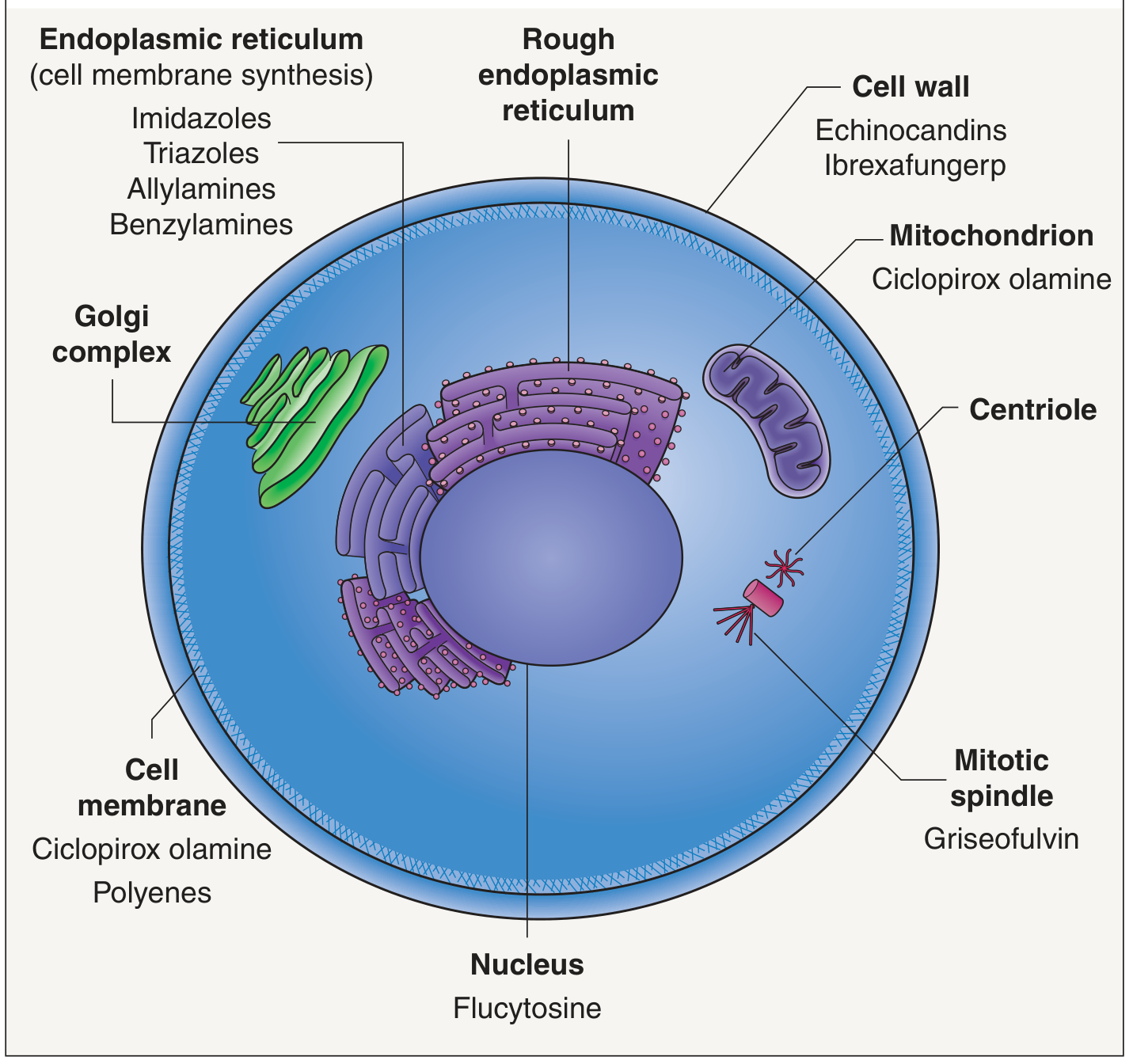
GROUP 1: POLYENE MACROLIDES
AMPHOTERICIN B
- Binds specifically to ergosterol in the fungal cell membrane
- Forms transmembrane pores (ion channels) in the membrane
- Pores cause leakage of K⁺, Mg²⁺, and other intracellular contents
- Results in depolarization and osmotic lysis → fungicidal
- Human cell membranes contain cholesterol (not ergosterol), so there is selective toxicity - but amphotericin B still has some affinity for cholesterol, explaining its toxicity
- Candida spp., Cryptococcus neoformans, Aspergillus spp.
- Endemic fungi: Histoplasma capsulatum, Blastomyces dermatitidis, Coccidioides immitis
- Mucor spp. (Mucormycosis)
- Resistance is rare
- Nearly insoluble in water; formulated as colloidal suspension with sodium deoxycholate for IV use
- NOT absorbed orally (used only topically/orally for GI candidiasis)
- Widely distributed; penetrates most tissues including pleural, peritoneal, synovial fluids
- Poor CSF penetration with IV administration → requires intrathecal injection for fungal meningitis
- Half-life biphasic: initial ~24 hours, terminal ~15 days
- Excreted very slowly; detected in urine for up to 7 weeks after stopping
| Formulation | Type | Dose (mg/kg/d) | Toxicity |
|---|---|---|---|
| Conventional AmB (AMB-d) | Colloidal suspension | 0.5-1 | High nephrotoxicity |
| AmB Lipid Complex (ABLC) | Lipid complex | 5 | Lower |
| AmB Colloidal Dispersion (ABCD) | Colloidal dispersion | 3-6 | Moderate |
| Liposomal AmB (L-AmB) | True liposomes | 3-6 | Lowest |
- Fever, chills, rigors, muscle spasms, vomiting, headache, hypotension - nearly universal
- Management: Slow infusion rate; premedicate with antipyretics, antihistamines, meperidine (for rigors), or corticosteroids
- Give a 1 mg test dose IV first to gauge severity
- Nephrotoxicity (most serious and clinically significant): occurs in almost all patients
- Azotemia (BUN/creatinine rise) - partly reversible (vasoconstriction/prerenal) and partly irreversible (tubular injury)
- Renal tubular acidosis - K⁺ and Mg²⁺ wasting (hypokalemia, hypomagnesemia)
- Risk reduced by sodium loading (normal saline infusion before each dose)
- Irreversible damage usually with >4 g cumulative dose
- Normocytic normochromic anemia: reduced erythropoietin production from damaged renal tubular cells
- Liver function abnormalities (occasional)
- Intrathecal administration: seizures, chemical arachnoiditis, serious neurological sequelae
- Drug of choice for severe, life-threatening systemic fungal infections
- Cryptococcal meningitis (with flucytosine)
- Invasive aspergillosis (now often superseded by voriconazole)
- Invasive candidiasis
- Mucormycosis (only effective option)
- Empirical therapy in febrile neutropenic patients
NYSTATIN
- Polyene macrolide, same mechanism as amphotericin B (binds ergosterol, forms pores)
- TOO TOXIC for parenteral use - used only topically
- Not absorbed from skin, mucous membranes, or GI tract → minimal systemic toxicity
- Available as creams, ointments, lozenges, vaginal suppositories
- Uses: Oropharyngeal candidiasis (thrush), vaginal candidiasis, intertriginous candidal infections, oral GI candidiasis (swish-and-swallow)
- Adverse effects: Unpleasant bitter taste (oral preparations)
GROUP 2: AZOLES
- Inhibit fungal cytochrome P450 enzyme CYP51 (also called lanosterol 14-α-demethylase)
- This enzyme is essential for converting lanosterol → ergosterol in the ergosterol biosynthesis pathway
- Result: depletion of ergosterol + accumulation of toxic methylated sterols → impaired cell membrane function → fungistatic (mostly)
- Triazoles have greater selectivity for fungal CYP than imidazoles → less human toxicity and fewer drug interactions
- Overexpression of efflux pumps (MDR1, CDR1/CDR2)
- Mutations in ERG11 gene (CYP51) reducing azole binding affinity
- Upregulation of ERG11
Pharmacokinetic Comparison Table
| Drug | Solubility | CSF:Serum | Half-life | Elimination | Route |
|---|---|---|---|---|---|
| Ketoconazole | Low | <0.1 | 7-10 h | Hepatic | Oral |
| Itraconazole | Low | <0.01 | 24-42 h | Hepatic | Oral, IV |
| Fluconazole | High | >0.7 | 22-31 h | Renal | Oral, IV |
| Voriconazole | High | >0.21 | 6 h | Hepatic | Oral, IV |
| Posaconazole | Low | - | 25 h | Hepatic | Oral, IV |
| Isavuconazole | High | - | 130 h | Hepatic | Oral, IV |
IMIDAZOLES
Ketoconazole
- First systemic azole; now largely replaced by triazoles
- Limited spectrum; poor CNS penetration; significant drug interactions and toxicity
- Adverse effects:
- Inhibits adrenal and gonadal steroid synthesis (CYP11A1, CYP17A1): gynecomastia, menstrual irregularities, impotence, adrenal insufficiency - unique among azoles
- Hepatotoxicity (most significant)
- GI intolerance
- Still used topically: seborrheic dermatitis, pityriasis versicolor (shampoo, cream)
Clotrimazole, Miconazole
- Used topically only - too toxic for systemic use
- Available OTC; used for vulvovaginal candidiasis, tinea infections, oral thrush (clotrimazole loches)
- Negligible systemic absorption; adverse effects rare
TRIAZOLES (The "Big Four")
1. Fluconazole
- Most widely used antifungal; excellent oral bioavailability and safety
- Unique features:
- Water-soluble → excellent distribution including CSF (CSF:serum >0.7 - best of all azoles)
- Renally excreted (only azole primarily excreted by kidneys); dose adjustment in renal failure required
- Available both oral and IV (same bioavailability - oral preferred when possible)
- Spectrum: Candida (most species), Cryptococcus neoformans
- Resistance: C. krusei (inherently resistant), C. glabrata (often resistant/dose-dependent)
- NOT active against Aspergillus
- Clinical Uses:
- Drug of choice for oropharyngeal and esophageal candidiasis
- Cryptococcal meningitis (maintenance/consolidation after AmB induction)
- Vaginal candidiasis (single 150 mg oral dose)
- Prophylaxis in immunocompromised patients (neutropenia, HIV)
- Coccidioidomycosis
- Adverse effects: generally well tolerated; GI upset, headache; hepatotoxicity (rare but reported); teratogenic (avoid in pregnancy)
- Drug interactions: CYP2C9 and CYP3A4 inhibitor → raises levels of warfarin, phenytoin, sulfonylureas, cyclosporine
2. Itraconazole
- Broadest spectrum among older azoles - covers Aspergillus (unlike fluconazole)
- Spectrum: Candida, Aspergillus, Cryptococcus, endemic mycoses (Histoplasma, Blastomyces, Sporothrix, Coccidioides)
- Pharmacokinetics: poor/variable absorption (requires acidic pH; absorption reduced by antacids, PPIs); highly lipophilic; poor CSF penetration
- Take capsules with food; oral solution (cyclodextrin vehicle) taken fasting - better absorbed
- Clinical Uses: Histoplasmosis, blastomycosis, aspergillosis (mild), dermatophytosis, onychomycosis, sporotrichosis
- Adverse effects: GI upset, hepatotoxicity; negative inotropic effect - contraindicated in heart failure; peripheral edema
- Drug interactions: CYP3A4 inhibitor; raises levels of digoxin, cyclosporine, warfarin, statins
3. Voriconazole
- Drug of choice for invasive aspergillosis (superior to amphotericin B in clinical trials)
- Extended spectrum: all Candida including fluconazole-resistant strains, Aspergillus, Fusarium, Scedosporium
- Good oral bioavailability; good CNS penetration; available oral and IV
- Adverse effects (unique):
- Hepatotoxicity (common, dose-related)
- Reversible visual disturbances: photopsia (flashes, color changes, blurred vision) - occurs in ~30% of patients, especially within first week; generally transient
- Photosensitivity (long-term): increases risk of skin cancers
- Peripheral neuropathy (with long-term use)
- Hallucinations and encephalopathy (rare)
- Drug interactions: potent inhibitor of CYP2C19, CYP2C9, CYP3A4; complex interactions; rifampicin markedly reduces voriconazole levels (contraindicated together)
4. Posaconazole
- Broadest spectrum triazole - includes Mucor (mucormycosis) and Rhizopus
- Used for prophylaxis of invasive fungal infections in high-risk patients (AML, HSCT)
- Treatment of refractory aspergillosis and mucormycosis
- Oral (tablet or suspension) and IV
- Adverse effects: QT prolongation, hepatotoxicity, GI intolerance
5. Isavuconazole
- Similar spectrum to posaconazole; licensed for invasive aspergillosis and mucormycosis
- Half-life ~130 hours (very long; less frequent dosing)
- Generally better tolerated than voriconazole (less photosensitivity, fewer visual disturbances)
- Causes QT shortening (unique - other azoles cause QT prolongation)
GROUP 3: ALLYLAMINES
TERBINAFINE
- Inhibits squalene epoxidase (an enzyme in the ergosterol biosynthesis pathway, upstream of azoles)
- Blocks conversion of squalene → squalene epoxide
- Results in: (1) depletion of ergosterol + (2) accumulation of squalene (which is toxic to fungi)
- Fungicidal (unlike azoles, which are mostly fungistatic)
- Well absorbed orally; 250 mg/day
- Highly keratophilic and lipophilic - concentrates in skin, nails, and hair follicles
- Long half-life allows drug to persist in nails for months after stopping
- Onychomycosis (nail fungal infection) - drug of choice; 250 mg/day for 6 weeks (fingernails) or 12 weeks (toenails); cure rate up to 90%
- More effective than griseofulvin or itraconazole for onychomycosis
- Tinea pedis, tinea cruris, tinea corporis (oral and topical)
GROUP 4: ECHINOCANDINS
Caspofungin, Micafungin, Anidulafungin
- Inhibit β-1,3-D-glucan synthase - the enzyme that synthesizes β-1,3-glucan, a key structural polysaccharide in the fungal cell wall
- Depletion of glucan compromises cell wall integrity → osmotic instability → fungicidal against Candida, fungistatic against Aspergillus
- Highly selective: β-1,3-glucan is absent from human cells → minimal human toxicity
- No action against Cryptococcus neoformans, zygomycetes (Mucor), or dermatophytes (they lack the target or have different cell wall composition)
- Available IV only (large cyclic peptides; not orally absorbed)
- Widely distributed; poor CNS penetration
- Metabolized in liver; excreted in urine and feces
- Caspofungin: 70 mg loading dose, then 50 mg/day
- Micafungin: 100-150 mg/day
- Anidulafungin: 200 mg loading dose, then 100 mg/day
- Invasive candidiasis (first-line, including candidemia)
- Esophageal candidiasis
- Invasive aspergillosis (alternative to voriconazole)
- Empirical antifungal therapy in febrile neutropenia (caspofungin)
- Treatment of candidiasis in ICU patients
- Caspofungin: infusion-related reactions (histamine-mediated flushing, rash), hepatotoxicity (mild elevation of LFTs)
- Micafungin: hepatotoxicity (increased risk in patients with hepatic impairment)
- Anidulafungin: diarrhea, hypokalemia; rare infusion reactions
- Echinocandins are safe in pregnancy compared to azoles
GROUP 5: PYRIMIDINE ANALOG
FLUCYTOSINE (5-Fluorocytosine, 5-FC)
- Prodrug; taken up by fungi via cytosine permease
- Converted intracellularly: flucytosine → 5-fluorouracil (5-FU) by cytosine deaminase
- 5-FU is incorporated into fungal RNA → disrupts RNA function → interferes with protein synthesis
- Also phosphorylated to 5-fluorodeoxyuridine monophosphate → inhibits thymidylate synthase → blocks DNA synthesis
- Selective: human cells lack cytosine deaminase → minimal conversion of 5-FC to 5-FU in human cells
- Well absorbed orally (>90%); peak levels 1-2 hours
- Excellent CSF penetration (CSF = 70-80% of serum)
- Excreted unchanged by kidneys (GFR); dose adjustment required in renal failure
- Half-life 3-4 hours (normal renal function)
- Removed by hemodialysis
- Combined with amphotericin B for cryptococcal meningitis (synergistic combination; standard of care)
- Never used as monotherapy (resistance develops rapidly)
- Bone marrow suppression: leukopenia, thrombocytopenia, anemia (due to conversion to 5-FU)
- Hepatotoxicity: elevated liver enzymes
- GI: nausea, vomiting, diarrhea
- Monitor serum levels: therapeutic range 25-100 mcg/mL; toxic at >100 mcg/mL
GROUP 6: GRISEOFULVIN
- Fungistatic against dermatophytes only (Trichophyton, Microsporum, Epidermophyton)
- Mechanism: binds to microtubules (tubulin polymerization) → disrupts mitotic spindle → arrests fungal cell division at metaphase
- Also: deposited in newly forming keratin of skin, hair, and nails → binds keratin → protects new keratin from fungal infection
- Orally administered (microcrystalline form, 1 g/day); absorption improved with fatty meals
- Concentrated in keratin-containing structures (skin, hair, nails)
- Metabolized by liver; induces CYP enzymes
- Drug only prevents infection of new keratin - old infected keratin must be shed
- Skin and hair: 2-6 weeks
- Nails: months (often 6-12 months for toenails); high relapse rate
- Allergic reactions: serum-sickness-like syndrome, urticaria
- Serious skin reactions (rarely)
- Lupus-like syndrome
- Hepatotoxicity
- Drug interactions: CYP inducer → reduces warfarin levels (anticoagulant effect decreases), reduces OCP efficacy; antagonized by phenobarbital
High-Yield Comparison Summary Table
| Drug | Mechanism | Fungicidal? | Key ADR | Key Use |
|---|---|---|---|---|
| Amphotericin B | Ergosterol pore formation | Yes | Nephrotoxicity, infusion reactions | Life-threatening infections, mucormycosis |
| Nystatin | Ergosterol pore formation | Yes | Taste (oral) | Topical candidiasis only |
| Fluconazole | Ergosterol synthesis (CYP51) | No (static) | Hepatotoxicity, teratogenicity | Candidiasis, cryptococcal meningitis |
| Itraconazole | Ergosterol synthesis (CYP51) | No (static) | Negative inotropy, GI | Aspergillosis, endemic mycoses, onychomycosis |
| Voriconazole | Ergosterol synthesis (CYP51) | No (static) | Visual disturbances, photosensitivity | Invasive aspergillosis (DOC) |
| Posaconazole | Ergosterol synthesis (CYP51) | No (static) | QT prolongation | Mucormycosis, prophylaxis |
| Terbinafine | Squalene epoxidase | Yes | Hepatotoxicity (rare) | Onychomycosis (DOC), dermatophytosis |
| Caspofungin | β-1,3-glucan synthase | Yes (Candida) | Well tolerated, mild LFT rise | Candidemia, aspergillosis |
| Flucytosine | DNA/RNA synthesis (5-FU) | No (static) | Bone marrow suppression | Cryptococcal meningitis (+AmB) |
| Griseofulvin | Mitotic spindle (microtubules) | No (static) | Lupus-like syndrome | Tinea capitis (dermatophytes only) |
Key High-Yield Points (KDT Exam Facts)
- Ergosterol is the main target of antifungal drugs (polyenes and azoles both target it at different steps)
- Amphotericin B - broadest spectrum, drug of choice for life-threatening fungal infections; the "gold standard" but major nephrotoxicity
- Fluconazole - best CSF penetration among azoles; renally excreted; drug of choice for Candida and Cryptococcus
- Voriconazole - drug of choice for invasive aspergillosis; causes visual disturbances (unique)
- Terbinafine - inhibits squalene epoxidase (not CYP51); fungicidal; drug of choice for onychomycosis
- Echinocandins - target fungal cell wall (not membrane); IV only; best tolerated; drug of choice for invasive candidiasis especially in ICU
- Flucytosine - NEVER used alone (rapid resistance); always combined with amphotericin B for cryptococcal meningitis
- Griseofulvin - only for dermatophytes (not yeasts); binds keratin; arrests mitosis; largely replaced now
- Nystatin - too toxic for systemic use; topical only
- Ketoconazole - only azole that inhibits steroid synthesis (used to treat Cushing's syndrome off-label); causes gynecomastia, adrenal suppression
- Amphotericin B + flucytosine = synergistic for cryptococcal meningitis
- Posaconazole is the only azole with reliable activity against Mucor (mucormycosis)
- Echinocandins have no activity against Cryptococcus (lacks β-1,3-glucan synthase target)
- All azoles are inhibitors of CYP enzymes → multiple drug interactions (ketoconazole > itraconazole > fluconazole)
- Rifampicin is a CYP inducer - contraindicated with voriconazole (dramatically reduces voriconazole levels)
Detsiled for git chapter patho refer robbins
Which part of the GIT chapter from Robbins do you want?
Gastrointestinal Tract Pathology
(Robbins & Cotran Pathologic Basis of Disease - Chapter 17)
PART I: ESOPHAGUS
Normal Anatomy
1. ESOPHAGEAL OBSTRUCTION & DYSMOTILITY
Functional Obstruction (Dysmotility)
- Nutcracker esophagus: High-amplitude, uncoordinated contractions of inner circular + outer longitudinal smooth muscle. Diagnosed by manometry.
- Diffuse esophageal spasm (corkscrew esophagus): Repetitive, simultaneous contractions of normal amplitude in the distal esophagus. Barium swallow shows corkscrew appearance.
- Lower esophageal sphincter (LES) dysfunction: High resting pressure or incomplete relaxation; may occur alone or with the above patterns.
Zenker (Pharyngoesophageal) Diverticulum
- Caused by impaired relaxation and spasm of the cricopharyngeus muscle
- Located immediately above the upper esophageal sphincter
- Develops after age 50; may reach several cm
- Accumulates food → regurgitation, halitosis, neck mass
Achalasia
- Failure of LES to relax during swallowing + absence of peristalsis in the esophageal body
- Progressive dysphagia, regurgitation, weight loss
- Caused by loss of inhibitory (nitrergic/VIP) neurons in the myenteric plexus
- Marked dilation of the esophagus above the LES ("bird beak" appearance on barium swallow)
Benign Strictures
- Fibrous thickening of submucosa; caused by chronic GERD, irradiation, or caustic injury
- Progressive dysphagia for solids; weight maintained
Esophageal Webs and Rings
- Webs: Semi-circumferential mucosal protrusions, usually upper esophagus; associated with GERD, graft-versus-host disease
- Plummer-Vinson syndrome: Webs + iron deficiency anemia + glossitis + cheilosis in females
- Rings (Schatzki rings): Circumferential lesions; more common than webs; related to GERD
2. ESOPHAGITIS AND RELATED DISORDERS
Lacerations (Mallory-Weiss Tear)
- Longitudinal mucosal tears at the gastroesophageal junction
- Due to severe retching/vomiting (classic after alcohol binge)
- Presents with hematemesis
- Usually heals spontaneously; rarely requires surgery
Chemical and Infectious Esophagitis
- Caustic injury: alkalis are more damaging than acids (penetrate deeper)
- Infectious: most common in immunocompromised patients
- Candida esophagitis: white pseudomembranes; dysphagia/odynophagia
- Herpes simplex: punched-out ulcers; intranuclear inclusions (Cowdry A bodies)
- CMV esophagitis: linear ulcers; intranuclear AND intracytoplasmic inclusions
Reflux Esophagitis (GERD)
- Decreased LES tone (alcohol, smoking, obesity, hiatal hernia)
- Increased abdominal pressure (pregnancy, obesity)
- Delayed gastric emptying
- Basal zone hyperplasia (>20% of epithelial thickness)
- Elongation of vascular papillae (>two-thirds of epithelial thickness)
- Intraepithelial eosinophils and neutrophils
- Erosions and ulceration in severe disease
Eosinophilic Esophagitis
- Allergic/immune-mediated; increasing incidence
- Dense eosinophil infiltration (>15 eos/HPF) in mid or proximal esophagus (vs. distal eosinophils in GERD)
- Endoscopy: rings, furrows, plaques, strictures ("ringed esophagus")
- Presents with dysphagia, food impaction, chest pain
Esophageal Varices
- Portal hypertension (most commonly cirrhosis) → increased pressure in portal system → collateral channels form including submucosal veins of the esophagus
- Varices can rupture → massive upper GI hemorrhage (life-threatening)
3. BARRETT ESOPHAGUS
- Salmon-colored (pink-red) mucosa replacing the pearly-white squamous lining proximally from GEJ
- Histology: Goblet cells (hallmark) - identify intestinal metaplasia
- Risk of dysplasia → adenocarcinoma: Barrett esophagus increases risk ~40-fold
4. ESOPHAGEAL TUMORS
Adenocarcinoma
- Arising in Barrett esophagus → sequence: metaplasia → dysplasia → adenocarcinoma
- Location: Distal esophagus/GEJ (nearly always)
- Epidemiology: Most common esophageal cancer in the United States and Western Europe; increasing incidence; more common in white males
- Risk factors: GERD, obesity, Barrett esophagus, smoking
- Molecular: ERBB2 amplification, TP53 mutations, chromosomal instability
- Morphology: Flat/raised patches → nodular mass → ulcerated/exophytic tumor (Fig 17.11A). Gland-forming adenocarcinoma with irregular lumens (Fig 17.12A).
- Clinical: Progressive dysphagia, weight loss; late presentation → poor prognosis
Squamous Cell Carcinoma (SCC)
- Location: Middle third of esophagus most frequently; also upper third
- Epidemiology: More common in developing countries (Iran, China, East Africa); in USA it is more common in urban areas and in Americans of African descent (8× more frequent than Northern Europeans)
- Risk factors: Alcohol + tobacco (synergistic), caustic injury, achalasia, Plummer-Vinson syndrome, tylosis (autosomal dominant palmoplantar hyperkeratosis), dietary nitrosamines, very hot beverages, nutritional deficiencies, HPV (in high-risk regions)
- Molecular: Three subtypes - Nrf2 pathway mutations (subtype 1); immune infiltration + caspase-7 cleavage (subtype 2); SMARCA4 + PIK3CA/PTEN mutations (subtype 3)
- Morphology: Stricture with ulcerated mass; nests of malignant squamous cells with keratin pearls (Fig 17.12B)
- Clinical: Dysphagia (progressive), weight loss, pain; invades mediastinum, trachea, aorta
PART II: STOMACH
Normal Anatomy
- Cardia/Antrum: mucin-secreting foveolar cells; antrum also contains G cells (secrete gastrin)
- Fundus/Body: Parietal cells (secrete H⁺ and intrinsic factor) + Chief cells (secrete pepsinogen/digestive enzymes)
5. GASTROPATHY AND ACUTE GASTRITIS
- Gastritis = mucosal inflammation with neutrophils present
- Gastropathy = mucosal injury with no or rare inflammatory cells

- Mucus + phospholipid layer (prevents direct epithelial contact)
- Bicarbonate secretion (creates neutral pH "unstirred layer")
- Physical barrier of surface epithelial cells
- Rapid epithelial turnover (every 3-7 days)
- Rich mucosal vasculature
- NSAIDs: Inhibit COX-1/2 → reduce prostaglandin E2 and I2 → impair mucus, bicarbonate, phospholipid secretion + vasoconstriction → ischemia
- Alcohol: Direct mucosal toxicity
- H. pylori: Bacterial virulence factors, urease-mediated ammonia production, inflammatory mediators
- Bile reflux: From duodenum into stomach (after surgery or pyloric dysfunction)
- Stress: Reduced mucosal blood flow → ischemia
- Mucosal edema, congestion, neutrophil infiltration
- Erosions: defects in mucosa without penetrating through muscularis mucosae
- Ulcers: deeper defects extending into or beyond muscularis mucosae
6. STRESS-RELATED MUCOSAL DISEASE (Stress Ulcers)
- Reduced mucosal blood flow → ischemia → breakdown of mucosal defenses
- Curling ulcers: Associated with burn injuries (duodenal/gastric)
- Cushing ulcers: Associated with raised intracranial pressure (gastric, duodenal, esophageal); very deep, risk of perforation; caused by vagal stimulation → excess acid secretion
7. CHRONIC GASTRITIS
A. Helicobacter pylori Gastritis
- H. pylori is a gram-negative, spiral-shaped, flagellated bacterium that colonizes the mucus overlying antral epithelium
- Urease produces ammonia → neutralizes local pH → allows bacterial survival
- CagA (cytotoxin-associated gene A): virulence factor; strains carrying CagA have higher risk of peptic ulcer and cancer
- VacA (vacuolating cytotoxin A): forms channels in epithelial membranes
- Inflammation: IL-8 production → neutrophil recruitment; chronic infiltrate with lymphocytes, plasma cells
- Host genetic factors (e.g., IL-10 polymorphisms) determine whether infection remains antral or progresses to pangastritis with atrophy and intestinal metaplasia → gastric cancer
- Organisms visible in superficial mucus on H&E; best seen with Warthin-Starry silver stain or immunostain
- Antrum preferred biopsy site
- Lamina propria: sheets of plasma cells + lymphocytes + macrophages + neutrophils
- Pit abscesses: Neutrophils accumulated in gland lumens
- Lymphoid aggregates with germinal centers = induced MALT (→ risk of MALToma/lymphoma)
- Thickened rugal folds in intense infection (can mimic carcinoma)
B. Autoimmune Atrophic Gastritis (Type A Gastritis)
- Autoimmune attack on parietal cells (fundus/body)
- Antibodies to parietal cells (anti-H⁺/K⁺ ATPase)
- Antibodies to intrinsic factor (blocks B12 binding or receptor binding)
- Loss of parietal cells → achlorhydria (no acid) → antral G-cell hyperplasia (tries to stimulate acid)
- Loss of intrinsic factor → failure to absorb vitamin B12 → pernicious anemia
- Spares the antrum (contrast with H. pylori which affects antrum predominantly)
- Fundus/body: atrophy of oxyntic glands, intestinal metaplasia
- Enterochromaffin-like (ECL) cell hyperplasia: Due to hypergastrinemia (G-cell hyperplasia → gastrin excess → stimulates ECL cells)
8. PEPTIC ULCER DISEASE
- ~95% are associated with H. pylori or NSAID use
- H. pylori → antral gastritis → increased gastrin → increased acid → duodenal/gastric ulcer
- Location: Duodenum > Stomach (4:1 ratio)
- Duodenal ulcers: usually first part of duodenum (D1), associated with H. pylori + hyperacidity
- Gastric ulcers: lesser curvature; associated with H. pylori + impaired mucosal defense
- Gross: Round/oval ulcer with sharply punched-out edges; base is clean (vs. ragged, heaped-up edges of malignant ulcers)
- Histology (4 zones from surface to base):
- Surface: fibrin and necrotic debris
- Second: Acute inflammatory cells (neutrophils)
- Third: Granulation tissue
- Deepest: Fibrous scar tissue (fibrosis)
- Surrounding mucosa shows evidence of chronic gastritis
- Hemorrhage (most common): erosion of gastroduodenal artery
- Obstruction: pyloric stenosis from scarring
- Perforation: acute peritonitis
- Sinister transformation: gastric ulcers rarely → carcinoma (duodenal ulcers do NOT)
9. HYPERTROPHIC GASTROPATHIES
Ménétrier Disease
- Rare; caused by excessive TGF-α secretion → foveolar cell hyperplasia (mucus-secreting cells)
- Massive hypertrophy of rugal folds (cerebroid appearance) in fundus/body; spares antrum
- Loss of parietal cells → achlorhydria
- Protein loss from hypertrophied mucosa → protein-losing enteropathy → hypoalbuminemia, edema
- Risk of gastric adenocarcinoma
Zollinger-Ellison Syndrome
- Caused by gastrin-secreting tumor (gastrinoma) - most often in pancreas or duodenum
- Excess gastrin → parietal cell hyperplasia → massive acid hypersecretion
- 60-90% of gastrinomas are malignant
- Results in: multiple peptic ulcers (often in unusual locations - second/third part of duodenum, jejunum), refractory to treatment
- Associated with MEN1 (Multiple Endocrine Neoplasia type 1)
10. GASTRIC TUMORS
Gastric Polyps
- Inflammatory/hyperplastic polyps (75% of all gastric polyps): associated with H. pylori gastritis; dysplasia risk 1-20% in large polyps
- Fundic gland polyps: Associated with PPI use and FAP (familial adenomatous polyposis)
- Gastric adenomas: True neoplastic polyps; risk factors = intestinal metaplasia, chronic gastritis
Gastric Adenocarcinoma
- Most common malignant gastric tumor
- Major cause of cancer death worldwide (especially East Asia)
- Sequence for intestinal type: normal mucosa → H. pylori gastritis → multifocal atrophic gastritis → intestinal metaplasia → dysplasia → carcinoma (Correa cascade)
- Molecular: Chromosomal instability (CIN), microsatellite instability (MSI), EBV-associated, and genomically stable subtypes
- Risk factors: H. pylori (most important), diet (smoked/pickled foods, nitrites), blood group A, Lynch syndrome, familial diffuse gastric cancer (CDH1/E-cadherin mutations)
- Intestinal type: Bulky, exophytic/ulcerating tumor; gland-forming; associated with intestinal metaplasia background
- Diffuse type: Poorly cohesive tumor cells; signet ring cells (mucin-filled cells that push the nucleus to periphery); infiltrates entire gastric wall → linitis plastica ("leather bottle stomach") - stomach loses distensibility
Gastric Lymphoma (MALT Lymphoma)
- B-cell lymphoma arising from MALT induced by H. pylori
- Eradication of H. pylori can induce regression of early-stage MALTomas
- Advanced disease: Chromosomal translocation t(11;18) → independent of H. pylori → requires chemotherapy/radiation
Gastrointestinal Stromal Tumor (GIST)
- Most common mesenchymal tumor of the GI tract
- Originates from interstitial cells of Cajal (pacemaker cells of the gut)
- KIT (CD117) mutation in ~75-80% → constitutive activation of receptor tyrosine kinase
- PDGFRA mutation in ~10%
- Treated with imatinib (Gleevec) - KIT inhibitor; revolutionized treatment
- Morphology: Spindle cells or epithelioid cells; immunohistochemistry shows CD117, CD34, DOG1 positivity
Gastric Neuroendocrine Tumors (Carcinoid)
- Arise from ECL cells; well-differentiated
- Types 1 and 2: associated with hypergastrinemia (autoimmune gastritis or ZES); usually benign
- Type 3: sporadic; NO hypergastrinemia; highest malignant potential
PART III: SMALL INTESTINE AND COLON
11. INTESTINAL OBSTRUCTION
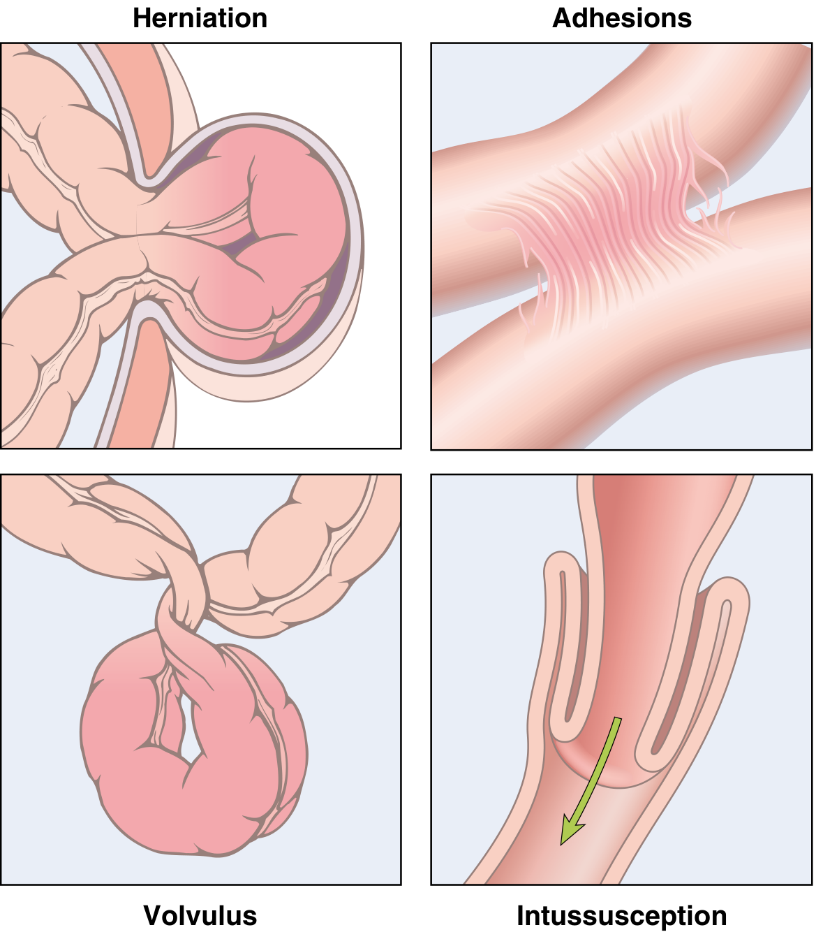
- Hernias (most common worldwide): Weakness in abdominal wall → external herniation of bowel (inguinal > femoral > umbilical) → incarceration → strangulation → infarction
- Adhesions: Post-surgical/inflammatory fibrous bridges → internal herniation; leading cause of obstruction in USA
- Volvulus: Twisting of bowel loop about its mesenteric attachment → both luminal and vascular obstruction; most common in sigmoid colon and cecum
- Intussusception: Telescoping of proximal bowel into adjacent distal segment; in children often triggered by Peyer's patch hypertrophy (viral illness); in adults suggests neoplasm as a lead point
12. ISCHEMIC BOWEL DISEASE
- Arterial thrombosis/embolism (SMA occlusion)
- Venous thrombosis
- Non-occlusive: low-flow states (heart failure, septic shock)
- Watershed areas most vulnerable: splenic flexure (junction of SMA/IMA territories) and rectosigmoid
- Mucosal infarction: Superficial; limited to mucosa; may recover
- Mural infarction: Mucosa + submucosa; hemorrhagic necrosis
- Transmural infarction: Full-thickness necrosis → perforation, peritonitis
- "Thumbprinting" on imaging = submucosal hemorrhage/edema
13. MALABSORPTION AND DIARRHEA
- Intraluminal digestion (luminal phase): pancreatic enzymes, bile acids
- Mucosal absorption (mucosal phase): epithelial transport
- Lymphatic transport (transport phase): chylomicrons
- Endocrine/neuroendocrine regulation
Celiac Disease (Celiac Sprue / Gluten-Sensitive Enteropathy)
- Immune-mediated enteropathy triggered by gluten (wheat, rye, barley)
- Genetic predisposition: HLA-DQ2 (up to 90% of cases) or HLA-DQ8
- Gluten → intestinal deamidation by tissue transglutaminase 2 (TG2) → negatively charged peptides → bind DQ2/DQ8 on APCs → activate CD4+ T cells (IFNγ, IL-21, IL-2) → villous atrophy
- CD4+ T cells + IL-15 → expand intraepithelial CD8+ T cells → destroy enterocytes
- Anti-TG2 IgA and anti-gliadin antibodies: diagnostically useful
- Villous atrophy (blunting/effacement of villi)
- Crypt hyperplasia (compensatory)
- Intraepithelial lymphocytosis (CD8+ T cells in surface epithelium - hallmark)
- Increased plasma cells, mast cells, eosinophils in lamina propria
- Complications: Refractory celiac disease, enteropathy-associated T-cell lymphoma (EATL), small intestinal adenocarcinoma
- Treatment: Strict gluten-free diet → histological recovery
14. INFECTIOUS ENTEROCOLITIS
| Organism | Site | Mechanism | Key Feature |
|---|---|---|---|
| Vibrio cholerae | Small intestine | CT toxin → cAMP → Cl⁻ secretion | "Rice-water" diarrhea; massive dehydration |
| Campylobacter | Colon | Invasion + toxin | Bloody diarrhea; precedes Guillain-Barré syndrome |
| Shigella | Left colon/ileum | Invasion via M cells | Bloody dysentery; reactive arthritis |
| Salmonella (non-typhoidal) | Colon + small intestine | Invasion | Watery/bloody diarrhea |
| Salmonella typhi | Small intestine | Systemic invasion via Peyer's patches | Typhoid fever - rose spots, bradycardia, "pea soup" diarrhea |
| Yersinia | Ileum, appendix, right colon | Invasion | Mimics appendicitis; mesenteric adenitis |
| ETEC | Small intestine | Heat-labile/stable toxins | Traveler's diarrhea |
| EHEC (O157:H7) | Colon | Shiga toxin | Bloody diarrhea → HUS (hemolytic uremic syndrome) |
| C. difficile | Colon | Toxin A (enterotoxin) + Toxin B (cytotoxin) | Pseudomembranous colitis after antibiotics |
Typhoid Fever - Morphology
- Peyer's patches swell → "button ulcers" along long axis of ileum
- Macrophage predominance (not neutrophils)
- Ulcers can perforate → peritonitis (complication)
- "Typhoid nodules" in spleen/liver
- Rose spots on skin (transient)
Pseudomembranous Colitis (C. difficile)
- Following antibiotic therapy → loss of normal flora → C. difficile overgrowth
- Toxin A + Toxin B → enterocyte injury
- Morphology: Yellow-green pseudomembranes (fibrin, mucus, neutrophils, necrotic cells) attached to damaged mucosa; "volcano" lesions
- Clinical: Profuse watery diarrhea, cramping, fever; can progress to toxic megacolon
15. INFLAMMATORY BOWEL DISEASE (IBD)
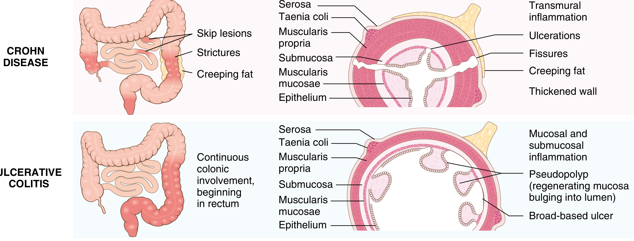
- Dysregulated immune response to intestinal microbiota in genetically susceptible individuals
- Key genetic loci: NOD2 (first gene identified for CD; involved in bacterial sensing in macrophages), IL-23R, ATG16L1 (autophagy)
- Proposed mechanism: Loss of immune tolerance to normal gut flora → mucosal immune activation → tissue damage
Comparison Table: Crohn Disease vs Ulcerative Colitis
| Feature | Crohn Disease | Ulcerative Colitis |
|---|---|---|
| Bowel region | Any part of GI tract (mouth to anus) | Colon only |
| Most common site | Terminal ileum + cecum (40% SI only) | Rectum (always involved) → extends proximally |
| Distribution | Skip lesions (patchy) | Continuous, diffuse |
| Depth of inflammation | Transmural | Limited to mucosa/submucosa |
| Granulomas | Yes (~35% of cases) | No |
| Fistulae/sinuses | Yes (common) | No |
| Strictures | Common | Rare |
| Perianal disease | Yes | No |
| Wall thickening | Marked (rubbery) | Normal |
| Cobblestone mucosa | Yes (ulcers + spared mucosa) | No |
| Creeping fat | Yes (mesenteric fat wraps bowel) | No |
| Pseudopolyps | Moderate | Marked |
| Ulcer type | Deep, knife-like, linear | Superficial, broad-based |
| Cancer risk | Moderate (if colonic involvement) | High (after 10 years, pancolitis) |
| Surgery curative? | No (recurs) | Yes (total colectomy) |
| Smoking | Worsens | Smoking is protective |
Crohn Disease - Morphology
- Skip lesions; terminal ileum most commonly affected
- Aphthous ulcers → elongated serpentine ulcers along bowel axis → cobblestone mucosa
- Fissures → perianal abscesses, fistulae (entero-enteric, enterovesical, enterovaginal, perianal)
- Creeping fat (mesenteric fat extends over serosal surface)
- Transmural inflammation; non-caseating granulomas (35%)
- Crypt abscesses, crypt distortion, pyloric metaplasia (in ileum)
Ulcerative Colitis - Morphology
- Always involves rectum; extends proximally in continuous fashion; may involve entire colon (pancolitis)
- Backwash ileitis: Superficial ileal inflammation in pancolitis
- Pseudopolyps (inflammatory polyps): Regenerating mucosa islands surrounded by deep ulcers - looks like polyps but they are not neoplastic
- Mucosa is hyperemic, granular; broad-based, superficial ulcers
- Inflammation limited to mucosa/submucosa; no transmural involvement, no granulomas
- Crypt abscesses and crypt distortion
Extraintestinal Manifestations (Both IBD)
- Joints: Peripheral arthritis, ankylosing spondylitis (HLA-B27 associated)
- Skin: Erythema nodosum, pyoderma gangrenosum
- Eyes: Uveitis, episcleritis
- Liver: Primary Sclerosing Cholangitis (PSC) - strongly associated with UC
- Blood: Anemia (blood loss, B12/folate deficiency)
Colitis-Associated Neoplasia
- IBD patients (especially UC) have increased risk of colorectal adenocarcinoma
- Sequence: Dysplasia-associated lesion or mass (DALM) → adenocarcinoma
- Surveillance colonoscopy with biopsies recommended after 8-10 years of pancolitis
16. DIVERTICULAR DISEASE
- Pseudodiverticula: Herniation of mucosa and submucosa through defects in muscularis propria (not true diverticula as they lack all wall layers)
- Most common in sigmoid colon; related to low-fiber diet (increased intraluminal pressure)
- Asymptomatic in most; Diverticulitis: infection/perforation of a diverticulum → left lower quadrant pain, fever, "left-sided appendicitis"
- Complications: abscess, perforation, fistula, obstruction
17. INTESTINAL POLYPS AND COLORECTAL NEOPLASIA
Polyp Types
| Type | Malignant Potential | Key Features |
|---|---|---|
| Hyperplastic polyp | None | Small; "sawtooth" luminal surface; rectosigmoid; most common polyp type |
| Inflammatory/juvenile polyp | Low (1-20% if large) | Associated with chronic gastritis/IBD |
| Peutz-Jeghers polyp | Low (hamartoma) | Smooth muscle bundles in stroma; multiple in small intestine; associated with mucocutaneous pigmentation + GI and extraintestinal cancer risk |
| Tubular adenoma | Low-intermediate | Pedunculated; tubular architecture |
| Tubulovillous adenoma | Intermediate | Mixed architecture |
| Villous adenoma | High (40% if >4 cm) | Sessile; finger-like projections; rectum; secretes mucus → hypokalemia |
Adenomas
- Epithelial neoplasms; present in ~30% of Western adults by age 60
- Hallmarks of epithelial dysplasia: nuclear hyperchromasia, elongation, stratification, prominent nucleoli
- Risk of malignant transformation correlates with: size >1 cm, villous architecture, and high-grade dysplasia
Familial Adenomatous Polyposis (FAP)
- Autosomal dominant mutation in APC (adenomatous polyposis coli) gene on chromosome 5q21
- 100s to 1000s of polyps throughout colon; virtually 100% risk of colon cancer by age 40 if untreated
- APC is a tumor suppressor that regulates WNT/β-catenin signaling; loss → excess β-catenin → nuclear transcription → cell proliferation
- Gardner syndrome: FAP + desmoid tumors + osteomas + epidermoid cysts
- Turcot syndrome: FAP + medulloblastoma (or HNPCC + glioblastoma)
- Treatment: prophylactic colectomy
Hereditary Non-Polyposis Colorectal Cancer (HNPCC / Lynch Syndrome)
- Autosomal dominant mutations in DNA mismatch repair (MMR) genes: MSH2, MLH1 (most common), MSH6, PMS2
- Leads to microsatellite instability (MSI) → accumulation of mutations
- Cancer risk: colorectal (right-sided, multiple primaries), endometrial (most common extracolonic), ovarian, gastric, urinary tract
- Fewer polyps than FAP but markedly increased cancer risk
- Testing: Immunohistochemistry for MMR proteins on tumor; genetic testing for germline mutations
18. COLORECTAL ADENOCARCINOMA
- Most common malignancy of the GI tract; 2nd leading cause of cancer death in the USA (after lung)
- Incidence peaks at 60-70 years; <20% occur before age 50 (but increasing in young adults)
- High risk: North America, Australia, Europe, Japan; Low risk: India, Africa, South America
-
APC/β-catenin (WNT) pathway - Chromosomal Instability (CIN) - "Classic" pathway
- Normal mucosa → APC mutation → adenoma → KRAS mutation → DCC loss → TP53 mutation → carcinoma
- APC loss → excess β-catenin → proliferation
- Most sporadic colon cancers (85%)
- Associated with FAP
-
Mismatch Repair Deficiency - Microsatellite Instability (MSI) pathway
- MMR gene mutation/silencing (MLH1 hypermethylation in sporadic; germline mutation in HNPCC)
- Rapid accumulation of mutations in microsatellite regions → MSI
- Associated with HNPCC; better prognosis; responds to immune checkpoint inhibitors (PD-L1 high)
- Right colon (cecum, ascending): Polypoid/exophytic fungating mass; rarely obstructs (wide lumen); presents with occult blood loss → anemia
- Left colon (descending, sigmoid): Annular, napkin-ring lesion → constricts lumen → obstruction, change in bowel habits
- Histology: Gland-forming adenocarcinoma; variable mucin production; necrotic centers; desmoplastic stroma
- Direct: penetrates bowel wall into adjacent structures
- Lymphatic: mesenteric lymph nodes
- Hematogenous: Liver (most common distant metastasis), then lungs
- Transcoelomic: peritoneal seeding
| Stage | Dukes | TNM | Description | 5-yr Survival |
|---|---|---|---|---|
| A | A | T1/T2 N0 M0 | Confined to bowel wall | >90% |
| B | B | T3/T4 N0 M0 | Through bowel wall | 70-80% |
| C | C | Any T N1 M0 | Lymph node positive | 30-50% |
| D | D | Any T any N M1 | Distant metastasis | <10% |
- Right colon: Fatigue, weakness (from anemia), occult blood, palpable mass
- Left colon: Change in bowel habits, blood in stool, crampy pain, obstruction
- Both: Weight loss, anorexia
- CEA (carcinoembryonic antigen): Used for monitoring recurrence after surgery (not a screening test)
Key Concepts Summary (Exam High-Yield Points)
| Topic | Key Fact |
|---|---|
| Barrett esophagus | Intestinal metaplasia of distal esophagus; goblet cells; 40× risk of adenocarcinoma |
| Esophageal cancer | Adenocarcinoma (West, distal) vs SCC (developing countries, mid-esophagus) |
| H. pylori | Most common cause of chronic gastritis; CagA is key virulence factor |
| Autoimmune gastritis | Anti-parietal cell + anti-intrinsic factor antibodies; pernicious anemia; spares antrum |
| Peptic ulcer | 4 zones: fibrin/necrosis → neutrophils → granulation tissue → fibrosis |
| Stress ulcers | Curling (burns) vs Cushing (head injury - very deep, perforate) |
| Ménétrier disease | TGF-α excess; rugal hypertrophy; protein-losing enteropathy |
| ZES | Gastrinoma; multiple ulcers; 60-90% malignant; MEN1 |
| GIST | KIT mutation; treated with imatinib; CD117+, CD34+, DOG1+ |
| Celiac disease | HLA-DQ2/DQ8; TG2; villous atrophy + crypt hyperplasia + IELs; gluten-free diet |
| Crohn disease | Skip lesions; transmural; granulomas; fistulae; any GI site; NOT cured by surgery |
| Ulcerative colitis | Continuous; rectum always; mucosal only; pseudopolyps; cured by colectomy; PSC association |
| Typhoid | Button ulcers along long axis of ileum; macrophage predominance |
| C. difficile | Pseudomembranous colitis; after antibiotics; Toxin A + B |
| FAP | APC mutation; 100s of polyps; 100% cancer risk; Gardner/Turcot syndrome |
| Lynch (HNPCC) | MSH2/MLH1 mutations; MSI; right colon + endometrial cancers |
| Colorectal cancer | Left = napkin-ring (obstruction); Right = polypoid (anemia); liver = first metastasis |
Similarly for the hepatobiliary system
Hepatobiliary System Pathology
(Robbins & Cotran Pathologic Basis of Disease - Chapter 18)
PART I: THE LIVER
Normal Anatomy & Physiology (Key Points)
1. LIVER FAILURE
- A: Acetaminophen overdose, Hepatitis A, Autoimmune hepatitis
- B: Hepatitis B
- C: Hepatitis C, Cryptogenic
- D: Drugs/toxins, Hepatitis D
- E: Hepatitis E; Esoteric causes (Wilson disease, Budd-Chiari syndrome, lymphoma)
- F: Fatty change (microvesicular) - fatty liver of pregnancy, valproate, tetracycline, Reye syndrome
- Coagulopathy: Reduced synthesis of coagulation factors (I, II, V, VII, IX, X) → bleeding tendency; PT/INR elevated
- Hepatic encephalopathy: Accumulation of nitrogenous wastes (ammonia) + false neurotransmitters → asterixis, confusion, coma
- Portal hypertension: Cirrhosis → increased resistance in portal venous system → esophageal varices, ascites, splenomegaly, hemorrhoids (caput medusae)
- Hepatorenal syndrome: Renal failure (reversible with liver transplant) in severe liver disease; decreased renal perfusion from renin/angiotensin activation in portal hypertension
- Portopulmonary hypertension: Increased pulmonary arterial pressure
- Jaundice: Inability to conjugate/excrete bilirubin
2. CIRRHOSIS
- Chronic hepatitis B
- Chronic hepatitis C
- Metabolic dysfunction-associated steatotic liver disease (MASLD)
- Alcohol-associated liver disease
- Gross: Smooth liver capsule → bumpy surface with depressed scar areas + bulging regenerative nodules
- Micro: Parenchymal nodules surrounded by dense fibrous bands (highlighted with Masson's trichrome stain for collagen)
- Micronodular cirrhosis (nodules <3 mm): Typically seen in alcoholic liver disease and hemochromatosis
- Macronodular cirrhosis (nodules >3 mm): Typically seen in viral hepatitis
- Thin incomplete scars with ductular reaction = morphologic regression of cirrhosis (can occur with treatment)
- Portal hypertension → esophageal varices (rupture = massive haematemesis), ascites, splenomegaly
- Hepatocellular failure → coagulopathy, encephalopathy, jaundice, hypoalbuminemia
- Hepatocellular carcinoma (increased risk in all forms of cirrhosis)
3. VIRAL HEPATITIS
Hepatitis Virus Comparison Table
| Feature | HAV | HBV | HCV | HDV | HEV |
|---|---|---|---|---|---|
| Virus type | ssRNA (picornavirus) | Partially dsDNA (hepadnavirus) | ssRNA (flavivirus) | Circular defective ssRNA | ssRNA |
| Transmission | Fecal-oral (contaminated food/water) | Parenteral, sexual, perinatal | Parenteral (blood-to-blood); intranasal cocaine | Parenteral (requires HBV) | Fecal-oral |
| Incubation | 2-6 weeks | 2-26 weeks | 4-26 weeks | Same as HBV | 4-5 weeks |
| Chronicity | Never | 5-10% | >80% | 10% co-infection; 90-100% superinfection | Immunocompromised only |
| Diagnosis | Serum IgM anti-HAV | HBsAg, HBcAb, HBsAb; PCR for HBV DNA | ELISA anti-HCV; PCR for HCV RNA | IgM/IgG antibodies; PCR HDV RNA | IgM/IgG; PCR HEV RNA |
| Carrier state | No | Yes | Yes | Yes | No |
Hepatitis A (HAV)
- Self-limited; no chronicity, no carrier state
- Most common cause of symptomatic acute hepatitis worldwide
- Shed in stool 2-3 weeks before AND 1 week after onset of jaundice (peak infectivity)
- Complications: Acute liver failure (~0.5%); cholestatic hepatitis (prolonged cholestasis)
- Prevention: HAV vaccine (excellent efficacy); passive immunization with Ig
Hepatitis B (HBV)
-
Serology interpretation:
- HBsAg: Current infection (acute or chronic); first marker to appear (4-8 weeks post-exposure)
- Anti-HBs: Recovery/immunity (protective); appears after HBsAg disappears
- Anti-HBc IgM: Acute HBV infection (the "window period" marker when HBsAg has cleared but anti-HBs not yet appeared)
- Anti-HBc IgG: Past infection or ongoing; persists for life
- HBeAg: Active viral replication; high infectivity
- Anti-HBe: Declining replication; lower infectivity
- HBV DNA: Most sensitive marker of active infection/replication (PCR)
-
"Window period": HBsAg has cleared but anti-HBs not yet detectable; only anti-HBc IgM is positive - do not miss this!
-
Course: 90-95% of immunocompetent adults recover fully; 5-10% develop chronic infection; neonates infected perinatally have 90% risk of chronicity
-
Complications of chronic HBV: Cirrhosis → HCC (HBV integrates into host genome; HCC can occur even without cirrhosis)
-
Morphology (acute): Ground-glass hepatocytes (HBsAg accumulation in ER); acidophil bodies (apoptotic hepatocytes = Councilman/Mallory bodies); lobular disarray; inflammation; ballooning degeneration
Hepatitis C (HCV)
- Most common cause of chronic liver disease in the USA (before MASLD surpassed it)
- High propensity for chronicity (>80%) due to rapid viral mutation (quasispecies) → evades immune clearance
- Mechanism of liver injury: Immune-mediated (CD8+ T cells attack infected hepatocytes) rather than direct viral cytotoxicity
- Morphology: Lymphoid follicles in portal tracts (characteristic); bile duct damage; steatosis; lobular inflammation
- Natural history: Chronic hepatitis → cirrhosis (20% over 20 years) → HCC
- Treatment: Direct-acting antivirals (DAAs) achieve >95% sustained virologic response (cure); e.g., sofosbuvir + ledipasvir
Hepatitis D (HDV)
- Defective virus - requires HBsAg envelope from HBV for its own packaging and transmission
- Co-infection (HBV + HDV simultaneously): Generally self-limited; better prognosis; <5% chronicity
- Superinfection (HDV in chronic HBV carrier): 90-100% chronicity; severe disease; rapidly progresses to cirrhosis
Hepatitis E (HEV)
- Fecal-oral transmission; large epidemics in South Asia, Africa, Mexico (contaminated water)
- Usually self-limited in healthy individuals (similar to HAV)
- Exception: Very high mortality in pregnant women (20-25% in third trimester) → acute liver failure
- Chronic HEV seen in immunocompromised patients (transplant recipients)
Clinicopathologic Syndromes of Viral Hepatitis
- Hepatocyte injury: ballooning degeneration, acidophil/Councilman bodies (apoptotic hepatocytes with pyknotic nuclei)
- Lobular disarray; Kupffer cell hyperplasia
- Portal tract inflammation: lymphocytes, plasma cells, macrophages
- Cholestasis: bile plugs in canaliculi
- Asymptomatic with recovery (most common)
- Acute symptomatic hepatitis with recovery: 4 phases - incubation → preicteric (nausea, fever, malaise) → icteric (jaundice, dark urine, pale stools) → convalescence
- Acute liver failure (~1% of symptomatic acute hepatitis)
- Chronic hepatitis (only with HBV, HCV, HDV)
- Carrier state (HBV, HCV)
4. AUTOIMMUNE HEPATITIS
- Typically young to middle-aged women
- Positive ANA (antinuclear antibody), anti-smooth muscle antibody (anti-SMA), anti-LKM1 (liver-kidney microsomal antibody), elevated IgG
- Type 1 (most common): ANA + anti-SMA positive; better prognosis
- Type 2: Anti-LKM1 positive; younger patients; more severe
- Morphology: Interface hepatitis (lymphoplasmacytic infiltrate at the portal-lobular interface), emperipolesis (lymphocytes within hepatocytes), rosette formation
- Treatment: Corticosteroids ± azathioprine → excellent response in most
5. DRUG- AND TOXIN-INDUCED LIVER INJURY (DILI)
- Intrinsic (predictable, dose-dependent): e.g., Acetaminophen - toxic metabolite NAPQI → zone 3 (centrilobular) necrosis; antidote is N-acetylcysteine (NAC)
- Idiosyncratic (unpredictable, not dose-dependent): Hypersensitivity reaction; majority of DILI; e.g., isoniazid, halothane, diclofenac
- Drugs can mimic any pattern of liver injury - must always be considered in differential diagnosis
- Temporal correlation between drug initiation and liver disease onset is required for diagnosis
6. STEATOTIC LIVER DISEASE
A. Alcohol-Associated Liver Disease (ALD)
- Spectrum of disease: Steatosis → Alcoholic Hepatitis → Fibrosis/Cirrhosis
- Risk: >80 g/day for males; females more susceptible at lower doses
- Only 10-15% of heavy drinkers develop cirrhosis (other factors: sex, genetics, comorbidities)
- Alcohol → acetaldehyde (toxic) + excess NADH → impaired fatty acid oxidation → steatosis
- Acetaldehyde → adducts with proteins → Mallory-Denk bodies
- Oxidative stress + mitochondrial dysfunction + cytokine release (TNF-α via TLR4/Kupffer cells)
- Intestinal permeability increases (especially with estrogen) → LPS enters portal blood → Kupffer cell activation
- Hepatic steatosis (fatty liver): Earliest change; macrovesicular fat accumulation (lipid droplets displace nucleus to periphery); reversible with abstinence
- Alcoholic hepatitis (steatohepatitis):
- Hepatocyte ballooning + necrosis
- Mallory-Denk bodies (dense eosinophilic intracytoplasmic inclusions of aggregated cytokeratin intermediate filaments)
- Neutrophil infiltration (lobular)
- Pericellular fibrosis ("chicken wire" fibrosis) around hepatocytes
- Megamitochondria (large eosinophilic inclusions)
- Cirrhosis: Micronodular → macronodular; end-stage
B. Metabolic Dysfunction-Associated Steatotic Liver Disease (MASLD) [formerly NAFLD/NASH]
- Most common liver disease worldwide; closely linked to metabolic syndrome (obesity, T2DM, hypertension, dyslipidemia)
- Spectrum: Steatosis (MASLD) → Steatohepatitis (MASH) → Fibrosis → Cirrhosis → HCC
- Insulin resistance → increased free fatty acid delivery to liver → hepatic steatosis
- "Multiple hits": oxidative stress + mitochondrial dysfunction + gut-derived endotoxins + adipokine imbalance → hepatocyte injury → inflammation → fibrosis
- Similar to ALD: macrovesicular steatosis, ballooned hepatocytes, Mallory-Denk bodies, lobular inflammation with neutrophils
- Key difference: MASLD/MASH predominantly affects zone 3 (centrilobular); ALD is similar
- Progressive fibrosis: pericellular ("chicken wire") → bridging → cirrhosis
7. INHERITED LIVER DISEASES
A. Hereditary Hemochromatosis
- Autosomal recessive mutation in HFE gene (chromosome 6p; C282Y most common mutation) → abnormal HFE protein → failure to reduce intestinal iron absorption → iron overload
- HFE normally interacts with transferrin receptor to sense body iron stores; mutant HFE fails to upregulate hepcidin (the iron-regulatory hormone) → excessive iron absorption continues unchecked
- Iron deposits primarily in: liver, pancreas, heart, joints, skin, pituitary/gonads
- Iron causes injury through: oxidative stress (Fenton reaction → free radical generation), direct organelle damage
- Micronodular cirrhosis (100%)
- Diabetes mellitus ("bronze diabetes") (75-80%)
- Abnormal skin pigmentation - bronze-gray skin (75-80%)
- Golden-brown hemosiderin granules in hepatocytes, Kupffer cells, bile duct cells
- Perls' Prussian blue stain confirms iron deposits
- Micronodular cirrhosis; iron also in pancreatic acinar cells, cardiac myocytes, adrenal cells
B. Wilson Disease
- Autosomal recessive mutation in ATP7B gene (chromosome 13q14) encoding a copper-transporting ATPase expressed in hepatocytes
- Defective copper transport → failure to incorporate copper into ceruloplasmin + failure to excrete copper in bile → copper accumulates in liver first, then brain, eyes, kidneys
- Liver: chronic hepatitis → cirrhosis; may present as acute liver failure
- Brain: Lenticular degeneration (basal ganglia) → tremors, rigidity, psychiatric disturbances, dysarthria, dysphagia
- Eyes: Kayser-Fleischer rings - golden-brown rings at periphery of cornea (Descemet membrane); seen on slit-lamp examination; virtually pathognomonic
- Kidneys: Fanconi syndrome (proximal tubular dysfunction → aminoaciduria, phosphaturia)
- Hemolytic anemia (copper released into blood from damaged hepatocytes)
- Liver: steatosis → hepatocyte necrosis → portal inflammation → cirrhosis
- Rhodanine stain (red-orange) or orcein stain: highlights copper deposits
- Electron microscopy: mitochondrial changes (distinctive early finding)
C. α1-Antitrypsin (A1AT) Deficiency
- Autosomal recessive mutation in SERPINA1 gene (chromosome 14q32)
- PiZZ genotype (most severe): Mutant Z protein misfolds and accumulates as aggregates in hepatocyte ER (instead of being secreted) → ER stress + apoptosis → liver disease
- Absent serum A1AT → uninhibited elastase activity in lungs → panacinar emphysema (lower lobes)
- PAS-positive, diastase-resistant intracytoplasmic globules in hepatocytes (periportal) - hallmark
- Ranges from neonatal hepatitis to cirrhosis in adults
8. CHOLESTATIC DISEASES
Bilirubin Metabolism & Jaundice
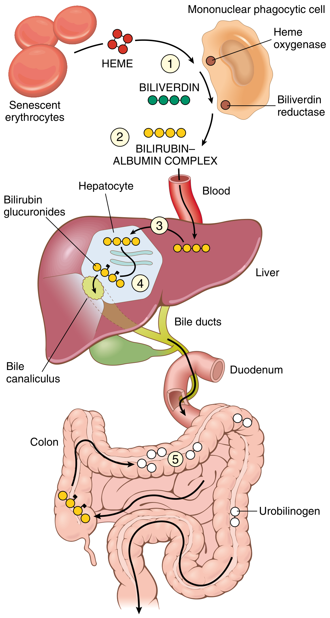
- Senescent RBCs degraded by macrophages (spleen/liver/bone marrow) → Heme → Biliverdin → Bilirubin (unconjugated, insoluble, albumin-bound)
- Hepatocyte uptake → conjugation with glucuronic acid (UGT1A1 enzyme) → bilirubin glucuronides (water-soluble, non-toxic)
- Excretion into bile → intestine → bacterial deconjugation → urobilinogen (colorless) → stercobilin (brown feces color)
- Enterohepatic circulation: ~20% urobilinogen reabsorbed → liver → re-excreted in bile; small amount in urine
| Type | Bilirubin | Urine bilirubin | Urobilinogen | Causes |
|---|---|---|---|---|
| Pre-hepatic (hemolytic) | Unconjugated ↑ | Negative (indirect not filtered) | ↑ | Hemolysis, ineffective erythropoiesis |
| Hepatocellular | Both ↑ | Positive | Variable | Hepatitis, cirrhosis, drug injury |
| Post-hepatic (obstructive/cholestatic) | Conjugated ↑ | Positive (dark urine) | ↓ or absent (pale stools) | Gallstones, carcinoma of head of pancreas, PSC, PBC |
- Gilbert syndrome: Mild UGT1A1 promoter mutation → reduced conjugation; benign; elevated unconjugated bilirubin especially with fasting/stress; no treatment needed
- Crigler-Najjar syndrome Type I: Complete absence of UGT1A1 → severe unconjugated hyperbilirubinemia → kernicterus → fatal unless treated (liver transplant)
- Crigler-Najjar Type II: Partial deficiency; responds to phenobarbital (induces residual UGT1A1)
- Dubin-Johnson syndrome: Defect in MRP2 (canalicular transport protein for conjugated bilirubin) → conjugated bilirubin regurgitated into blood; black-pigmented liver (lipofuscin-like pigment); benign
- Rotor syndrome: Similar to Dubin-Johnson but no liver pigmentation; benign
Primary Biliary Cholangitis (PBC)
- T-cell-mediated attack on bile duct epithelium
- Hallmark: Antimitochondrial antibody (AMA) directed against PDC-E2 (E2 component of pyruvate dehydrogenase complex) - present in 95% of cases
- Retention of bile salts → secondary hepatocellular injury → cirrhosis
- "Florid duct lesion": Dense lymphocytic + granulomatous infiltrate destroying small bile ducts
- Poorly formed granulomas in portal tracts centered on bile ducts
- Portal lymphoplasmacytic inflammation with ductular reaction
- Progressive: bile duct loss → ductopenia → biliary-type cirrhosis
Primary Sclerosing Cholangitis (PSC)
- Inflammatory bowel disease (70%) - especially ulcerative colitis (IBD precedes PSC in most cases)
- 65% ANCA-positive; AMA typically negative
- Concentric periductal fibrosis ("onion-skin" fibrosis) obliterating medium-sized ducts
- Alternating strictures and dilations of bile ducts → "beads on a string" pattern on MRCP/cholangiography
- Progressive bile duct loss → biliary cirrhosis
| Feature | PBC | PSC |
|---|---|---|
| Sex | 90% female | 70% male |
| Age | ~50 years | ~30 years |
| Ducts affected | Small intrahepatic | Large intra + extrahepatic |
| Serology | AMA positive (95%) | ANCA positive (65%), AMA negative |
| Radiology | Normal | "Beads on a string" |
| Associations | Sjögren, scleroderma | IBD (UC, 70%) |
| Malignancy risk | HCC | Cholangiocarcinoma |
9. HEPATIC VASCULAR DISORDERS
Budd-Chiari Syndrome (Hepatic Vein Thrombosis)
- Thrombosis of 2 or 3 hepatic veins → impaired hepatic venous outflow → congestion + necrosis
- Causes: Polycythemia vera (most common), myeloproliferative disorders, hypercoagulable states (factor V Leiden, antiphospholipid syndrome, paroxysmal nocturnal hemoglobinuria, oral contraceptive use), pregnancy, HCC invasion
- Morphology: Severe centrilobular congestion and necrosis; marked hepatomegaly; ascites; "nutmeg liver" (mottled appearance)
- Clinical: Abdominal pain, hepatomegaly, ascites; Doppler US shows absent hepatic vein flow; chronic cases → cirrhosis
Passive Congestion (Right Heart Failure / Cardiac Cirrhosis)
- Chronic right-sided heart failure → elevated central venous pressure → hepatic venous congestion
- "Nutmeg liver": Alternating areas of red congestion (centrilobular) and yellow fat (periportal)
- Morphology: Centrilobular (zone 3) sinusoidal dilation and congestion; with severe/prolonged cases → centrilobular necrosis → fibrosis → cardiac cirrhosis
Portal Vein Obstruction and Thrombosis
- Non-cirrhotic portal hypertension; causes: neonatal omphalitis/umbilical vein infection, intraabdominal sepsis, pancreatitis, hypercoagulable states, HCC invasion
- Clinical: portal hypertension, varices, splenomegaly WITHOUT significant liver dysfunction (liver parenchyma is intact)
Sinusoidal Obstruction Syndrome (SOS, formerly Veno-Occlusive Disease)
- Obstruction of hepatic sinusoids and terminal hepatic venules (not large veins)
- Causes: Bone marrow transplant conditioning regimens (cyclophosphamide + whole body radiation), pyrrolizidine alkaloids (herbal teas)
- Morphology: Sinusoidal fibrin deposition → fibrosis of terminal hepatic venules
- Clinical: Jaundice, hepatomegaly, weight gain, ascites within weeks of BMT
10. LIVER TUMORS
Benign Liver Tumors
- Most common benign liver tumor; incidental finding; no malignant potential
- Blood-filled vascular spaces lined by endothelium; "light-up" on contrast MRI
- Benign hepatocyte tumor; strongly associated with oral contraceptive use in young women; also anabolic steroids
- Molecular subtypes: HNF1A-mutated (steatotic), β-catenin-activated (risk of HCC), inflammatory
- Risk of rupture and hemorrhage (especially during pregnancy)
- May undergo malignant transformation (β-catenin-activated subtype)
- Morphology: Well-demarcated, pale yellow-tan mass; no bile ducts, no portal tracts; hepatocytes in sheets and cords
- Non-neoplastic hyperplastic response to a pre-existing vascular anomaly; not a true neoplasm
- Central stellate scar with thick-walled blood vessels (pathognomonic on imaging)
- No malignant potential; not hormone-dependent; no risk of rupture
Hepatocellular Carcinoma (HCC)
- 5th most common cancer globally; 3rd most common cause of cancer death
-
85% occur in Asia and sub-Saharan Africa (HBV endemic areas)
- Pronounced male predominance (up to 8:1 in high-incidence areas)
- In Asia: peak 20-40 years (perinatal HBV); In West: older adults (cirrhosis from HCV/MASLD)
- Chronic HBV (most important globally; HCC can occur without cirrhosis via HBV DNA integration)
- Chronic HCV (virtually only in cirrhotic livers)
- Aflatoxin B1 (Aspergillus-contaminated crops; synergizes with HBV; causes TP53 R249S mutation)
- Alcohol-associated cirrhosis
- MASLD/MASH (even without cirrhosis)
- Hemochromatosis, Wilson disease, α1-AT deficiency (increased risk)
- Most HCC arise in cirrhotic livers (but 15-20% in non-cirrhotic liver)
- Driver mutations: β-catenin activating mutations (40%), TERT promoter mutations (50-60%), TP53 inactivating mutations (up to 60%), HBx protein disrupts tumor suppression
- Precursor lesions: Large cell change, small cell change, dysplastic nodules (low grade → high grade → early HCC)
- Special subtype: Fibrolamellar HCC - adolescents/young adults; no underlying cirrhosis; DNAJB1::PRKACA fusion gene; fibrous bands between tumor cells; better prognosis
- Gross: Single large mass, multifocal nodules, or diffusely infiltrating
- Cut surface: green (bile-secreting), yellow-white (fatty), hemorrhagic
- Micro: Hepatocytes in trabecular/acinar pattern; bile production (pathognomonic); vascular invasion is common (portal vein)
- CD10, Hep Par-1, Glypican-3 (GPC3), AFP immunostaining useful for diagnosis
- Often asymptomatic until advanced stage (right upper quadrant pain, weight loss, hepatomegaly, ascites)
- Alpha-fetoprotein (AFP): Tumor marker - elevated in 50-70% of HCC; >400 ng/mL strongly suggests HCC
- Surveillance: Ultrasound ± AFP every 6 months in cirrhotic patients
- Spread: intrahepatic (via portal vein), lung, regional lymph nodes, adrenals
- Poor prognosis if not detected early; treatment: resection, ablation, sorafenib, immunotherapy, liver transplant (Milan criteria)
Cholangiocarcinoma (Malignant Biliary Tumors)
- Adenocarcinoma of biliary epithelium; intrahepatic or extrahepatic (Klatskin tumor at hilum)
- Risk factors: PSC (most important in West), liver flukes (Clonorchis sinensis, Opisthorchis viverrini - endemic in Southeast Asia), choledochal cysts, hepatolithiasis, chronic biliary inflammation
- Morphology: Dense desmoplastic stroma; CK7+ and CK19+ (biliary markers); often aggressive perineural invasion
- Clinical: Obstructive jaundice (Klatskin), elevated ALP/GGT; CA 19-9 tumor marker; very poor prognosis
Metastases to Liver
- Most common hepatic tumors overall (much more common than primary liver tumors in Western countries)
- Most frequent primaries: colon (via portal vein), stomach, pancreas, breast, lung
- Multiple nodules; "bull's-eye" appearance on imaging (central necrosis)
PART II: THE GALLBLADDER
11. CHOLELITHIASIS (GALLSTONES)
- Affect 10-20% of adults in Western countries; >95% of biliary tract disease is attributable to gallstones
| Feature | Cholesterol Stones | Pigment Stones |
|---|---|---|
| Composition | >50% cholesterol crystals | Calcium bilirubinate (black or brown) |
| Color | Yellow to pale green | Black (sterile) or Brown (infected) |
| Prevalence | 80% of stones in Western countries | 20% |
| Radiopaque? | No (radiolucent) | Black: Yes (radiopaque); Brown: variable |
| Risk factors | "4 Fs": Fat, Female, Forty, Fertile; OCP; pregnancy; obesity; metabolic syndrome; rapid weight loss; gallbladder stasis | Hemolytic anemia, biliary infection, Crohn disease (terminal ileal disease → reduced bile salt reabsorption), cirrhosis |
| Mechanism | Cholesterol supersaturation of bile + nucleation factor + gallbladder stasis | Unconjugated bilirubin precipitation (hemolysis) or bilirubin from bacterial deconjugation |
- Pain (biliary colic): right upper quadrant colicky pain after fatty meals (cystic duct obstruction)
- Inflammation: Cholecystitis (acute or chronic)
- Pancreatitis: Stone impacted at ampulla of Vater → biliary pancreatitis
- Empyema, jaundice (common bile duct obstruction), gallbladder carcinoma (long-term), Mirizzi syndrome, gallstone ileus (rare)
12. CHOLECYSTITIS
Acute Cholecystitis
- Usually triggered by obstruction of the cystic duct by a stone → bile stasis → mucosal injury → secondary inflammation
- Chemical initially (prostaglandins, bile salts), bacterial superinfection later (E. coli, Klebsiella, anaerobes)
- Morphology: Enlarged, tense gallbladder; fibrinous exudate on serosa; turbid bile with fibrin and pus; stone in cystic duct/neck; wall edematous and hyperemic; neutrophil infiltrate
- Severe: Gangrenous cholecystitis (green-black necrosis + perforation); Empyema (pus-filled gallbladder); Emphysematous cholecystitis (gas-forming organisms - diabetics)
- In severely ill patients: major surgery, trauma, burns, sepsis, prolonged TPN
- Mechanism: gallbladder ischemia + bile stasis (no enteric stimulation → no CCK → no gallbladder contraction)
- More insidious; higher rate of gangrene and perforation (delayed diagnosis in critically ill); higher mortality
Chronic Cholecystitis
- Associated with gallstones in >90% of cases; may develop from repeated acute attacks or insidiously
- Rokitansky-Aschoff sinuses: Herniation of mucosa into the muscular layer (due to increased intraluminal pressure) - characteristic
- Morphology: Thickened fibrotic wall; variable lymphocytic infiltrate; mucosal atrophy
- Porcelain gallbladder: Extensive calcification of the gallbladder wall in chronic cholecystitis; historically associated with cancer risk (now debated)
13. GALLBLADDER CARCINOMA
- Most common malignancy of the extrahepatic biliary tract
- Gallstones present in 95% of cases - but only 1-2% of patients with stones develop cancer; common thread is chronic inflammation
- Risk factors: Female sex, gallstones, chronic biliary infection (Asia: liver flukes, bacteria), gallbladder adenoma, anomalous pancreatic ductal junction
- Molecular: EGFR/HER2 amplification, RAS mutation, TP53 mutation (up to 50%)
- Mainly adenocarcinoma (most common at the fundus)
- Firm, poorly circumscribed mass OR diffuse infiltration (mimics chronic cholecystitis)
- Precursor lesions: flat dysplastic lesions (biliary intraepithelial neoplasia), papillary-tubular neoplasm (intracholecystic)
- Desmoplastic stroma; perineural and vascular invasion; CK7/CK19+
14. BILIRUBIN METABOLISM DISORDERS (Key Syndromes)
| Syndrome | Defect | Bilirubin | Clinical | Liver |
|---|---|---|---|---|
| Gilbert | UGT1A1 promoter (↓ activity) | Unconjugated ↑ (mild) | Benign; intermittent jaundice with fasting/stress | Normal |
| Crigler-Najjar I | UGT1A1 absent | Unconjugated markedly ↑ | Kernicterus, fatal without transplant | Normal |
| Crigler-Najjar II | UGT1A1 partial | Unconjugated ↑ | Responds to phenobarbital | Normal |
| Dubin-Johnson | MRP2 (canalicular transporter) defect | Conjugated ↑ | Benign; black liver on gross | Black pigment |
| Rotor | OATP1B1/B3 transporter defect | Conjugated ↑ | Benign; no liver pigment | Normal |
High-Yield Key Concepts Summary
| Topic | Exam Pearl |
|---|---|
| Cirrhosis | Parenchymal nodules + fibrous bands; portal hypertension + liver failure |
| HBV window period | Anti-HBc IgM ONLY positive (HBsAg cleared, anti-HBs not yet appeared) |
| HCV chronicity | >80% chronic; lymphoid follicles in portal tracts on biopsy |
| HDV superinfection | 90-100% chronicity; most severe |
| HEV in pregnancy | 20-25% maternal mortality in third trimester |
| Alcoholic hepatitis | Mallory-Denk bodies; neutrophil infiltrate; AST:ALT >2:1 |
| MASLD | ALT > AST; zone 3 steatosis; linked to metabolic syndrome |
| Hemochromatosis | HFE gene; bronze diabetes; Prussian blue stain; arthropathy of MCP joints |
| Wilson disease | ATP7B gene; Kayser-Fleischer rings; low ceruloplasmin |
| α1-AT deficiency | PAS+ diastase-resistant globules in hepatocytes; liver disease = protein accumulation |
| PBC | AMA (anti-PDC-E2); middle-aged women; florid duct lesion; Sjögren's association |
| PSC | Beads on string; 70% male; 70% IBD (UC); onion-skin fibrosis; cholangiocarcinoma risk |
| Budd-Chiari | Hepatic vein thrombosis; nutmeg liver; PCV most common cause |
| HCC | AFP; HBV + HCV + Aflatoxin; TERT + β-catenin + TP53 mutations; fibrolamellar = young adults |
| Cholesterol stones | 4 Fs; radiolucent; 80% of all stones |
| Pigment stones (black) | Hemolysis; radiopaque |
| Gallbladder carcinoma | Stones in 95%; female > male; fundus; poor prognosis (<10% 5-yr survival) |
| Cholelithiasis complication | Biliary pancreatitis (stone at ampulla of Vater) |
| Rokitansky-Aschoff sinuses | Mucosal herniation into muscularis; chronic cholecystitis |
| Gilbert syndrome | Commonest hereditary bilirubin disorder; benign; UGT1A1 promoter mutation |
| Dubin-Johnson | Black liver; conjugated hyperbilirubinemia; benign |
Listen Serous cyst adenoma of ovary Fibroadenoma Dermoid cyst Dysgerminoma Ca of breast RCC Granular contracted kidney Ca of Urinary bladder Seminoma Leiomyoma (subserosa +submucosa +intramural) Endometrial hyperpalsia Carcinoma cervix Chroinic cholecystitis Carcinoma of stomach Carcinoma of stomach (pre pyloric) CA SMALL intestine Ca caecum Ca colon Micronodular cirrhosis Miliary TB LIPOMA ARTHEROSCLEROSIS LEFT VENTRICULAR HYPER TROPY these are my nuseum soecimen topic so detailwd explanation for my practical examination thats why i able to give all answer
Listen Serous cyst adenoma of ovary Fibroadenoma Dermoid cyst Dysgerminoma Ca of breast RCC Granular contracted kidney Ca of Urinary bladder Seminoma Leiomyoma (subserosa +submucosa +intramural) Endometrial hyperpalsia Carcinoma cervix Chroinic cholecystitis Carcinoma of stomach Carcinoma of stomach (pre pyloric) CA SMALL intestine Ca caecum Ca colon Micronodular cirrhosis Miliary TB LIPOMA ARTHEROSCLEROSIS LEFT VENTRICULAR HYPER TROPY these are my nuseum soecimen topic so detailwd explanation for my practical examination thats why i able to give all answer
Listen Serous cyst adenoma of ovary Fibroadenoma Dermoid cyst Dysgerminoma Ca of breast RCC Granular contracted kidney Ca of Urinary bladder Seminoma Leiomyoma (subserosa +submucosa +intramural) Endometrial hyperpalsia Carcinoma cervix Chroinic cholecystitis Carcinoma of stomach Carcinoma of stomach (pre pyloric) CA SMALL intestine Ca caecum Ca colon Micronodular cirrhosis Miliary TB LIPOMA ARTHEROSCLEROSIS LEFT VENTRICULAR HYPER TROPY these are my nuseum soecimen topic so detailwd explanation for my practical examination thats why i able to give all answer
MUSEUM SPECIMEN GUIDE FOR PRACTICAL EXAMINATION
(Robbins & Cotran Pathologic Basis of Disease)
FEMALE REPRODUCTIVE SYSTEM
1. SEROUS CYSTADENOMA OF OVARY
- Unilocular or multilocular cyst - a single large cyst or several cysts divided by thin septa
- Wall: Thin, smooth, glistening inner surface
- Size: Can range from small (few cm) to very large (filling the abdomen)
- Contents: Watery, straw-colored (serous) fluid
- Surface: Smooth outer surface; may have small papillary projections on inner wall (if borderline, more papillary projections)
- Bilateral in ~15-20% of cases
- Cyst lined by a single layer of flattened to cuboidal epithelium resembling fallopian tube epithelium (ciliated cells)
- Psammoma bodies (laminated calcific concentric rings) - very characteristic, seen especially in borderline/malignant types
- Most common ovarian tumor - serous type
- 75% are benign or borderline; 25% malignant
- Benign: age 20-45; carcinoma: older age
- Risk factors for serous carcinoma: nulliparity, BRCA1/BRCA2 mutations
- BRCA1 increases ovarian cancer risk to 20-60% by age 70
- Psammoma bodies = calcified concentric rings - hallmark of papillary serous tumors
- Bilateral ovarian involvement suggests malignancy
- High-grade serous carcinoma: TP53 mutations; Low-grade: KRAS/BRAF mutations
2. DERMOID CYST (Mature Cystic Teratoma of Ovary)
- Unilocular cyst lined by skin-like gray-white wrinkled epidermis
- Contents: Hair (most striking feature), sebaceous/greasy material (yellow-white cheesy material)
- Protruding hair shafts visible from the cyst wall
- Tooth structures and areas of calcification commonly found in the wall
- Rokitansky's protuberance (dermoid plug): a solid nodule projecting into the cyst cavity - classic feature; teeth, bone, cartilage may be embedded here
- Size: Usually 5-15 cm
- Bilateral in 10-15% of cases
- One ovary usually affected
- Cyst wall: stratified squamous epithelium with sebaceous glands, hair follicles, sweat glands (skin adnexa)
- Other germ layers also present: cartilage, bone, thyroid tissue, neural tissue
- Goblet cells, bronchial epithelium may be seen
- Most common germ cell tumor of ovary - mature cystic teratoma
- Karyotype: 46,XX - arise from oocyte after 1st meiotic division
- Malignant transformation in 1% - most commonly to Squamous Cell Carcinoma
- Struma ovarii = teratoma where >50% thyroid tissue (can cause hyperthyroidism)
- Complications: torsion (most common), rupture (chemical peritonitis), malignant change
- Treatment: cystectomy (ovary-conserving)
3. DYSGERMINOMA
- Solid tumor - this is the key distinguishing feature from most other ovarian tumors
- Large, bulky mass - can fill the abdomen (range: barely visible to very large)
- Cut surface: Solid, yellow-white to gray-pink ("fish-flesh"), soft and fleshy in consistency
- Lobulated appearance - divided into lobules by fibrous septa
- 80-90% unilateral
- Smooth outer surface, may show areas of necrosis/hemorrhage in larger tumors
- Large vesicular cells (polyhedral) with clear cytoplasm, well-defined cell borders
- Large, centrally placed regular nuclei with prominent nucleoli
- Cells grow in sheets or cords separated by scant fibrous stroma
- Stroma infiltrated by lymphocytes (like seminoma)
- Non-caseating granulomas may be present
- IHC: KIT+, OCT3/4+, NANOG+ (stem cell markers); PLAP+
- 50% of all malignant germ cell tumors of ovary
- Age: 2nd and 3rd decade of life; 75% in teens/young adults
- Some arise in patients with gonadal dysgenesis/pseudohermaphroditism
- 15% have elevated hCG (if syncytiotrophoblasts present)
- Marker: Isochromosome 12p (i12p) - same as testicular seminoma
- KIT mutations in 30-50% - potential therapeutic target
- Highly radiosensitive and chemosensitive
- Unilateral, capsule-intact tumor: >90% 10-year survival after salpingo-oophorectomy
- Even with spread, often curable with chemotherapy
4. FIBROADENOMA (of Breast)
- Well-circumscribed, discrete, rubbery nodule - very clearly demarcated from surrounding breast tissue
- Gray-white in color
- Bulges above the cut surface (stands out clearly)
- Slit-like spaces (gland-like structures) visible on cut surface
- Size: Usually 1-3 cm; can be very large ("giant fibroadenoma")
- Capsule: Well-encapsulated
- Consistency: Rubbery/firm but not hard; mobile on palpation (clinically called "breast mouse")
- In older women: stroma becomes densely hyalinized, may show calcification
- Two patterns:
- Pericanalicular: Stroma surrounds patent oval/rounded ducts
- Intracanalicular: Stroma compresses and distorts ducts into curved/slit-like clefts
- Stroma: delicate, loose, often myxoid (resembles intralobular stroma)
- Epithelium: double-layered (inner epithelial + outer myoepithelial cells)
- Driver mutation: MED12 gene (also seen in uterine leiomyoma)
- Most common benign breast tumor in women under 35
- "Breast mouse" = clinically - mobile, non-tender, discrete lump
- Multiple and bilateral in young women
- Hormonally responsive: grows in pregnancy, regresses after menopause
- MED12 mutation in 2/3 of fibroadenomas
- NOT a premalignant lesion (no significant increase in breast cancer risk)
- Rarely undergoes infarction during pregnancy
- Giant fibroadenoma in adolescent = juvenile fibroadenoma
5. CARCINOMA OF BREAST (Invasive Breast Carcinoma)
- Irregular, stellate/star-shaped mass with poorly defined margins - classic "crab-claw" or spiculated appearance
- Rock-hard consistency (scirrhous/stony hard) - due to desmoplastic stroma
- Gray-white cut surface with yellow chalky streaks (necrotic areas or elastosis)
- Gritty sensation when cut with knife (calcium deposits)
- Skin changes if advanced: dimpling/peau d'orange (orange peel appearance), nipple retraction
- Size: Variable; screening detects small tumors <2 cm
- Fixity: Adherent to pectoralis fascia or skin in advanced disease
- Cords, nests, glands, or sheets of malignant epithelial cells
- Dense desmoplastic stroma surrounding tumor cells
- Pleomorphic nuclei, prominent nucleoli, mitotic figures
- Lymphovascular invasion may be seen
- Luminal A: ER+/PR+, HER2-, low grade - best prognosis
- Luminal B: ER+, HER2+ or high Ki-67
- HER2: HER2 overexpressed (gene amplified)
- Triple Negative (TNBC): ER-, PR-, HER2- - worst prognosis; associated with BRCA1 mutations
- Risk factors: female sex, age, early menarche/late menopause, nulliparity, exogenous estrogens, obesity, BRCA1/BRCA2/PALB2/TP53 mutations
- Familial breast cancer: 1/4 to 1/3 of all cases
- BRCA1: chromosome 17q21; BRCA2: chromosome 13q12
- Spread: axillary lymph nodes first (lateral and central tumors); internal mammary nodes (medial tumors)
- Peau d'orange: lymphatic obstruction causing skin edema
- Nipple retraction: Cooper's ligament involvement
- Paget's disease of nipple: large pale Paget cells in epidermis of nipple = underlying ductal carcinoma
6. RENAL CELL CARCINOMA (RCC)
- Location: Usually at one pole of the kidney (upper pole more common), within the renal cortex
- Spherical/ovoid mass - clearly demarcated, often with a pseudocapsule
- Cut surface: Bright yellow or orange-yellow color (due to lipid-laden clear cells) - most characteristic feature
- Variegated appearance: Yellow areas with foci of white (necrosis), red-brown (hemorrhage), gray (fibrosis), cystic areas
- Tumor thrombus in renal vein - classic and important finding; may extend into IVC and right atrium
- Size: Often large by the time of diagnosis (>10 cm) as it is silent
- Compressed adjacent renal parenchyma (pseudocapsule)
- Large cells with clear cytoplasm (glycogen and lipid washed out in processing)
- Cells arranged in nests/trabeculae surrounded by rich sinusoidal vasculature
- Nuclei: small to large with nucleoli
- Delicate fibrovascular stroma
- Classic triad: hematuria + loin pain + palpable flank mass (only 10% of cases)
- Most reliable sign: painless hematuria
- "Great mimic" in medicine - paraneoplastic syndromes: polycythemia (erythropoietin), hypercalcemia, hypertension, Cushing syndrome, feminization
- Metastases: lungs (>50%), bones (33%), lymph nodes, liver, brain
- In 15% of new patients, metastases already present at diagnosis
- Genetics: VHL gene mutation (chromosome 3p25) in clear cell RCC - leads to HIF-1 overactivation and angiogenesis (VEGF upregulation)
- VHL syndrome: bilateral, multiple clear cell RCCs
- Treatment: radical nephrectomy; VEGF inhibitors (sunitinib) + immune checkpoint inhibitors for metastatic disease
- 5-year survival: ~70% overall; ~100% without metastases
7. GRANULAR CONTRACTED KIDNEY (Nephrosclerosis / Hypertensive Kidney)
- Small, shrunken kidney - reduced in size bilaterally; weight 110-130 g (normal ~150 g)
- Cortical surface: Fine, even granularity - described as "grain leather" or "finely granular surface" - the hallmark feature
- Granules = small cortical scars (collapsed areas) alternating with tiny nodules (preserved parenchyma) = irregular bumpy surface but with FINE granules
- Color: Pale, gray-brown
- Cut surface: Thinned cortex
- Capsule: Adherent (cannot be stripped off easily)
- Bilateral involvement
Differentiate from large nodular kidneys of ARPKD or the coarse scarring of chronic pyelonephritis (irregular, asymmetric scarring with blunted calyces).
- Hyaline arteriolosclerosis - thickened arteriolar walls, hyaline pink deposits, narrowed lumens
- Fibroelastic hyperplasia of interlobular arteries
- Glomerulosclerosis - collapsed GBM, Bowman space collagen
- Tubular atrophy and interstitial fibrosis
- Alternating zones of atrophy with better-preserved parenchyma
- Cause: chronic hypertension (also aging, more common in Africans)
- Pathogenesis: HTN → arteriolar thickening → ischemia → glomerulosclerosis
- Fine granular surface = cortical scars + bulging preserved parenchyma
- Two processes: medial/intimal thickening + hyalinization of arterioles
- Rarely causes uremia unless malignant phase develops
- Malignant hypertension (5% of hypertensives): rapid rise in BP → flea-bitten kidney (petechial hemorrhages) on gross; onion-skin hyperplastic arteriolitis on microscopy
8. CARCINOMA OF URINARY BLADDER (Urothelial Carcinoma)
- Two main patterns:
- Papillary (exophytic): Frond-like, cauliflower-like projections arising from mucosa - looks like a sea anemone or cauliflower floating into the lumen
- Flat/Sessile (invasive): Solid, ulcerated, infiltrating mass with necrosis and hemorrhage
- Location: Most common at trigone (posterior wall, near ureteric orifices)
- Multifocal - multiple tumors often present (field effect/polyclonal origin)
- Hemorrhagic areas common
- In advanced cases: tumor may obstruct ureteric orifice → hydronephrosis
- Low-grade papillary: Papillary fronds lined by urothelium with mild-moderate atypia; cells maintain polarity
- High-grade papillary: Marked cytologic atypia, loss of polarity, frequent mitoses
- Invasive carcinoma: Nests/cords of malignant cells invading lamina propria and/or muscularis propria (detrusor muscle)
- CIS (carcinoma in situ): Flat lesion, cytologically malignant cells confined to urothelium without invasion
- Most common symptom: painless hematuria (intermittent)
- Frequency, urgency, dysuria may accompany
- Male:Female ratio = 3:1; age 50-80 years
- Risk factors: cigarette smoking #1 (3-7 fold increased risk; 50-80% of male bladder cancers), industrial exposure to aryl amines (2-naphthylamine), aniline dyes, Schistosoma haematobium (causes squamous cell carcinoma)
- Squamous cell carcinoma: associated with schistosomiasis/chronic infection
- Key prognostic factor: depth of invasion (T staging)
- Non-muscle invasive = Ta/T1 (lamina propria): 98% 10-year survival; treat with TUR + BCG
- Muscle-invasive (T2+): 30% 5-year mortality; requires radical cystectomy
- High recurrence rate (70%) for non-muscle-invasive tumors
- BCG (Bacillus Calmette-Guérin): intravesical immunotherapy for high-risk non-invasive tumors
MALE REPRODUCTIVE SYSTEM
9. SEMINOMA (of Testis)
- Testis enlarged but maintains its ovoid shape
- Well-circumscribed mass that may replace the entire testis
- Cut surface: Homogeneous, pale/cream-white to gray, lobulated appearance - described as "fish flesh"
- Soft, fleshy consistency
- NO hemorrhage or necrosis in pure seminoma (unlike embryonal carcinoma) - important distinguishing feature
- Fibrous septa may divide the tumor into lobules
- Tunica albuginea usually intact (not breached in early cases)
- Large polyhedral cells with clear cytoplasm (glycogen-rich)
- Well-defined cell borders
- Centrally placed nuclei with prominent nucleoli
- Cells arranged in sheets divided by fibrous septa
- Lymphocytic infiltrate in stroma (characteristic)
- Non-caseating granulomas may be present
- IHC: KIT+, OCT3/4+, NANOG+, PLAP+; Cytokeratin-
- Peak age: 30-40 years; most common testicular cancer
- Risk factors: cryptorchidism (#1), Klinefelter syndrome (for mediastinal GCTs), infertility, prior GCT in contralateral testis
- Germ cell neoplasia in situ (GCNIS) = precursor; 90% of invasive GCTs are preceded by GCNIS
- Marker: Isochromosome 12p (i12p) - found in virtually all GCTs
- Serum markers: AFP = NEGATIVE (AFP positive = non-seminomatous); hCG mildly elevated in 15% (if syncytiotrophoblasts); LDH may be elevated
- Spread: Para-aortic lymph nodes FIRST (not inguinal, because testes descend from retroperitoneum)
- Radiosensitive - treated with radiation to para-aortic nodes (Stage I/IIA)
- Excellent prognosis: Stage I - 99% cure rate
- Spermatocytic tumor: older men (>65 years), does NOT metastasize, not related to GCNIS
UTERUS
10. LEIOMYOMA (Fibroid) - Subserosal + Submucosal + Intramural
- Well-circumscribed, discrete, round/spherical nodules
- Firm, gray-white cut surface
- Whorled pattern on cut section - smooth muscle bundles arranged in interlacing whorls - MOST CHARACTERISTIC feature
- Multiple tumors usually (rarely single)
- Three locations (MUST know for exam):
- Intramural: Within the myometrium - most common type; uterus uniformly enlarged
- Submucosal: Just beneath endometrium; projects into uterine cavity; causes heavy menstrual bleeding; can become pedunculated and prolapse through cervix
- Subserosal: Just beneath serosa; projects outward; can become pedunculated; may undergo torsion
- Size: Variable - mm to massive tumors filling pelvis
- Large tumors may show yellow-brown to red softening (degeneration)
- Fresh: pale gray-white
- Degenerated: yellow (hyaline), red (red degeneration in pregnancy), green/black (necrosis)
- Bundles of smooth muscle cells resembling normal myometrium
- Cells uniform in size, oval nuclei, long slender bipolar cytoplasmic processes (cigar-shaped nuclei)
- Scarce mitotic figures (KEY: <5 mitoses/10 HPF)
- No necrosis, no significant atypia
- Surrounded by compressed pseudocapsule
- Most common tumor in women (20-25% of women over 35)
- Mutation: MED12 gene (70% of cases); also chromosome 12q14 rearrangements
- Hormone-dependent: grows under estrogen stimulation; regresses post-menopause
- Symptoms: abnormal uterine bleeding (submucosal type), urinary frequency (compresses bladder), infertility, pressure symptoms
- Pregnancy complications: increased spontaneous abortion, malpresentation, postpartum hemorrhage; red degeneration = hemorrhagic infarction during pregnancy (acute pain)
- Malignant transformation to leiomyosarcoma: EXTREMELY RARE (<1 in 1000)
- Leiomyosarcoma: >10 mitoses/10 HPF + coagulative tumor cell necrosis + significant atypia
11. ENDOMETRIAL HYPERPLASIA
- Thickened, spongy endometrium - normally endometrium is 1-3 mm; in hyperplasia it may be 5-10+ mm
- Polypoid/velvety surface - irregular, sometimes polypoid projections
- Pale pink, soft, thick endometrial lining
- No specific unique gross features that distinguish it from carcinoma - requires biopsy/curettage for diagnosis
-
Hyperplasia without atypia:
- Increased gland-to-stroma ratio
- Variable gland size and shape; may be dilated
- Some intervening stroma preserved
- Nuclei: round, uniform, basally oriented - NO atypia
- Rarely progresses to carcinoma (1-3%)
-
Atypical hyperplasia (Endometrioid Intraepithelial Neoplasia/EIN):
- Back-to-back glands with minimal intervening stroma
- Nuclear atypia: Enlarged, rounded, prominent nucleoli
- Loss of polarity
- ~30% progress to endometrial carcinoma
- PTEN mutations in >20% of cases
- Obesity (peripheral conversion of androgens to estrogens in fat)
- Menopause
- Polycystic ovarian syndrome (PCOS)
- Functioning granulosa cell tumor of ovary
- Exogenous estrogen therapy (HRT without progesterone)
- Presentation: abnormal uterine bleeding (postmenopausal or irregular menstrual)
- PTEN tumor suppressor gene loss - most important molecular alteration (>20% hyperplasia, 30-80% endometrial carcinoma)
- Cowden syndrome (germline PTEN mutation): high incidence of endometrial carcinoma
- Without atypia: 1-3% progress to carcinoma
- With atypia (EIN): 30% progress to carcinoma; treat with hysterectomy
- Treatment: progesterone therapy (for hyperplasia without atypia); hysterectomy (for atypia)
12. CARCINOMA CERVIX
- Two main patterns:
- Exophytic (fungating): Cauliflower-like mass projecting from the ectocervix - more visible on examination
- Endophytic (infiltrative): Barrel-shaped cervix - tumor grows within cervical stroma, enlarging the cervix; less visible; worse prognosis
- Location: Most commonly at transformation zone (squamocolumnar junction)
- Ulceration with necrosis in advanced tumors
- Friable tissue that bleeds easily on touch
- In advanced disease: parametrial spread, bladder/rectal invasion, vesicovaginal fistula
- Squamous cell carcinoma: Nests and tongues of malignant squamous epithelium invading cervical stroma; may be keratinizing (keratin pearls) or non-keratinizing
- Adenocarcinoma: Malignant glandular epithelium with large hyperchromatic nuclei, mucin-depleted (dark) glands
- Koilocytic atypia in adjacent CIN: nuclear enlargement + perinuclear halo = HPV cytopathic effect
- LSIL (CIN1): Immature cells in lower 1/3 of epithelium; koilocytes present; usually regresses spontaneously
- HSIL (CIN2/3): Immature cells in upper 2/3 to full thickness; high risk of progression to invasive carcinoma
- HPV types: High-risk = 16, 18 (16 = squamous; 18 = adenocarcinoma)
- HPV encodes E6 (inactivates p53) and E7 (inactivates Rb) → cell cycle deregulation
- Peak age of invasive carcinoma: 45-50 years
- Screening: Pap smear (detects precursor lesions)
- Vaccine: HPV vaccine (quadrivalent: 6, 11, 16, 18) prevents infection
- Spread: direct extension to parametrium, rectum, bladder; lymphatics to pelvic nodes
- Staging is CLINICAL (FIGO staging)
- Koilocyte = diagnostic of HPV infection; nuclear enlargement + perinuclear halo
- p16 overexpression on IHC = surrogate marker for high-risk HPV integration
HEPATOBILIARY SYSTEM
13. CHRONIC CHOLECYSTITIS
- Gallstones present in the lumen (90%+ cases) - may be single large stone or multiple faceted stones; cholesterol stones (yellow) or pigment stones (dark brown/black)
- Thickened gallbladder wall - gray-white, fibrotic
- Bile: Green-yellow mucoid bile in lumen
- Serosa: Usually smooth and glistening; may be dull from fibrosis; dense adhesions may be present (sequelae of prior acute inflammation)
- Mucosa: Generally preserved; may appear flattened
- Wall: Variably thickened, opaque gray-white (fibrosis)
- Occasionally: Porcelain gallbladder = extensive dystrophic calcification of wall (associated with increased risk of gallbladder carcinoma)
- Chronic inflammatory infiltrate: Lymphocytes, plasma cells, macrophages in mucosa and subserosal fibrous tissue
- Subepithelial and subserosal fibrosis
- Rokitansky-Aschoff sinuses = outpouchings of mucosal epithelium through the gallbladder wall into the muscularis/serosa - pathognomonic of chronic cholecystitis
- Xanthogranulomatous cholecystitis: Foamy macrophages (xanthoma cells) in thickened wall (triggered by rupture of Rokitansky-Aschoff sinuses + biliary phospholipid accumulation)
- Rokitansky-Aschoff sinuses: diagnostic feature; sinus contains bile
- 90%+ cases have cholelithiasis; may occur without stones (acalculous)
- Porcelain gallbladder: calcification of wall; associated with carcinoma risk
- "5F" risk factors for gallstones: Female, Fat (obese), Forty, Fertile, Fair
- Cholesterol stones: radiolucent, yellow, smooth, often single; pure cholesterol
- Pigment stones: black = hemolytic anemia; brown = infection/bile stasis
- Complication: Mucocele (obstruction by stone + mucus accumulation), Empyema, perforation, carcinoma
- Mirizzi syndrome: stone in Hartmann's pouch compressing common bile duct
14. MICRONODULAR CIRRHOSIS
- Liver enlarged initially (fatty liver/hepatitis stage), later shrunken and firm in end-stage
- Capsular surface: Converted from smooth to uniformly bumpy with small nodules
- Nodule size: ALL nodules uniformly <3 mm (pinhead to millet-grain sized) - this is the KEY feature of micronodular cirrhosis
- Fibrous bands: Thin bands separating the nodules (not always clearly visible grossly)
- Color: Tan to yellow (fatty change) or brown-yellow
- Consistency: Firm, tough (due to fibrosis)
- Depressed areas = fibrous scars; elevated areas = regenerative nodules
Macronodular cirrhosis: Nodules >3 mm (up to several cm) - post-viral hepatitis (HBV/HCV). Mixed cirrhosis: both types.
- Parenchymal nodules surrounded by dense bands of fibrous tissue (collagen)
- Nodules contain hepatocytes (regenerative); loss of normal lobular architecture
- Vascular distortion - portal-to-portal and portal-to-central fibrous bridges
- Variable degrees of: fatty change, Mallory-Denk bodies (alcoholic), hepatocyte ballooning, inflammation
- Most common cause of micronodular cirrhosis: Alcoholic liver disease (also hemochromatosis, primary biliary cirrhosis early)
- Pathogenesis: chronic hepatocyte injury → stellate cell activation → collagen deposition → fibrosis → nodule formation
- Complications: portal hypertension → esophageal varices (hemorrhage), splenomegaly, ascites, caput medusae; hepatic encephalopathy (shunting); hepatorenal syndrome; hepatocellular carcinoma (HCC) risk
- Laboratory: elevated PT (synthetic failure), low albumin, raised bilirubin, raised AST/ALT
- Reversibility: thin incomplete scars may regress if cause removed (e.g., sobriety)
- Child-Pugh score: classifies severity; A=good, B=moderate, C=poor prognosis
GASTROINTESTINAL SYSTEM
15. CARCINOMA OF STOMACH
- Fungating/Polypoid mass projecting into lumen, OR
- Ulcerating mass with raised, irregular, everted (rolled-out) edges - most common for advanced carcinoma
- Borrmann classification:
- Type I: Polypoid/fungating
- Type II: Ulcerated with raised margins (most common)
- Type III: Ulcerated with infiltrating margins
- Type IV: Diffuse infiltrating (linitis plastica)
- Prepyloric location: Tumor at pyloric antrum/prepyloric region; causes gastric outlet obstruction
- Ulcer characteristics (malignant vs benign ulcer):
- Malignant: Irregular edges, raised/heaped-up everted borders, nodular floor, necrotic base; folds DO NOT radiate to crater
- Benign peptic ulcer: Regular round/oval, flat/punched-out edges, smooth floor; folds radiate to crater
- Diffuse infiltrating adenocarcinoma (signet ring cell type)
- Stomach wall is uniformly thickened and rigid
- Stomach cannot distend
- Cut section: thickened, rubbery gray-white wall
- Folds are absent or obliterated
- Intestinal type: Gland-forming adenocarcinoma; tumor cells form recognizable tubules/glands
- Diffuse type: Discohesive cells, often with signet ring cells (large mucin vacuole pushing nucleus to periphery); no gland formation; diffuse infiltration of wall
- H. pylori infection → chronic atrophic gastritis → intestinal metaplasia → dysplasia → carcinoma (Correa cascade)
- Risk factors: H. pylori, diet (smoked/salted foods, nitrites), atrophic gastritis, intestinal metaplasia, pernicious anemia, partial gastrectomy (remnant stomach)
- Most common site: Antrum and lesser curvature (intestinal type); Fundus/body (diffuse type)
- CDH1 mutation: familial diffuse gastric carcinoma (E-cadherin loss)
- Virchow's node: left supraclavicular node (Troisier's sign)
- Sister Mary Joseph nodule: periumbilical nodule (peritoneal spread)
- Krukenberg tumor: bilateral ovarian metastases (signet ring cells)
- Blumer's shelf: rectal/pelvic peritoneum deposit (felt on rectal exam)
- Irish node: left axillary node
- Staging by AJCC T-N-M system; surgical resection is mainstay
16. CARCINOMA OF SMALL INTESTINE
- Circumferential/annular "napkin ring" constriction - most classic presentation
- Or polypoid/fungating mass projecting into lumen
- Ulcerated surface with necrosis
- Causes intestinal obstruction due to annular constriction
- Adjacent bowel: dilated proximally (obstruction), collapsed distally
- Adenocarcinoma (most common in small intestine): Duodenum > jejunum > ileum
- Carcinoid tumor (neuroendocrine tumor): Most common in ileum; causes carcinoid syndrome when it metastasizes (flushing, diarrhea, bronchospasm, right heart valve disease)
- Most common small intestinal malignancy: adenocarcinoma (in duodenum/jejunum)
- Most common site for carcinoid tumor: ileum
- Risk factors for adenocarcinoma: Crohn's disease, FAP, celiac disease, Lynch syndrome
- Carcinoid syndrome: hepatic metastases → 5-HT (serotonin) reaches systemic circulation
- Carcinoid: argentaffinoma; Zollinger-Ellison syndrome (gastrinoma in duodenum)
- Presentation: obstruction, bleeding, perforation
17. CARCINOMA OF CAECUM (Right-Sided Colon Cancer)
- Large, polypoid/fungating mass projecting into the large lumen of the cecum
- Cauliflower-like growth pattern
- Ulcerated surface
- Does NOT cause obstruction early (cecal lumen is large and contents are liquid)
- Iron deficiency anemia is the usual presentation (occult bleeding from the polypoid mass)
- Less likely to be annular
- Right-sided colon cancers: polypoid/fungating, present with occult blood/anemia, larger tumors, better prognosis
- Left-sided colon cancers (sigmoid/rectum): annular/constricting "napkin ring," present with obstruction, change in bowel habits, fresh rectal bleeding
- Microsatellite instability (MSI-H): more common in right-sided/cecal cancers; Lynch syndrome
- KRAS mutation in ~40-50% of colorectal cancers
- Spread: direct → adjacent organs; lymphatic → regional nodes; hematogenous → liver first (via portal vein), then lungs
- CEA (carcinoembryonic antigen): tumor marker used for follow-up (not screening)
- FAP: APC mutation; >100 adenomatous polyps; 100% risk of carcinoma by age 40
- Lynch syndrome (HNPCC): mismatch repair gene mutation (MLH1, MSH2); right-sided predominance
18. CARCINOMA OF COLON (Left-Sided / Sigmoid)
- Annular/circumferential "napkin ring" or "apple core" constriction - the classic gross appearance
- Encircles the bowel wall circumferentially
- Causes obstruction: Luminal narrowing leads to change in bowel habits, obstipation, complete obstruction
- Ulcerated mucosa within the constriction
- Proximal bowel dilated (obstruction); distal bowel collapsed
- Wall: thickened, rigid, indurated
- Serosa: Puckered, retracted at the tumor site
- Dukes A = confined to mucosa/submucosa
- Dukes B = through muscularis (no nodes)
- Dukes C = lymph node metastases
- Dukes D = distant metastases
- Polyp-carcinoma sequence: Adenomatous polyp → dysplasia → carcinoma (APC → KRAS → TP53 cascade)
- Most colorectal cancers arise from adenomatous polyps
- Villous adenoma has highest malignant potential; tubular adenoma lowest
- Colonoscopy screening detects and removes polyps before malignant transformation
- Liver is the most common site of distant metastasis (portal venous drainage)
- Second most common cause of cancer death in Western world
- Synchronous tumors (multiple primary CRCs) possible; must examine entire colon
SOFT TISSUE / SYSTEMIC SPECIMENS
19. MILIARY TUBERCULOSIS
- Lung is studded with innumerable small white/gray nodules
- Nodules are uniformly sized (~1-2 mm), resembling millet seeds scattered throughout both lungs
- Nodules are discrete, firm, slightly raised
- Distribution: Random throughout all lobes and segments - no particular locus
- Color: Gray-white (pale, chalky) or yellow-white
- Total lung weight: Increased; lungs may feel heavier and firmer
- Cut surface: Same miliary nodules throughout the parenchyma
- Also seen in: liver (gray-white nodules on cut surface), spleen (grey-white spots on cut surface - "sago spleen"), meninges, kidneys, adrenals
- Epithelioid cell granulomas with central caseation necrosis
- Granuloma components: Langerhans giant cells (horseshoe-shaped nuclei), epithelioid macrophages, lymphocytic rim, central caseous necrosis
- Acid-fast bacilli (ZN stain): may be found in macrophages/necrotic center
- Granulomas are at the same stage of evolution (synchronous) as they all arrived via blood at the same time
- Cause: hematogenous spread of TB - can occur during primary progressive TB or reactivation
- Risk groups: immunocompromised (HIV, steroids, malnutrition, diabetes), infants/elderly
- Organs affected: lungs, liver, spleen, bone marrow, meninges, kidneys, adrenals
- "Simon's foci" - apical granulomas in lung from primary bacteremia (can reactivate)
- Mantoux test may be negative in miliary TB (anergy)
- Diagnosis: high-resolution CT chest (snowstorm pattern), bone marrow biopsy, fundoscopy (choroidal tubercles), liver biopsy; sputum AFB/culture
- Choroidal tubercles (eye) = pathognomonic of miliary TB
- Treatment: standard HRZE regimen; corticosteroids for TB meningitis
20. LIPOMA
- Soft, fluctuant, doughy/compressible mass - consistency of normal fat
- Well-encapsulated with a thin fibrous capsule
- Yellow color throughout - identical to normal fat
- Lobulated surface (fat lobules)
- Size: Usually 5-10 cm; can be larger
- Location: Subcutis of proximal extremities (thigh, shoulder, upper back) most common; also trunk
- Mobility: Freely mobile under skin
- Painless clinically
- Well-encapsulated mass of mature adipocytes - identical to normal fat
- Thin fibrous capsule
- Uniform cell size; peripheral nuclei; clear vacuolated cytoplasm (lipid)
- No atypia, no mitoses (if these are present → liposarcoma)
- Chromosomal rearrangements involving chromosome 12q (HMGA2 dysregulation)
- Most common benign soft tissue tumor in adults
- Multiple lipomas = lipomatosis (can involve a limb)
- Dercum's disease = painful multiple lipomas
- Madelung's disease = multiple symmetric lipomas of neck/shoulder
- Familial multiple lipomatosis = autosomal dominant
- Liposarcoma (malignant): most common sarcoma of adulthood; deep-seated; retroperitoneum or proximal extremity; MDM2 amplification in well-differentiated type; FUS::DDIT3 fusion in myxoid type
- Hibernoma: benign tumor of brown fat; brown-tan color; characteristic "mulberry cells"
CARDIOVASCULAR SYSTEM
21. ATHEROSCLEROSIS
- Intimal surface shows progressive changes:
- Fatty streaks: Yellow, flat or slightly raised streaks on intima (earliest visible lesion; seen even in children); composed of lipid-laden macrophages (foam cells)
- Fibrous plaques: Raised, white/pale yellow, firm plaques with well-defined borders; pearly white/glistening fibrous cap; MOST CHARACTERISTIC of established atherosclerosis
- Complicated plaques:
- Calcification: Hard, chalky deposits within plaque (gritty sensation)
- Ulceration: Break in fibrous cap with eroded surface (thrombogenic)
- Thrombosis: Overlying thrombus (red-brown/maroon) on an ulcerated plaque
- Hemorrhage into plaque
- Aneurysm formation (especially abdominal aorta)
- Distribution: Most severe at: abdominal aorta > coronary arteries > popliteal arteries > descending thoracic aorta > internal carotid arteries > Circle of Willis vessels
- Lower limbs: Atheromatous plaques cause pale/white patches on intima
- Fibrous cap: Dense fibrous tissue + smooth muscle cells + collagen
- Atheromatous core (necrotic lipid core): Foam cells, cholesterol crystals (cholesterol clefts), necrotic debris, calcification
- Shoulder region: Macrophages, T cells (most vulnerable to rupture)
- Intimal thickening
- Fibrous cap: smooth muscle cells, collagen, proteoglycans
- Core: cholesterol clefts (needle-shaped empty spaces), foam cells (lipid-laden macrophages), necrotic debris, calcification
- Inflammatory infiltrate: macrophages, T lymphocytes
- Response to injury hypothesis (Virchow/Ross): endothelial injury → lipoprotein accumulation → monocyte adhesion → foam cell formation → SMC migration and proliferation → fibrous cap
- Most important risk factors: hypercholesterolemia (LDL), smoking, hypertension, diabetes; plus genetics, male sex, age
- Stable plaque: thick fibrous cap, small lipid core, calcified
- Unstable/vulnerable plaque: thin fibrous cap, large lipid core, many inflammatory cells → rupture → acute coronary syndrome
- Plaque rupture → thrombus → MI/stroke/sudden death
- Abdominal aortic aneurysm: most common true aneurysm; atherosclerotic; below renal arteries; risk of rupture
- Monckeberg's medial calcification: calcification of tunica media of muscular arteries; does NOT cause luminal obstruction; incidental
22. LEFT VENTRICULAR HYPERTROPHY (LVH) / Hypertensive Heart Disease
- Heart is enlarged/heavy - normal adult heart ~300-350 g; in LVH may exceed 500 g (can weigh 600-800+ g)
- Left ventricular wall thickened - normally up to 1.2-1.5 cm; in LVH may exceed 2.0 cm
- Concentric hypertrophy: LV wall is thick, LV cavity is REDUCED/NORMAL in size (not dilated) - this is the initial stage
- Eccentric hypertrophy (late/dilated phase): LV cavity becomes dilated (heart failure stage)
- Papillary muscles: Prominent, bulging (also hypertrophied)
- Interventricular septum: Thickened
- Left atrium: Enlarged (due to impaired diastolic filling from stiff LV)
- The right heart is normal or minimally affected
- Ventricles on cut section: LV wall clearly thicker than RV; normal ratio LV:RV = 3:1; in LVH this increases
- Increase in transverse diameter of myocytes (myocyte hypertrophy - not hyperplasia, since cardiac cells are terminally differentiated)
- Nuclear enlargement - large, irregular, "box-car" nuclei (hallmark histologic feature)
- Variable perivascular and interstitial fibrosis
- No inflammatory infiltrate in pure hypertrophy
- Diagnostic criteria: LV hypertrophy (concentric) + clinical/pathologic evidence of hypertension in other organs (kidneys, blood vessels)
- Compensatory → maladaptive: initially useful, eventually leads to diastolic dysfunction, then systolic dysfunction → CHF
- LV wall >2 cm + heart weight >500 g = significant hypertrophy
- Concentric hypertrophy: wall thickens, cavity stays same/reduces - increased wall:radius ratio
- Eccentric hypertrophy: wall thickens + cavity dilates - seen in volume overload (aortic regurgitation, mitral regurgitation) or end-stage
- Complications: diastolic heart failure (stiff ventricle, impaired filling), atrial fibrillation (left atrial dilation), IHD risk, sudden cardiac death
- Can regress with effective antihypertensive treatment
- Echo: most sensitive diagnostic tool for LVH (increased LV mass index)
QUICK COMPARISON TABLE FOR VIVA
| Specimen | Key Gross Feature | Key Microscopy | Key Viva Fact |
|---|---|---|---|
| Serous Cystadenoma | Thin-walled unilocular cyst, watery fluid, smooth inner lining | Tubal-like ciliated epithelium; Psammoma bodies | Most common ovarian tumor |
| Dermoid Cyst | Hair + sebaceous material + teeth/calcification | Skin adnexa + 3 germ layer tissue | 1% malignant change → SCC |
| Dysgerminoma | Solid, gray-white, fleshy, lobulated | Clear cells, lymphocytic stroma, i12p | Ovarian counterpart of seminoma |
| Fibroadenoma | Well-encapsulated rubbery gray-white, bulges out | MED12 mutation; peri/intracanalicular pattern | "Breast mouse"; most common benign breast tumor |
| Ca Breast | Stellate, rock-hard, gray-white, scirrhous | Desmoplastic stroma; ductal nests | BRCA1/2; Triple negative = worst prognosis |
| RCC | Bright yellow polar cortical mass with pseudocapsule; renal vein thrombus | Clear cell with clear cytoplasm | VHL gene; "great mimic"; yellow = key |
| Granular Contracted Kidney | Small, bilateral, finely granular ("grain leather") cortex | Hyaline arteriolosclerosis; glomerulosclerosis | Benign nephrosclerosis; HTN |
| Ca Bladder | Papillary/cauliflower OR ulcerating sessile mass | Urothelial carcinoma; graded by atypia | Painless hematuria; smoking #1 risk |
| Seminoma | Enlarged testis; homogeneous cream-white fleshy lobulated; NO hemorrhage | Sheets of clear cells + lymphocytes | AFP negative; radiosensitive; i12p |
| Leiomyoma | Multiple gray-white whorled nodules in uterine wall | Smooth muscle bundles, scarce mitoses | Most common tumor in women; MED12 |
| Endometrial Hyperplasia | Thickened spongy endometrium | Increased gland:stroma ratio; PTEN mutation | Unopposed estrogen; PTEN loss |
| Ca Cervix | Exophytic (cauliflower) or endophytic (barrel) mass at os | SCC nests or adenocarcinoma glands | HPV 16/18; E6/E7 inactivate p53/Rb |
| Chronic Cholecystitis | Thickened wall + gallstones | Rokitansky-Aschoff sinuses; fibrosis | >90% have gallstones |
| Micronodular Cirrhosis | Uniformly small (<3mm) nodules throughout liver | Parenchymal nodules + fibrous bands | Alcoholic liver disease cause |
| Ca Stomach | Ulcerated heaped-up everted margins (or linitis plastica) | Intestinal glands OR signet ring cells | H. pylori; Virchow's node; Krukenberg |
| Ca Small Intestine | Annular constriction or polypoid mass | Adenocarcinoma glands (or carcinoid) | Rare; carcinoid = ileum; 5-HT syndrome |
| Ca Caecum | Polypoid/fungating cauliflower mass | Adenocarcinoma | Presents with occult blood/anemia; MSI |
| Ca Colon | Annular "napkin ring" constriction | Adenocarcinoma | Obstruction; polyp-carcinoma sequence |
| Miliary TB | Uniform tiny 1-2 mm gray-white nodules throughout lung | Caseating granulomas with Langhans cells | Hematogenous spread; choroidal tubercles |
| Lipoma | Yellow, soft, lobulated, encapsulated | Mature adipocytes, thin capsule | Most common soft tissue tumor |
| Atherosclerosis | Fibrous plaques on aortic intima; ulceration; thrombus | Fibrous cap + atheromatous lipid core + foam cells | Plaque rupture → MI; VHL mutation → VEGF |
| LVH | Heavy heart (>500g); thick LV wall (>2cm); concentric | Myocyte hypertrophy; "box-car" nuclei; fibrosis | HTN; concentric; regresses with treatment |
- When you pick up a specimen, first identify: What organ? → Normal or abnormal? → What type of lesion?
- Always note: size, color, consistency, surface, cut surface, any special features
- For any cystic ovarian specimen: note whether it is unilocular/multilocular, wall thickness, contents, and any papillary projections
- For GI specimens: always describe the mucosal pattern, any mass, and most importantly the shape of the mass (polypoid vs. annular)
- For solid organ tumors: note the cut surface color (yellow = RCC; gray-white = most carcinomas; cream-white fleshy = seminoma)
- Connect gross appearance to "what would the examiner ask" → pathogenesis, complications, markers, staging