
Sui
- Sui (the blockchain/cryptocurrency)?
- Sui generis (legal/Latin term)?
- Sui as a name or character?
- Something else entirely?
Sui in obs
- Suicide/Suicidal ideation in Obstetrics - mental health in pregnancy/postpartum
- SUI (Stress Urinary Incontinence) in Obstetrics - pelvic floor dysfunction related to childbirth
- Something else - a specific abbreviation used in your curriculum?
What does 'Sui in obs' refer to?
stress urinary incontinence pelvic floor anatomy urethra
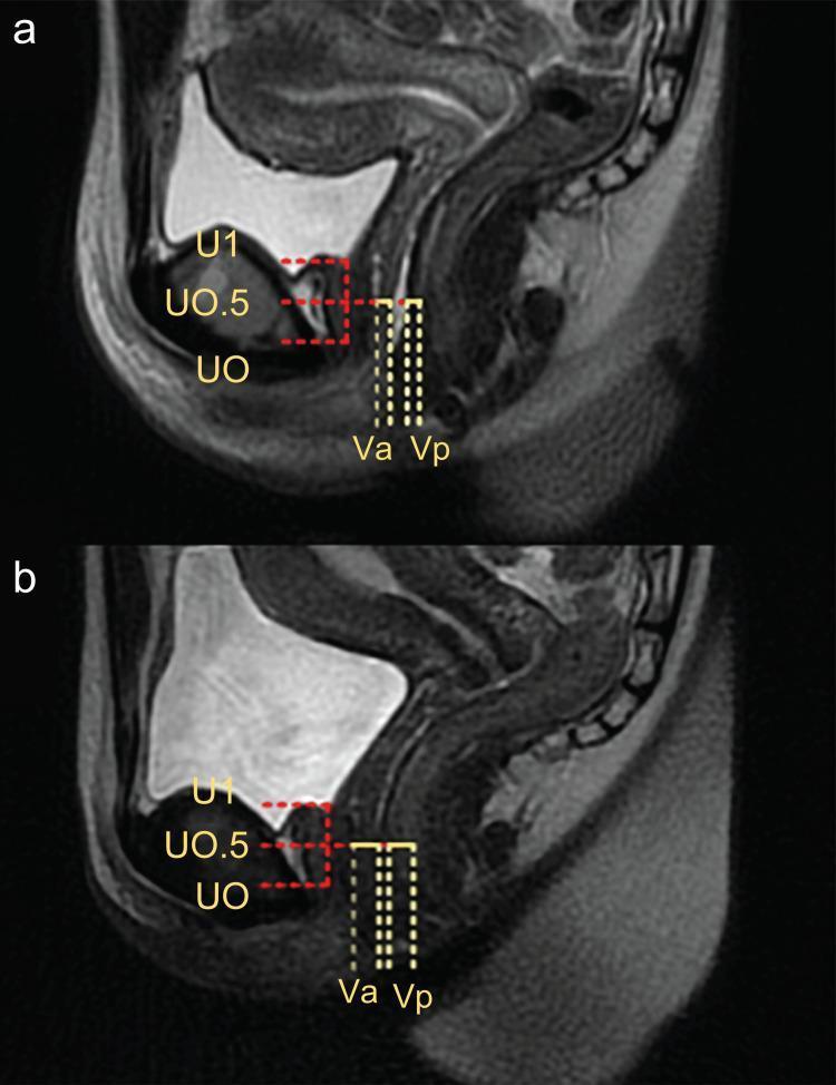
This diagnostic image consists of two sagittal T2-weighted magnetic resonance imaging (MRI) scans of the female pelvic floor, comparing anatomy before (a) and after (b) treatment for stress urinary incontinence (SUI). The images focus on the mid-urethra and vaginal wall interface. Key anatomical landmarks are labeled: U1 represents the upper end of the urethra (vesicourethral boundary), U0 marks the lower end (urethral opening), and U0.5 indicates the midway point. In image (a), representing the baseline (T0), the anterior (Va) and posterior (Vp) vaginal walls measured at the level of U0.5 appear thinner with a combined thickness of approximately 8 mm. In image (b), representing the post-treatment state (T3), there is a visible increase in the combined thickness of the Va and Vp to approximately 12 mm, suggesting tissue remodeling or swelling. A red dotted box highlights the adipose tissue space and urethral morphology, which shifts from an elliptical shape in (a) to a more circular configuration in (b). The image illustrates the anatomical changes associated with laser therapy in the management of pelvic floor disorders.
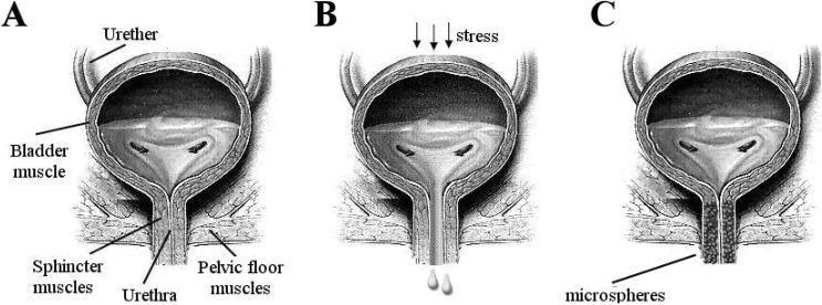
This medical anatomical diagram illustrates the pathophysiology and treatment of stress urinary incontinence (SUI) through three coronal views of the human bladder and urethra (A, B, and C). Diagram A represents normal anatomy and function, showing a healthy bladder muscle, intact ureters, and a closed urethra supported by strong sphincter and pelvic floor muscles. Diagram B depicts the mechanism of stress urinary incontinence, where external 'stress' (indicated by downward arrows) causes involuntary urine leakage through a weakened urethral opening and compromised pelvic floor support. Diagram C demonstrates a therapeutic intervention using urethral bulking agents. It shows the injection of 'microspheres' into the periurethral tissues, which facilitates complete coaptation (closure) of the urethral lumen to prevent leakage. Key labels include the bladder muscle, ureters, urethra, sphincter muscles, and pelvic floor muscles. The illustration is designed for educational use in urology to explain urinary control mechanisms and the clinical application of polymeric microspheres as bulking agents.
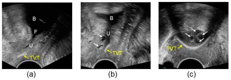
Pelvic floor sonography images demonstrating the management of recurrent stress urinary incontinence using a combination of a Tension-free Vaginal Tape (TVT) and a bulking agent (Bulkamid). Image (a) is a sagittal view showing the bladder (B) and urethra (U) under straining. It reveals proximal urethral funneling (F) and a TVT positioned too distally, failing to provide support. Image (b) shows the same sagittal view following the injection of bulking agent depots (white arrows). The depots are positioned between the bladder neck and the TVT, effectively closing the urethral lumen during straining to restore continence. Image (c) provides an axial view of the urethra, illustrating four hypoechoic bulking agent depots (white arrows) arranged circularly around the urethral lumen to achieve coaptation. The echogenic TVT (yellow arrow) is visible in its relation to the stabilized urethral structure. These images illustrate the use of functional ultrasound to assess implant positioning and surgical outcomes in urogynecology.
Stress Urinary Incontinence (SUI) in Obstetrics
Definition
Pathophysiology
- The vaginal wall acts as a "hammock" supporting the bladder neck and urethra via fibromuscular attachments to the pelvic sidewall (levator ani)
- Loss of this hammock support causes SUI
- SUI results from connective tissue laxity in the vagina and its supporting ligaments (pubourethral, cardinal/uterosacral, arcus tendineus fascia pelvis)
- Pelvic floor muscles act in three directions to close the urethra - forward pull of pubococcygeus + backward pull of levator plate
Obstetric Risk Factors
| Risk Factor | SUI Risk |
|---|---|
| Pregnancy | ~50% of pregnant women report SUI symptoms |
| Vaginal delivery | 2x increased risk vs. cesarean (2016 meta-analysis) |
| Parity | Strong independent risk factor for SUI specifically |
| Obesity | Strong risk - pudendal nerve injury, levator ani atrophy |
| Diabetes | Independent risk factor |
| Smoking/COPD | Chronic increased abdominal pressure |
| Age <50 years | SUI predominates (vs. urgency UI which rises >50 yrs) |
- Vaginal birth causes urethral injury (muscular and neurologic) via pudendal nerve damage and pelvic floor trauma
- Cesarean delivery appears protective against subsequent SUI
- ~50% of pregnant women report SUI symptoms, but most resolve after birth - however, recurrence within 5 years is high
Diagnosis
- Clinical evaluation: History + provocative cough/stress test
- Urodynamic studies (UDS): NOT required for uncomplicated straightforward SUI before surgery (VALUE trial: office evaluation alone non-inferior to UDS at 12 months). UDS IS recommended when:
- Mixed symptoms (SUI + urgency)
- Prior incontinence surgery
- Neurologic conditions
- Difficulty with bladder emptying
- Post-void residual (PVR): Should be checked
Treatment
Conservative (First-Line - Offer to All)
- Pelvic Floor Muscle Training (PFMT) - Kegel exercises, ideally supervised by a physiotherapist for at least 3 months
- Weight loss - 10% weight loss leads to >50% improvement in SUI symptoms
- Lifestyle: Reduce caffeine, alcohol; fluid optimization via bladder diary
- Bladder training: Timed voiding with progressive interval increase
- Biofeedback, electrical stimulation, pessaries, urethral plugs
Pharmacologic
- Duloxetine (SNRI) - increases efferent output at Onuf's nucleus in sacral spinal cord, raising urethral sphincter tone. Licensed in Europe (not USA) for SUI. Side effects: nausea, fatigue, dizziness
- Alpha-adrenergic agonists (ephedrine, pseudoephedrine) - not FDA-approved for SUI, rarely used
- Estrogen - systemic HRT is contraindicated (WHI and HERS trials showed it WORSENS incontinence); local/vaginal estrogen may help urgency symptoms short-term
Surgical (for failed conservative treatment)
| Procedure | Notes |
|---|---|
| Mid-urethral sling (MUS) - TVT/TOT | Gold standard surgical treatment; ~77% success at 12 months; shift from Burch colposuspension |
| Burch colposuspension | Open or laparoscopic; now less used since MUS arrival |
| Urethral bulking agents | For mild SUI or those unfit for surgery; no proven long-term efficacy |
| Artificial urinary sphincter (AUS) | Gold standard in males; ~80% continence rate |
Diagram - SUI Mechanism
Key Points for Exams
- SUI = leakage on exertion/coughing/sneezing; no detrusor contraction (differentiates from urgency UI)
- Vaginal delivery = 2x risk of SUI vs. cesarean
- 50% of pregnant women get SUI symptoms
- First-line treatment = PFMT for ≥3 months
- Systemic estrogen worsens SUI (WHI trial)
- Standard surgical treatment = mid-urethral sling
- UDS not needed in uncomplicated SUI before surgery