
Explain endocrine disorders
endocrine system glands diagram overview
diabetes mellitus pathophysiology insulin resistance
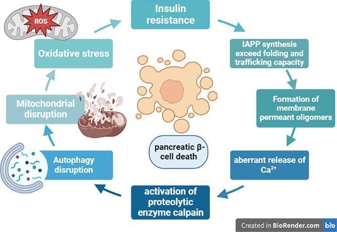
A pathophysiology diagram illustrating the vicious cycle of pancreatic beta-cell dysfunction and death involving Human Islet Amyloid Polypeptide (hIAPP). The process is depicted as a circular flow starting with 'Insulin resistance', which leads to increased 'IAPP synthesis exceeding folding and trafficking capacity'. This triggers the 'formation of membrane permeant oligomers', resulting in the 'aberrant release of Calcium (Ca2+)'. High calcium levels cause the 'activation of proteolytic enzyme calpain'. This leads to cellular degradation pathways, including 'autophagy disruption' and 'mitochondrial disruption' (depicted by a damaged mitochondrion), which generates 'Reactive Oxygen Species (ROS)' and 'oxidative stress'. Oxidative stress subsequently feeds back into increasing insulin resistance, completing the loop. Central to the diagram is an illustration of an hIAPP aggregate labeled 'pancreatic beta-cell death', highlighting the ultimate clinical consequence of this signaling pathway. This resource is relevant for endocrinology and the molecular study of Type 2 Diabetes Mellitus.
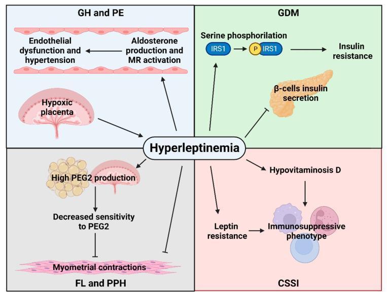
A pathophysiology diagram illustrating the central role of hyperleptinemia in the development of maternal obstetric complications, divided into four quadrants by condition. 1) GH and PE (Gestational Hypertension and Pre-eclampsia): Hyperleptinemia is linked to placental hypoxia and increased aldosterone production/MR activation, leading to endothelial dysfunction and hypertension. 2) GDM (Gestational Diabetes Mellitus): The diagram shows hyperleptinemia inducing serine phosphorylation of IRS1, leading to insulin resistance, while simultaneously inhibiting beta-cell insulin secretion. 3) FL and PPH (Failed Labor and Postpartum Hemorrhage): Hyperleptinemia stimulates high PGE2 production from adipose tissue and the placenta, which correlates with decreased sensitivity to PGE2 and subsequent inhibition of myometrial contractions. 4) CSSI (C-section Surgical Site Infection): Hyperleptinemia contributes to hypovitaminosis D and leptin resistance, both resulting in an immunosuppressive phenotype. Arrows indicate stimulatory pathways, while T-bars represent inhibitory actions, providing a comprehensive overview of hormonal and molecular signaling in obese pregnancies.
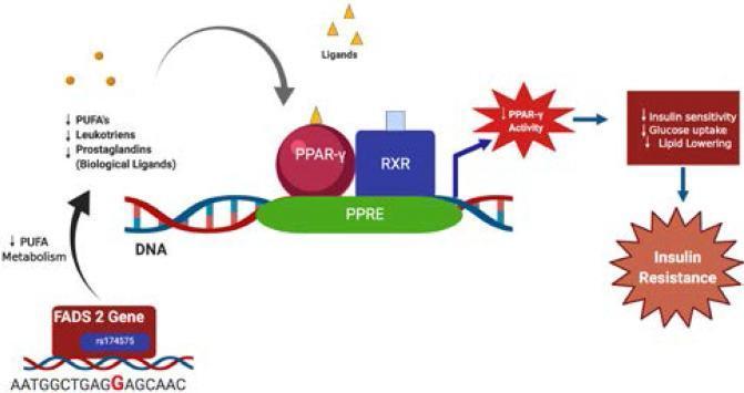
This pathophysiology diagram illustrates the suggestive molecular mechanism linking the FADS2 gene SNP rs174575 (G allele) to insulin resistance. The pathway begins with the FADS2 gene and the highlighted rs174575 variant, which leads to decreased polyunsaturated fatty acid (PUFA) metabolism. This reduction results in lower levels of biological ligands, specifically PUFAs, leukotrienes, and prostaglandins. These ligands normally bind to the Peroxisome Proliferator-Activated Receptor-gamma (PPAR-γ). In this model, altered ligand availability leads to diminished PPAR-γ activity. The diagram shows the PPAR-γ/RXR heterodimer complex bound to the Peroxisome Proliferator Response Element (PPRE) on a DNA sequence. The downstream functional consequences of decreased PPAR-γ activity are listed as reduced insulin sensitivity, decreased glucose uptake, and decreased lipid lowering. This sequence culminates in the development of insulin resistance, represented as a final starburst outcome. The illustration utilizes standard biochemical symbols, including a DNA double helix, geometric shapes for proteins (RXR, PPAR-γ), and flow arrows to indicate regulatory relationships in a cellular signaling context relevant to metabolic syndrome and Type 2 Diabetes Mellitus.
Endocrine Disorders: A Comprehensive Overview
The Endocrine Glands and Their Hormones
| Gland | Key Hormones | Main Actions |
|---|---|---|
| Hypothalamus | TRH, CRH, GnRH, GHRH, somatostatin, dopamine | Controls anterior pituitary secretion |
| Anterior Pituitary | TSH, ACTH, GH, LH, FSH, prolactin | Regulate downstream glands |
| Posterior Pituitary | ADH (vasopressin), oxytocin | Water reabsorption, uterine contractions |
| Thyroid | T3, T4 | Metabolism, growth, cardiac rate |
| Parathyroids | PTH | Calcium and phosphate regulation |
| Adrenal Cortex | Cortisol, aldosterone, androgens | Stress response, sodium balance |
| Adrenal Medulla | Epinephrine, norepinephrine | Fight-or-flight response |
| Pancreas (islets) | Insulin, glucagon | Glucose homeostasis |
| Gonads | Estrogen, testosterone, progesterone | Reproduction, secondary sex characteristics |
1. Diabetes Mellitus
Type 1 Diabetes
- Mechanism: Autoimmune destruction of pancreatic beta cells leads to absolute insulin deficiency.
- Onset: Typically in childhood/adolescence, though can occur at any age.
- Features: Polyuria, polydipsia, polyphagia, weight loss, risk of diabetic ketoacidosis (DKA).
- Treatment: Lifelong insulin replacement (basal-bolus regimens).
Type 2 Diabetes
- Mechanism: Progressive insulin resistance combined with relative insulin secretory deficiency. The beta-cell dysfunction cycle involves amyloid polypeptide (hIAPP) aggregation, oxidative stress, mitochondrial dysfunction, and ultimately beta-cell death - as illustrated below.
- Risk factors: Obesity, sedentary lifestyle, family history, older age.
- Features: Often asymptomatic early; hyperglycemia detected on screening. Long-term complications include retinopathy, nephropathy, neuropathy, and cardiovascular disease.
- Treatment: Lifestyle modification, metformin first-line, then GLP-1 agonists, SGLT-2 inhibitors, sulfonylureas, or insulin as needed.
Diagnosis
- Fasting blood glucose >125 mg/dL
- 2-hour or random glucose ≥200 mg/dL
- HbA1c ≥6.5%
2. Thyroid Disorders
Hyperthyroidism
- Most common cause: Graves' disease (autoimmune stimulation of TSH receptor).
- Other causes: Toxic multinodular goiter, toxic adenoma, thyroiditis.
- Symptoms: Tremor, anxiety, irritability, diarrhea, weight loss, sweating, palpitations, insomnia, exophthalmos (in Graves'), tachycardia, warm moist skin.
- Investigations: Suppressed TSH, elevated free T4 and/or T3; radioactive thyroid scan helps differentiate causes.
- Treatment: Radioactive iodine ablation (treatment of choice in adults with Graves'); antithyroid drugs (PTU, methimazole) for children or as bridge therapy; subtotal thyroidectomy for toxic adenoma.
Hypothyroidism
- Most common cause: Hashimoto thyroiditis (chronic autoimmune thyroiditis) - lymphocytic infiltration destroys thyroid parenchyma.
- Other causes: Thyroid ablation (post-radioiodine or post-surgery), iodine deficiency (most preventable cause of intellectual disability globally), congenital thyroid absence.
- Symptoms: Weight gain, fatigue, cold intolerance, constipation, dry skin, bradycardia, myxedema in severe cases, depression.
- Investigations: Elevated TSH (best screening test), low free T4.
- Treatment: Levothyroxine replacement, titrated to normalize TSH.
3. Adrenal Disorders
Cushing's Syndrome (Hypercortisolism)
- Most common cause: Long-term exogenous glucocorticoid use.
- Endogenous causes:
- Cushing's disease: ACTH-secreting pituitary adenoma (most common endogenous cause, ~70%)
- Adrenal adenoma/carcinoma: autonomous cortisol production (~10%)
- Ectopic ACTH from neuroendocrine tumors (e.g., small cell lung carcinoma)
- Clinical features: Central obesity, buffalo hump, moon face, proximal muscle weakness, easy bruising, purple striae, hirsutism, hypertension, osteoporosis, hyperglycemia, menstrual irregularities.
- Diagnosis: 24-hour urinary free cortisol, late-night salivary cortisol, 1mg overnight dexamethasone suppression test; MRI pituitary for Cushing's disease.
- Treatment: Surgical removal of the causative lesion (transsphenoidal surgery for pituitary adenoma, adrenalectomy for adrenal tumor). Steroidogenesis inhibitors (metyrapone, ketoconazole) as adjuncts.
Addison's Disease (Primary Adrenal Insufficiency)
- Mechanism: Destruction of the adrenal cortex (most commonly autoimmune in developed countries); results in deficiency of cortisol AND aldosterone.
- Symptoms: Fatigue, weight loss, hyperpigmentation (elevated ACTH stimulates melanocytes), postural hypotension, hyponatremia, hyperkalemia.
- Crisis: Addisonian crisis is a life-threatening emergency with severe hypotension, vomiting, and altered consciousness - treated with IV hydrocortisone and saline.
- Diagnosis: Low morning cortisol; failed cortisol rise on ACTH stimulation test; elevated ACTH.
- Treatment: Lifelong hydrocortisone + fludrocortisone replacement.
Primary Hyperaldosteronism (Conn's Syndrome)
- Excess aldosterone from adrenal adenoma or bilateral hyperplasia.
- Features: Hypertension (often resistant), hypokalemia, metabolic alkalosis.
- Diagnosis: Elevated aldosterone-to-renin ratio; CT adrenals; adrenal vein sampling.
- Treatment: Adrenalectomy for adenoma; mineralocorticoid antagonists (spironolactone) for hyperplasia.
Pheochromocytoma
- Catecholamine-secreting tumor of adrenal medulla (or extra-adrenal paraganglioma).
- Features: Episodic hypertension, headache, diaphoresis, palpitations ("5 Ps").
- Diagnosis: Plasma or urine metanephrines; CT/MRI adrenals.
- Treatment: Surgical resection after pre-operative alpha-blockade (phenoxybenzamine) then beta-blockade.
4. Pituitary Disorders
Acromegaly
- Caused by a GH-secreting pituitary adenoma in adults (after growth plate closure). In children (before closure), the result is gigantism.
- Features: Enlargement of hands, feet, facial features (prognathism, frontal bossing), macroglossia, carpal tunnel syndrome, sleep apnea, hypertension, diabetes, arthritis, and increased cardiovascular mortality.
- Diagnosis: Elevated IGF-1; failure to suppress GH after oral glucose load; MRI pituitary.
- Treatment: Transsphenoidal surgery first-line; somatostatin analogues (octreotide, lanreotide); pegvisomant (GH receptor antagonist); radiosurgery.
Hypopituitarism
- Deficiency of one or more pituitary hormones from tumors, surgery, radiation, or infarction.
- Features depend on which hormones are lost (GH deficiency: fatigue, reduced muscle mass; gonadotropin deficiency: infertility, hypogonadism; TSH deficiency: secondary hypothyroidism; ACTH deficiency: secondary adrenal insufficiency).
- Treatment: Replacement of deficient hormones.
Prolactinoma
- Most common pituitary tumor.
- Features: Galactorrhea, amenorrhea/irregular periods in women; erectile dysfunction, reduced libido in men; headache, visual field defects (bitemporal hemianopia) if large.
- Treatment: Dopamine agonists (cabergoline, bromocriptine) are first-line and highly effective.
Diabetes Insipidus
- Central DI: ADH deficiency (head trauma, pituitary surgery, tumors).
- Nephrogenic DI: Renal resistance to ADH.
- Features: Massive polyuria (>3L/day), polydipsia, dilute urine (low osmolality).
- Treatment: Central DI treated with desmopressin (synthetic ADH); nephrogenic DI treated with thiazide diuretics and low-salt diet.
5. Parathyroid Disorders
Primary Hyperparathyroidism
- Excessive PTH from adenoma (85%) or hyperplasia.
- Results in hypercalcemia: "bones, stones, groans, and psychic moans" - bone pain/osteoporosis, renal calculi, GI symptoms (nausea, constipation), neuropsychiatric symptoms.
- Treatment: Parathyroidectomy for symptomatic cases.
Hypoparathyroidism
- Most common cause: Inadvertent parathyroid removal during thyroidectomy.
- Results in hypocalcemia: Tetany, perioral tingling, Chvostek's sign, Trousseau's sign, seizures.
- Treatment: Calcium and vitamin D supplementation; PTH analogues in refractory cases.
6. Gonadal Disorders (Hypogonadism)
- Male hypogonadism: Low testosterone from primary testicular failure (Klinefelter syndrome, orchitis) or secondary (pituitary/hypothalamic). Features: Decreased libido, erectile dysfunction, infertility, osteoporosis, loss of body hair.
- Female hypogonadism: Primary ovarian insufficiency (Turner syndrome, autoimmune) or secondary. Features: Amenorrhea, infertility, hot flashes, osteoporosis.
- Treatment: Hormone replacement therapy (testosterone in men, estrogen/progesterone in women).
General Principles of Endocrine Pathology
| Category | Examples |
|---|---|
| Hormone excess | Cushing's, acromegaly, hyperthyroidism, primary hyperaldosteronism |
| Hormone deficiency | Hypothyroidism, Addison's disease, hypoparathyroidism, hypogonadism |
| Receptor defects | Nephrogenic DI, testicular feminization (androgen insensitivity) |
| Tumors | Pituitary adenoma, pheochromocytoma, carcinoid, MEN syndromes |
| Autoimmune destruction | Graves' disease, Hashimoto's, Type 1 diabetes, Addison's |
Key Diagnostic Tools
- Hormone levels: Basal and stimulated/suppressed (e.g., ACTH stimulation, dexamethasone suppression, oral glucose tolerance)
- HbA1c: Reflects average glucose over 3 months
- TSH: Best first-line screen for thyroid dysfunction
- Imaging: MRI pituitary, CT/MRI adrenals, thyroid ultrasound
- Functional tests: 24h urine catecholamines/metanephrines, urinary free cortisol
- Costanzo Physiology 7th Edition - Table 9.2 (endocrine glands summary)
- Goldman-Cecil Medicine International Edition - Endocrine Disorders
- Rosen's Emergency Medicine - Adrenal disorders
- Swanson's Family Medicine Review - Thyroid disease
- The Washington Manual of Medical Therapeutics - Acromegaly
- Henry's Clinical Diagnosis and Management by Laboratory Methods - Cushing's syndrome
Everyone disease add this 1. Definition 2. Pathophysiology (step-by-step →) 3. Risk Factors 4. Signs & Symptoms 5. Complications 6. Disease pattern 7. Medical Management 8. Nursing Diagnosis (Top 3) 9. Nursing Interventions
thyroid disease Graves hyperthyroidism hypothyroidism
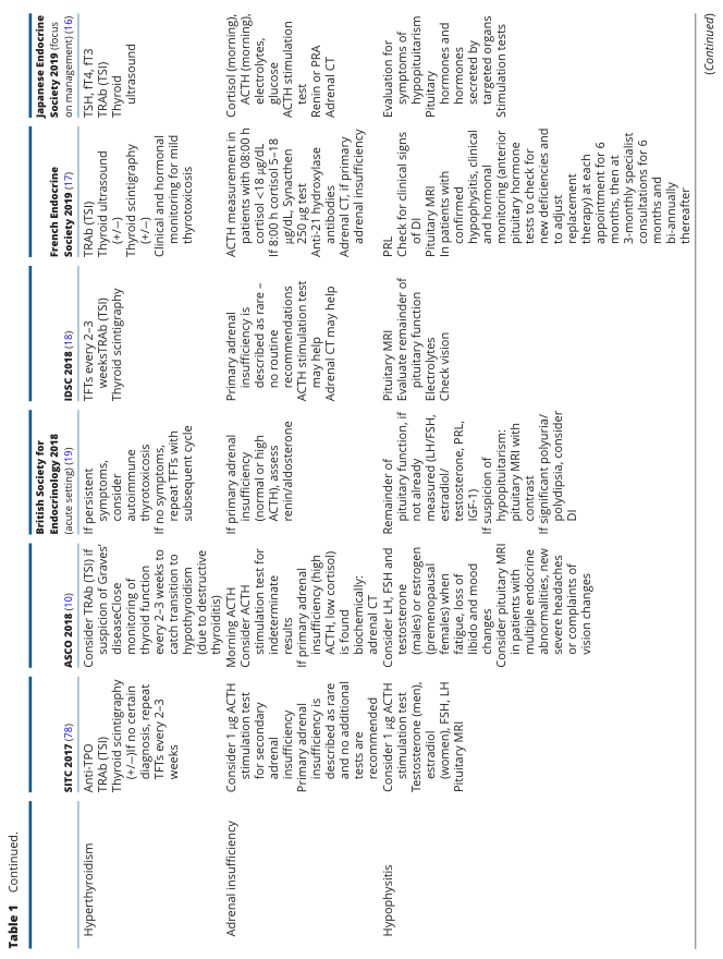
ENDOCRINE DISORDERS - Complete Structured Reference
1. DIABETES MELLITUS
1. Definition
2. Pathophysiology (Step-by-Step)
- Genetic susceptibility (HLA-DR3, HLA-DR4 alleles) triggers immune dysregulation
- Environmental trigger (viral infection, e.g., Coxsackievirus) activates autoreactive T cells
- T cells attack pancreatic beta cells (insulitis)
- Progressive beta-cell destruction → absolute insulin deficiency
- No insulin → glucose cannot enter cells → hyperglycemia
- Fat breakdown → free fatty acids → ketone bodies → DKA risk
- Genetic predisposition + obesity/sedentary lifestyle
- Excess free fatty acids + adipocytokines (TNF-α, IL-6) from adipose tissue → insulin resistance
- Pancreatic beta cells compensate by secreting more insulin (hyperinsulinemia)
- Over time, beta-cell exhaustion occurs (hIAPP aggregation → oxidative stress → beta-cell death)
- Relative insulin deficiency + persistent insulin resistance → sustained hyperglycemia
- Chronic hyperglycemia → AGE formation, PKC activation, polyol pathway disturbances → end-organ damage
3. Risk Factors
- Type 1: Family history, HLA genetics, autoimmune conditions, viral triggers
- Type 2: Obesity (BMI >30), physical inactivity, family history, age >45, hypertension, dyslipidemia, gestational diabetes history, polycystic ovary syndrome (PCOS), ethnicity (South Asian, African, Hispanic)
4. Signs & Symptoms
| Feature | Type 1 | Type 2 |
|---|---|---|
| Onset | Acute/rapid | Insidious |
| Classic symptoms | Polyuria, polydipsia, polyphagia, weight loss | Often asymptomatic early |
| Other features | Fatigue, blurred vision, DKA (nausea, vomiting, fruity breath) | Recurrent infections, slow wound healing, fatigue, paresthesia |
5. Complications
- Diabetic retinopathy - leading cause of blindness in working-age adults
- Diabetic nephropathy - leading cause of end-stage renal disease
- Diabetic neuropathy - peripheral (glove-and-stocking sensory loss), autonomic (gastroparesis, orthostatic hypotension)
- Coronary artery disease, stroke, peripheral arterial disease
- Diabetic ketoacidosis (Type 1), hyperosmolar hyperglycemic state (Type 2), hypoglycemia
6. Disease Pattern
- Type 1: Lifelong; variable control; honeymoon period early; worsens with stress/illness
- Type 2: Progressive; early reversibility with lifestyle change; complications appear after years of poor control; follows a metabolic syndrome pattern
7. Medical Management
- Type 1: Basal-bolus insulin regimen; continuous glucose monitoring (CGM); carbohydrate counting
- Type 2:
- 1st line: Metformin + lifestyle modification
- Add-on: GLP-1 receptor agonists (liraglutide, semaglutide), SGLT-2 inhibitors (empagliflozin), DPP-4 inhibitors, sulfonylureas
- Insulin when oral agents insufficient
- Targets: HbA1c <7%, fasting glucose 80-130 mg/dL, BP <130/80 mmHg
- Monitoring: HbA1c every 3 months, annual eye/kidney/foot exam
8. Top 3 Nursing Diagnoses
- Imbalanced Nutrition: More or Less Than Body Requirements related to inability to use glucose
- Risk for Unstable Blood Glucose Level related to inadequate insulin or non-adherence
- Deficient Knowledge related to disease management, insulin administration, diet
9. Nursing Interventions
- Monitor blood glucose as ordered (fasting, post-prandial, before/after insulin)
- Administer insulin or oral hypoglycemics on time; rotate injection sites
- Teach carbohydrate counting, portion control, and glycemic index foods
- Assess for hypoglycemia signs (shakiness, diaphoresis, confusion); keep glucose gel/juice at bedside
- Inspect feet daily - assess for ulcers, calluses, color, temperature, pulses
- Educate on sick-day rules and when to seek emergency care (DKA symptoms)
- Monitor HbA1c trends; coordinate with dietitian and diabetes educator
2. HYPOTHYROIDISM
1. Definition
2. Pathophysiology (Step-by-Step)
- Triggering cause (autoimmune/surgical/iodine deficiency/radiation)
- Hashimoto's: CD4+ T cells sensitized to thyroid antigens → lymphocytic infiltration of thyroid gland
- Inflammatory destruction of follicular cells → reduced T3/T4 synthesis
- Low T3/T4 → hypothalamus senses deficit → increased TRH
- Elevated TRH → anterior pituitary releases more TSH (TSH rises - hallmark of primary hypothyroidism)
- Thyroid gland unable to respond adequately → persistent hormone deficiency
- Reduced T3/T4 → slowed metabolism systemically: reduced cardiac output, decreased gut motility, accumulation of glycosaminoglycans (myxedema), cold intolerance
3. Risk Factors
- Female sex (5-10x more common)
- Age >60 years
- Personal/family history of autoimmune disease
- Previous thyroid surgery or radioiodine therapy
- Iodine deficiency (global)
- Lithium or amiodarone use
- Down syndrome, Turner syndrome
4. Signs & Symptoms
- Metabolic: Weight gain, cold intolerance, fatigue
- Cardiovascular: Bradycardia, diastolic hypertension, pericardial effusion
- GI: Constipation
- Skin/Hair: Dry coarse skin, brittle nails, hair loss, periorbital edema, non-pitting edema (myxedema)
- Neurological: Depression, cognitive slowing, carpal tunnel syndrome
- Reproductive: Menorrhagia, anovulation, infertility
- Severe (myxedema coma): Hypothermia, stupor, respiratory failure
5. Complications
- Myxedema coma (life-threatening; precipitated by infection, cold exposure, sedatives)
- Cardiovascular disease (hypercholesterolemia, atherosclerosis)
- Infertility and pregnancy complications
- Peripheral neuropathy
- Cretinism (congenital hypothyroidism - intellectual disability, growth retardation)
6. Disease Pattern
- Chronic, insidious onset; often diagnosed incidentally on TSH screening
- Gradual worsening over months to years if untreated
- Excellent response to treatment; lifelong medication required in most cases
7. Medical Management
- Levothyroxine (T4): Oral, once daily, weight-based starting dose (~1.6 mcg/kg/day)
- Taken on empty stomach, 30-60 min before food
- TSH monitored every 6-8 weeks until stable, then annually
- Adjust dose with pregnancy (need increases ~30-50%)
- Myxedema coma: IV levothyroxine 300-500 mcg loading dose + IV hydrocortisone + supportive care
8. Top 3 Nursing Diagnoses
- Activity Intolerance related to fatigue and decreased metabolic rate
- Constipation related to decreased GI motility
- Deficient Knowledge related to lifelong medication adherence
9. Nursing Interventions
- Administer levothyroxine at the same time each day; teach not to take with calcium, iron, or antacids (impair absorption)
- Monitor vital signs - watch for bradycardia and hypothermia
- Assess bowel function daily; encourage fiber and fluid intake
- Apply extra blankets; maintain warm environment (poor thermoregulation)
- Monitor for medication overdose signs (palpitations, tremor, weight loss = hyperthyroid symptoms)
- Educate: never stop medication abruptly; carry medical ID
- Screen for depression; refer to mental health support if needed
3. HYPERTHYROIDISM
1. Definition
2. Pathophysiology (Step-by-Step)
- Graves' disease: Autoimmune production of TSH receptor-stimulating antibodies (TSI/TRAb)
- TSI binds and continuously activates TSH receptor on thyroid follicular cells
- Unregulated T3/T4 synthesis and secretion (not subject to normal negative feedback)
- Excess T3/T4 in circulation → hypermetabolic state
- T3 upregulates beta-adrenergic receptors → enhanced catecholamine sensitivity → tachycardia, tremor, diaphoresis
- Increased oxygen consumption, heat production, protein catabolism, gut motility
- TSI cross-reacts with orbital fibroblasts → Graves' ophthalmopathy (proptosis, lid lag)
3. Risk Factors
- Female sex
- Age 20-40 years (peak for Graves')
- Family history of thyroid autoimmunity
- Smoking (especially for Graves' ophthalmopathy)
- High iodine intake (can precipitate toxic nodular goiter)
- Stress, pregnancy, postpartum period
- Medications: amiodarone, interferon-alpha, lithium withdrawal
4. Signs & Symptoms
- Metabolic: Weight loss despite increased appetite, heat intolerance, diaphoresis
- Cardiovascular: Tachycardia, palpitations, atrial fibrillation, wide pulse pressure
- Neurological: Tremor, anxiety, irritability, insomnia, emotional lability
- GI: Diarrhea, increased bowel frequency
- Musculoskeletal: Proximal muscle weakness
- Skin: Warm, moist, smooth skin; pretibial myxedema (Graves')
- Eyes: Proptosis/exophthalmos, lid lag, lid retraction, diplopia (Graves' only)
- Goiter: Diffuse enlargement (Graves'), nodular (toxic MNG)
5. Complications
- Thyroid storm (thyrotoxic crisis) - life-threatening hyperpyrexia, extreme tachycardia, altered consciousness; precipitated by surgery, infection
- Atrial fibrillation and heart failure
- Osteoporosis (excess T3 stimulates osteoclasts)
- Infertility; pregnancy complications (preterm birth, fetal growth restriction)
- Ophthalmopathy causing vision loss (Graves')
6. Disease Pattern
- Graves' disease: relapsing-remitting; remission possible with anti-thyroid drugs (~30-40%)
- Toxic adenoma/MNG: progressive without treatment; does not self-remit
7. Medical Management
- Anti-thyroid drugs (ATDs): Methimazole (first-line), PTU (in pregnancy first trimester, thyroid storm)
- Block thyroid peroxidase → reduce hormone synthesis
- Beta-blockers (propranolol): Symptomatic relief (tremor, palpitations) - do not reduce hormone levels
- Radioactive iodine (RAI - I-131): Ablates thyroid tissue; treatment of choice for adults with Graves'
- Thyroidectomy: For large goiter, suspected malignancy, failure of other therapies
- Thyroid storm: PTU + Lugol's iodine + propranolol + hydrocortisone + cooling measures
8. Top 3 Nursing Diagnoses
- Decreased Cardiac Output related to increased heart rate and cardiac workload
- Imbalanced Nutrition: Less Than Body Requirements related to hypermetabolism
- Anxiety related to excess catecholamine stimulation and disease process
9. Nursing Interventions
- Monitor heart rate, rhythm, and blood pressure continuously; report tachycardia >100 bpm or new arrhythmias
- Provide high-calorie, high-protein diet (4,000-5,000 kcal/day if severely hypermetabolic); small frequent meals
- Provide a cool, quiet, low-stimulation environment; limit visitors
- Administer methimazole/PTU with food to reduce GI upset; monitor for agranulocytosis (fever, sore throat → STAT WBC)
- Teach eye care for Graves' ophthalmopathy: lubricating eye drops, elevate HOB, wear dark glasses
- Educate: do not stop ATDs abruptly; report palpitations, fever, jaundice (PTU-related hepatotoxicity)
- Pre-RAI: ensure not pregnant; post-RAI: teach radiation precautions (distance from children/pregnant women for 1 week)
4. CUSHING'S SYNDROME
1. Definition
2. Pathophysiology (Step-by-Step)
- Trigger: pituitary ACTH-secreting adenoma (Cushing's disease), adrenal adenoma/carcinoma, ectopic ACTH tumor (e.g., small cell lung cancer), or exogenous steroid use
- Excess cortisol in circulation
- Cortisol promotes: gluconeogenesis (hyperglycemia), lipolysis and fat redistribution (central obesity, buffalo hump, moon face), protein catabolism (muscle wasting, thin skin, striae)
- Cortisol suppresses immune function and inflammatory response
- Excess cortisol acts on mineralocorticoid receptors → sodium retention → hypertension, hypokalemia
- Cortisol inhibits osteoblasts + increases osteoclast activity → osteoporosis
- Feedback loop broken: in Cushing's disease, pituitary adenoma resists normal feedback; cortisol stays elevated
3. Risk Factors
- Long-term corticosteroid use (most common)
- Pituitary adenoma (ACTH-secreting)
- Adrenal tumor (adenoma, carcinoma)
- Ectopic ACTH-producing tumors (small cell lung cancer, carcinoid tumors)
- Female sex, age 20-50 years (endogenous forms)
- MEN1 syndrome
4. Signs & Symptoms
- Classic triad: Central obesity + moon face + buffalo hump
- Easy bruising, purple abdominal striae (>1 cm wide)
- Proximal muscle weakness (difficulty rising from chair)
- Hypertension, hyperglycemia
- Hirsutism, menstrual irregularities, decreased libido
- Thin, fragile skin; poor wound healing
- Osteoporosis (back pain, vertebral fractures)
- Psychiatric symptoms: depression, anxiety, emotional lability
- Plethoric (reddish) face; acne
5. Complications
- Type 2 diabetes mellitus
- Pathological fractures (osteoporosis)
- Severe infections (immunosuppression)
- Adrenal crisis after treatment (sudden cortisol withdrawal)
- Cardiovascular disease (hypertension, atherosclerosis)
- Avascular necrosis of the hip
6. Disease Pattern
- Exogenous: dose- and duration-dependent; reversible with dose reduction/tapering
- Endogenous: chronic, progressive; cyclical in some cases; requires surgical intervention
7. Medical Management
- Exogenous: Gradual steroid tapering (never stop abruptly - risk of adrenal crisis)
- Cushing's disease (pituitary): Transsphenoidal adenomectomy (first-line)
- Adrenal Cushing: Adrenalectomy (laparoscopic preferred)
- Ectopic ACTH: Treat the primary tumor; steroidogenesis inhibitors if unresectable
- Medical steroidogenesis inhibitors: Metyrapone, ketoconazole, osilodrostat, pasireotide
- Post-operative: hydrocortisone replacement (adrenal insufficiency expected)
8. Top 3 Nursing Diagnoses
- Risk for Infection related to cortisol-induced immunosuppression
- Disturbed Body Image related to physical changes (moon face, weight gain, striae)
- Risk for Injury (Fractures) related to osteoporosis and muscle weakness
9. Nursing Interventions
- Strict aseptic technique for all procedures; monitor for early infection signs (temperature, WBC)
- Limit visitors with active infections; private room if possible
- Assist with ambulation; fall prevention: bed rails up, non-slip footwear, call bell in reach
- Assess skin daily for breakdown, bruising, wound healing; use gentle tape on fragile skin
- Provide psychological support; acknowledge changes in appearance; encourage verbalization
- Monitor blood glucose regularly; administer insulin as ordered
- Educate post-surgical patients on steroid replacement therapy - never skip doses; carry medical ID
5. ADDISON'S DISEASE (Primary Adrenal Insufficiency)
1. Definition
2. Pathophysiology (Step-by-Step)
- Triggering cause: autoimmune (most common, ~80% in developed countries), tuberculosis, metastatic cancer, HIV, bilateral adrenalectomy
- Autoimmune: Anti-21-hydroxylase antibodies → lymphocytic destruction of adrenal cortex
- Progressive destruction of all three cortical zones → loss of cortisol, aldosterone, and adrenal androgens
- Low cortisol → hypothalamus/pituitary lose negative feedback → elevated ACTH and MSH
- Elevated MSH → stimulates melanocytes → hyperpigmentation (skin creases, buccal mucosa)
- Low aldosterone → loss of sodium and water retention → hyponatremia, hypovolemia, hypotension
- Low aldosterone → decreased potassium excretion → hyperkalemia
- Low cortisol → hypoglycemia (decreased gluconeogenesis), fatigue, inability to mount stress response
- Addisonian crisis: Acute severe deficiency (triggered by infection, trauma, surgery) → circulatory collapse
3. Risk Factors
- Autoimmune polyendocrine syndromes (Type 1: Addison's + hypoparathyroidism + mucocutaneous candidiasis; Type 2: Addison's + Type 1 DM + autoimmune thyroiditis)
- Tuberculosis (most common cause globally)
- HIV/AIDS
- Bilateral adrenal hemorrhage (meningococcemia - Waterhouse-Friderichsen syndrome)
- Long-term steroid therapy (relative adrenal insufficiency upon abrupt withdrawal)
- Female sex, autoimmune history
4. Signs & Symptoms
- Hallmark: Hyperpigmentation (bronze skin, darkening of scars, palmar creases, buccal mucosa)
- Weakness, fatigue, lethargy
- Orthostatic hypotension, dizziness
- Weight loss, anorexia, nausea, vomiting
- Salt craving (due to aldosterone deficiency)
- Hyponatremia, hyperkalemia (electrolyte disturbances)
- Hypoglycemia
- Depression, irritability
- Crisis: Severe hypotension, abdominal pain, fever, altered consciousness
5. Complications
- Addisonian crisis (adrenal crisis) - life-threatening cardiovascular collapse
- Electrolyte disturbances (fatal hyperkalemia)
- Chronic fatigue and disability if undertreated
- Increased susceptibility to infections
- Growth retardation in children
6. Disease Pattern
- Chronic; insidious onset (symptoms appear after >90% of adrenal cortex is destroyed)
- Crisis can be precipitated by any physiological stress
- Requires lifelong hormone replacement; good quality of life with proper management
7. Medical Management
- Chronic replacement:
- Hydrocortisone 15-25 mg/day in 2-3 divided doses (higher morning dose to mimic diurnal rhythm)
- Fludrocortisone 0.05-0.1 mg/day (mineralocorticoid replacement)
- Sick-day rule: Double/triple hydrocortisone dose during illness, injury, or minor procedures
- Addisonian crisis: IV hydrocortisone 100 mg bolus, then 50-100 mg every 6-8 hours + IV normal saline + glucose + treat precipitating cause
- Regular monitoring: electrolytes, blood pressure, weight, ACTH, renin
8. Top 3 Nursing Diagnoses
- Deficient Fluid Volume related to aldosterone deficiency and sodium wasting
- Risk for Imbalanced Electrolytes related to hyponatremia and hyperkalemia
- Fatigue related to cortisol deficiency and hypoglycemia
9. Nursing Interventions
- Monitor vital signs hourly in crisis; assess for orthostatic hypotension with each position change
- Administer IV fluids (0.9% saline) and IV hydrocortisone as ordered; monitor response
- Monitor serum sodium, potassium, and glucose frequently; report abnormals
- Provide high-sodium diet; encourage liberal fluid intake (non-crisis)
- Educate: double corticosteroid dose during illness/fever/surgery; carry injectable hydrocortisone kit for emergencies
- Teach patient/family to recognize adrenal crisis: extreme weakness, vomiting, hypotension → call 911 immediately
- Ensure patient wears medical alert ID at all times
- Educate: never stop steroids abruptly; this is a lifelong condition
6. PHEOCHROMOCYTOMA
1. Definition
2. Pathophysiology (Step-by-Step)
- Neoplastic transformation of chromaffin cells (adrenal medulla) - sporadic or hereditary (RET, VHL, SDHB mutations)
- Tumor stores and episodically secretes large amounts of epinephrine and/or norepinephrine
- Catecholamines bind alpha-1 receptors → intense peripheral vasoconstriction → severe hypertension
- Catecholamines bind beta-1 receptors → increased heart rate, contractility → palpitations, tachycardia
- Metabolic effects: stimulate glycogenolysis and lipolysis → hyperglycemia
- Episodes triggered by tumor manipulation, physical activity, anesthesia induction, certain foods (tyramine) or drugs (metoclopramide, glucagon)
- Persistent catecholamine excess → left ventricular hypertrophy, catecholamine cardiomyopathy
3. Risk Factors
- MEN2A/MEN2B (RET mutations) - bilateral pheochromocytoma
- von Hippel-Lindau disease (VHL mutations)
- Neurofibromatosis type 1 (NF1)
- Hereditary paraganglioma-pheochromocytoma syndrome (SDHB/SDHD mutations)
- Age 40-60 years (sporadic); younger in hereditary forms
- Family history
4. Signs & Symptoms
- Severe headache
- Diaphoresis (drenching sweats)
- Palpitations
- Hypertension (episodic or sustained), resistant to standard treatment
- Pallor or flushing during paroxysms
- Tremor, anxiety, sense of doom
- Chest or abdominal pain
- Weight loss
- Hyperglycemia
5. Complications
- Hypertensive crisis and stroke
- Catecholamine-induced cardiomyopathy
- Cardiac arrhythmias
- Multi-organ failure (crisis)
- Malignant transformation (~10% of adrenal tumors are malignant; higher in extra-adrenal)
6. Disease Pattern
- Paroxysmal or sustained; attacks last minutes to hours
- Rule of 10s: 10% bilateral, 10% extra-adrenal, 10% malignant, 10% in children (traditional teaching)
- Surgical cure possible in most benign cases
7. Medical Management
- Pre-operative alpha-blockade (MANDATORY, minimum 10-14 days before surgery):
- Phenoxybenzamine (irreversible alpha-blocker) is first-line; alternative: doxazosin
- Purpose: dilate vasculature, normalize BP, restore intravascular volume
- Beta-blockade (ONLY after adequate alpha-blockade - never first; causes hypertensive crisis):
- Propranolol or atenolol for heart rate control
- Definitive treatment: Laparoscopic adrenalectomy
- Malignant/unresectable: Metaiodobenzylguanidine (MIBG) therapy, chemotherapy (cyclophosphamide + vincristine + dacarbazine)
- Monitor metanephrines post-operatively to confirm cure
8. Top 3 Nursing Diagnoses
- Risk for Hypertensive Crisis related to catecholamine surges
- Anxiety related to unpredictable paroxysmal episodes
- Deficient Knowledge related to pre-operative preparation and post-operative care
9. Nursing Interventions
- Monitor BP every 15-30 minutes during paroxysms; have IV phentolamine or nitroprusside ready for hypertensive crisis
- Instruct to avoid triggers: strenuous activity, emotional stress, tyramine-rich foods (aged cheese, wine), abdominal palpation
- Pre-operative: administer alpha-blockers as scheduled; monitor for postural hypotension; encourage high-salt, high-fluid intake to expand volume
- Maintain a calm, low-stimulation environment; reduce unnecessary procedures
- Post-adrenalectomy: monitor for hypotension (catecholamine drop after removal); IV fluids ready
- Monitor blood glucose; check for hyperglycemia post-op
- Educate on genetic screening recommendations if hereditary form suspected
7. PRIMARY HYPERALDOSTERONISM (CONN'S SYNDROME)
1. Definition
2. Pathophysiology (Step-by-Step)
- Adrenal adenoma (unilateral, ~35%) or bilateral adrenal hyperplasia (~65%) produces excess aldosterone autonomously
- Aldosterone binds mineralocorticoid receptors in principal cells of collecting ducts
- Upregulates Na+/K+-ATPase and epithelial sodium channels (ENaC) → increased sodium reabsorption
- Na+ retention → water follows → expanded intravascular volume → hypertension
- Sodium reabsorption is coupled to potassium and hydrogen ion secretion → hypokalemia and metabolic alkalosis
- Elevated aldosterone suppresses renin (negative feedback from volume expansion) → low plasma renin - diagnostic hallmark
- Persistent hypertension → end-organ damage (cardiac hypertrophy, renal damage)
3. Risk Factors
- Age 30-50 years
- Female sex (slightly)
- Resistant hypertension (on 3+ antihypertensives)
- Spontaneous or diuretic-resistant hypokalemia
- Adrenal incidentaloma
- Family history of early-onset hypertension or stroke
4. Signs & Symptoms
- Hypertension (often resistant to multiple drugs)
- Hypokalemia: Muscle weakness, fatigue, cramps, palpitations
- Polyuria and polydipsia (hypokalemia impairs ADH response)
- Metabolic alkalosis (can worsen hypokalemia symptoms)
- Often NO edema (aldosterone escape phenomenon)
- Headaches
5. Complications
- Stroke and cardiovascular events (higher risk than matched hypertensive patients)
- Left ventricular hypertrophy and heart failure
- Chronic kidney disease
- Atrial fibrillation
- Hypokalemia-induced arrhythmias
6. Disease Pattern
- Chronic and progressive without treatment
- Adenoma: cured by surgery; hyperplasia: managed lifelong with medication
- Screening recommended in all patients with resistant hypertension
7. Medical Management
- Screening: Aldosterone-to-renin ratio (ARR) >30-40 is positive
- Confirmatory tests: Salt loading test, fludrocortisone suppression test
- Imaging: CT adrenals; adrenal vein sampling (AVS) to lateralize
- Unilateral adenoma: Laparoscopic adrenalectomy → often curative
- Bilateral hyperplasia: Spironolactone (first-line MR antagonist) or eplerenone (fewer side effects); potassium supplementation
- Control BP with additional antihypertensives as needed
8. Top 3 Nursing Diagnoses
- Risk for Electrolyte Imbalance related to hypokalemia and metabolic alkalosis
- Decreased Cardiac Output related to dysrhythmias from hypokalemia
- Deficient Knowledge related to hypertension management and medication adherence
9. Nursing Interventions
- Monitor serum potassium, sodium, and ABGs; replace potassium IV or orally as ordered
- Continuous cardiac monitoring; watch for hypokalemia-induced ECG changes (flat T waves, U waves, prolonged QT)
- Monitor blood pressure in both arms; record lying and standing pressures
- Administer spironolactone with food (reduces GI upset); warn about gynecomastia, decreased libido
- Encourage potassium-rich foods (bananas, oranges, potatoes, spinach)
- Avoid potassium-wasting diuretics (furosemide, thiazides)
- Pre-operatively: ensure potassium normalized before adrenalectomy; post-op: monitor for hypoaldosteronism (hyperkalemia, hypotension)
8. ACROMEGALY
1. Definition
2. Pathophysiology (Step-by-Step)
- Pituitary somatotroph adenoma (>95% of cases) secretes GH autonomously
- GH acts directly on tissues AND stimulates liver to produce IGF-1 (insulin-like growth factor-1)
- IGF-1 mediates most somatic growth effects: stimulates chondrocytes and periosteal bone growth
- In adults (closed epiphyses): bone grows in width not length → acral enlargement (hands, feet, jaw)
- Excess GH + IGF-1 → soft tissue proliferation → coarsening of facial features, macroglossia, organomegaly
- GH is anti-insulin → insulin resistance → hyperglycemia/diabetes in ~25%
- Expanding pituitary tumor → compresses optic chiasm → bitemporal hemianopia; compresses normal pituitary → hypopituitarism
- Excess GH → cardiomyopathy, colon polyp growth, sleep apnea (macroglossia + soft tissue)
3. Risk Factors
- Sporadic (most common - somatic GNAS mutations)
- MEN1 syndrome (Multiple Endocrine Neoplasia Type 1)
- Carney complex
- McCune-Albright syndrome
- Familial isolated pituitary adenoma (FIPA)
4. Signs & Symptoms
- Acral changes: Enlarged hands and feet (ring/shoe size increase)
- Facial changes: Prognathism (jaw protrusion), frontal bossing, widened nose, macroglossia, widely spaced teeth
- Soft tissue: Coarsened skin, increased sweating, oily skin
- Carpal tunnel syndrome (bilateral)
- Joint pain (arthropathy); kyphosis
- Sleep apnea
- Hypertension, cardiomegaly
- Headaches, visual field defects (bitemporal hemianopia)
- Hyperglycemia/diabetes (~25%)
- Menstrual irregularity, decreased libido
- Colonic polyps
5. Complications
- Cardiovascular: cardiomegaly, hypertension, heart failure, arrhythmias - leading cause of death
- Sleep apnea: respiratory failure
- Diabetes mellitus
- Colon cancer (from polyps)
- Osteoarthritis and vertebral fractures
- Vision loss (optic chiasm compression)
- Hypopituitarism (compression of normal pituitary)
6. Disease Pattern
- Very slow, insidious progression (average 8-10 years from first symptom to diagnosis)
- Mortality 2-3x higher than general population if untreated
- Surgical cure possible in microadenomas; macroadenomas often need multimodal treatment
7. Medical Management
- Surgery: Transsphenoidal adenomectomy (first-line) - aim for GH <1 ng/mL and normal IGF-1
- Somatostatin analogues: Octreotide LAR, lanreotide (suppress GH secretion) - used if surgery fails or as pre-op to shrink tumor
- GH receptor antagonist: Pegvisomant (blocks peripheral GH action; very effective at normalizing IGF-1)
- Dopamine agonists: Cabergoline (modest GH reduction; used in mild cases)
- Radiation: Stereotactic radiosurgery (Gamma Knife) if surgery + medical therapy fail
- Monitoring: Oral glucose suppression test (GH should fall to <1 ng/mL), IGF-1 every 6 months
8. Top 3 Nursing Diagnoses
- Disturbed Body Image related to progressive physical changes
- Impaired Physical Mobility related to joint pain, arthropathy, and muscle weakness
- Risk for Ineffective Airway Clearance related to macroglossia and sleep apnea
9. Nursing Interventions
- Assess visual fields regularly; report any new visual changes immediately
- Encourage verbalization of feelings about body image; provide psychological support and referrals
- Assess joint mobility; refer to physiotherapy; recommend low-impact exercise (swimming, walking)
- Monitor blood glucose pre- and post-meals; administer insulin or oral hypoglycemics as ordered
- Assess sleep pattern; refer for polysomnography; ensure CPAP compliance if prescribed
- Monitor cardiac status (ECG, BP, heart sounds) - acromegalic cardiomyopathy screening
- Post-operative (transsphenoidal): Nasal packing/drip pad care; watch for CSF leak (clear nasal drainage); monitor sodium (risk of DI or SIADH post-op); neurological checks hourly
9. DIABETES INSIPIDUS
1. Definition
2. Pathophysiology (Step-by-Step)
- Central DI: Damage to hypothalamus/posterior pituitary (trauma, surgery, tumors, granulomas, autoimmune) → reduced ADH synthesis/secretion
- Nephrogenic DI: Mutations in V2 receptor (X-linked) or aquaporin-2 channel; or acquired from lithium, hypercalcemia, hypokalemia → kidney cannot respond to ADH
- ADH normally binds V2 receptors in collecting duct → inserts aquaporin-2 channels → water reabsorption
- Without functional ADH signaling → collecting duct remains impermeable to water
- Massive dilute urine output (>3-20 liters/day; urine osmolality <300 mOsm/kg)
- Serum osmolality rises → thirst center activated → polydipsia
- If patient cannot drink enough (unconscious, infant) → severe hypernatremia → neurological damage
3. Risk Factors
- Central DI: Pituitary/hypothalamic surgery, head trauma, brain tumors (craniopharyngioma), meningitis, histiocytosis X, sarcoidosis, postpartum pituitary infarction (Sheehan's syndrome)
- Nephrogenic DI: Lithium use (most common acquired cause), demeclocycline, aminoglycosides, hypercalcemia, severe hypokalemia, sickle cell disease, amyloidosis, X-linked genetic mutation (AVP receptor gene)
4. Signs & Symptoms
- Massive polyuria (3-20+ liters/day), dilute urine (pale, colorless)
- Intense polydipsia, preference for ice-cold water
- Nocturia (sleep disruption)
- Dehydration signs: dry mouth, dry skin, tachycardia, hypotension (if fluid intake insufficient)
- Hypernatremia: irritability, confusion, seizures (in dehydrated patients)
- Children: irritability, poor feeding, failure to thrive
5. Complications
- Severe hypernatremia → cerebral dehydration → neurological damage, coma
- Bladder distension and hydronephrosis (from chronic massive urine volumes)
- Growth retardation (children)
- Social and psychological impact (constant need to drink and urinate)
6. Disease Pattern
- Central DI post-surgery can be transient (resolves), permanent, or triphasic (DI → SIADH → DI)
- Nephrogenic from lithium may partially reverse on stopping lithium; genetic forms are lifelong
- Well-controlled with treatment; complications rare if managed adequately
7. Medical Management
- Central DI: Desmopressin (DDAVP) - intranasal, oral, or subcutaneous; take at bedtime and in morning
- Nephrogenic DI: Remove causative drug if possible; thiazide diuretics (paradoxically reduce urine output by promoting mild volume depletion); NSAIDs (indomethacin) as adjunct; amiloride for lithium-induced
- Fluid replacement: Hypotonic fluids (0.45% saline or dextrose 5% water) for hypernatremia - correct slowly (≤0.5 mEq/L/hour to prevent cerebral edema)
- Monitor serum sodium, urine osmolality, fluid balance
8. Top 3 Nursing Diagnoses
- Risk for Deficient Fluid Volume related to excessive urinary losses
- Disturbed Sleep Pattern related to nocturia
- Deficient Knowledge related to DDAVP administration and fluid management
9. Nursing Interventions
- Strict intake and output monitoring (hourly urine output); weigh patient daily
- Monitor serum sodium and urine osmolality closely; report sodium >145 mEq/L
- Ensure free access to oral fluids at all times; never restrict water without physician order
- Administer DDAVP at prescribed times; teach intranasal technique; watch for water intoxication from excessive DDAVP (headache, confusion, hyponatremia)
- Monitor for signs of dehydration: skin turgor, mucous membranes, BP, HR, urine color
- In hospitalized/unconscious patients: IV fluid replacement titrated to urine output
- Educate: take DDAVP consistently; do not over-drink beyond thirst; carry water at all times; medical ID
10. PRIMARY HYPERPARATHYROIDISM
1. Definition
2. Pathophysiology (Step-by-Step)
- Parathyroid adenoma (85%) or hyperplasia (15%) secretes PTH autonomously (ignores normal feedback)
- Excess PTH acts on bone → activates osteoclasts → increased bone resorption → calcium released into blood
- Excess PTH acts on kidneys → increases calcium reabsorption (tubular) and activates 1-alpha hydroxylase → more active vitamin D (calcitriol)
- Calcitriol increases calcium absorption from gut
- Net result: hypercalcemia
- Excess PTH also increases phosphate excretion (phosphaturia) → hypophosphatemia
- Hypercalciuria (excess calcium filtered) → renal calculi
- Chronic PTH excess → cystic bone disease (osteitis fibrosa cystica in severe cases)
3. Risk Factors
- Age >50 years; female sex (post-menopausal women most affected)
- History of neck radiation
- MEN1, MEN2A syndromes
- Prolonged lithium use (raises calcium set-point)
- Low calcium/vitamin D intake (stimulates parathyroid gland)
- Family history
4. Signs & Symptoms
- Bones: Bone pain, fragility fractures, subperiosteal bone resorption (radial aspect of middle phalanx on X-ray), brown tumors
- Stones: Renal calculi (calcium oxalate/phosphate), nephrocalcinosis, polyuria
- Groans: Nausea, vomiting, constipation, anorexia, peptic ulcer disease (pancreatitis)
- Psychic moans: Depression, anxiety, cognitive impairment, fatigue, weakness
- Hypertension (from calcium-mediated vasoconstriction)
- Most modern cases (>80%) are asymptomatic - discovered incidentally on routine calcium
5. Complications
- Nephrolithiasis, renal failure
- Osteoporosis and pathological fractures
- Hypercalcemic crisis: calcium >14 mg/dL → confusion, coma, cardiac arrhythmias
- Pancreatitis
- Band keratopathy (calcium deposits in cornea)
6. Disease Pattern
- Often asymptomatic and chronic; detected on routine labs
- Progressive if untreated; surgical cure in most cases (>95% after parathyroidectomy)
- Post-operative hypocalcemia ("hungry bone syndrome") in severe pre-operative bone disease
7. Medical Management
- Symptomatic or meets surgical criteria: Parathyroidectomy (removal of adenoma or 3.5 glands in hyperplasia)
- Surgical criteria (asymptomatic): Calcium >1 mg/dL above upper limit of normal, T-score <-2.5, age <50, eGFR <60, 24h urine calcium >400 mg
- Medical management (unsuitable for surgery):
- Cinacalcet (calcimimetic - increases calcium-sensing receptor sensitivity → reduces PTH) - reduces calcium but not fracture risk
- Bisphosphonates (alendronate) - protects bone density
- Adequate hydration; avoid thiazide diuretics, lithium, and immobilization
- Hypercalcemic crisis: IV saline hydration + furosemide + calcitonin + bisphosphonates (zoledronic acid) + consider dialysis
8. Top 3 Nursing Diagnoses
- Risk for Injury related to osteoporosis and pathological fractures
- Imbalanced Nutrition: Less Than Body Requirements related to nausea and anorexia
- Impaired Urinary Elimination related to renal calculi and polyuria
9. Nursing Interventions
- Fall prevention protocol: bed rails, non-slip socks, assist with mobility, remove trip hazards
- Encourage 2-3 liters of fluid daily to prevent kidney stones; monitor urine for blood or gravel
- Strain all urine if stones suspected; send for stone analysis
- Monitor serum calcium, phosphate, and PTH levels; report calcium >12 mg/dL
- Post-parathyroidectomy: monitor for hypocalcemia (Chvostek's sign, Trousseau's sign, perioral tingling, tetany); have IV calcium gluconate at bedside
- Encourage weight-bearing exercise for bone health (within safe limits)
- Educate: avoid prolonged bed rest; maintain adequate hydration; take vitamin D as prescribed
11. PROLACTINOMA
1. Definition
2. Pathophysiology (Step-by-Step)
- Monoclonal expansion of lactotroph cells in anterior pituitary → prolactinoma
- Tumor secretes excess prolactin autonomously
- Reproductive effects: Elevated prolactin → inhibits GnRH pulsatility → reduced LH/FSH → hypogonadism
- In women: anovulation, amenorrhea, infertility; estrogen deficiency → bone loss
- In men: reduced testosterone → erectile dysfunction, decreased libido, infertility
- Local effects (large macroadenomas): Compresses optic chiasm → bitemporal hemianopia; headache; compresses normal pituitary → hypopituitarism
- Galactorrhea: Prolactin directly stimulates mammary gland milk production (independent of pregnancy/estrogen)
3. Risk Factors
- Female sex (microadenomas more common)
- Age 20-40 years
- MEN1 syndrome
- Drugs that reduce dopamine (dopamine is the physiological inhibitor of prolactin): antipsychotics (haloperidol, risperidone), metoclopramide, domperidone, some antidepressants, opioids, methyldopa, verapamil
- Primary hypothyroidism (elevated TRH also stimulates prolactin release)
4. Signs & Symptoms
- Galactorrhea (milky nipple discharge - not related to pregnancy)
- Oligomenorrhea or amenorrhea
- Infertility
- Decreased libido, vaginal dryness
- Decreased libido, erectile dysfunction
- Infertility (decreased sperm)
- Gynecomastia (rarely galactorrhea)
- Headaches, visual field defects more common (macroadenoma)
- Bitemporal hemianopia
- Frontal headache
- Signs of other pituitary hormone deficiency
5. Complications
- Osteoporosis (from estrogen/testosterone deficiency)
- Infertility
- Vision loss (optic nerve compression)
- Pituitary apoplexy (hemorrhage into adenoma - sudden headache, visual loss, adrenal crisis)
6. Disease Pattern
- Microadenomas (<10mm): rarely grow; often stable or regress; excellent prognosis
- Macroadenomas (>10mm): may grow and compress surrounding structures; require more aggressive treatment
- Responds very well to dopamine agonists (>90% normalize prolactin, >50% tumor shrinkage)
7. Medical Management
- First-line: Dopamine agonists
- Cabergoline (preferred - twice weekly dosing, better tolerated, more effective): normalizes prolactin in ~85%, shrinks tumor in >50%
- Bromocriptine: Older agent; taken daily; more side effects (nausea)
- Discontinuation trial: After 2 years of normal prolactin + MRI showing <50% residual tumor → trial of stopping
- Surgery: Transsphenoidal adenomectomy for drug-intolerant patients or macroadenomas causing rapid vision loss
- Radiation: Stereotactic radiosurgery for resistant cases
- Confirm serum prolactin >300 ng/mL (virtually diagnostic); MRI pituitary for anatomy
8. Top 3 Nursing Diagnoses
- Sexual Dysfunction related to hypogonadism from hyperprolactinemia
- Disturbed Body Image related to galactorrhea and reproductive dysfunction
- Risk for Injury related to visual field defects in macroadenoma
9. Nursing Interventions
- Take cabergoline/bromocriptine with food to minimize nausea; take bromocriptine at bedtime initially
- Monitor for orthostatic hypotension (common with dopamine agonists)
- Assess visual fields at each visit; report any worsening to physician immediately
- Provide compassionate counseling for infertility and sexual dysfunction; refer to reproductive endocrinologist if fertility is desired
- Bone health: assess bone density (DEXA), encourage calcium and vitamin D, weight-bearing exercise
- Educate: medication is not a permanent cure in most cases; regular MRI follow-up; do not stop medication without medical advice
- Pre-pregnancy counseling: cabergoline is generally stopped once pregnancy confirmed; monitor for tumor enlargement during pregnancy
Quick Reference Summary Table
| Disorder | Key Hormone Problem | Hallmark Sign | First-Line Treatment |
|---|---|---|---|
| Type 1 Diabetes | Insulin deficiency (absolute) | DKA, thin body | Insulin |
| Type 2 Diabetes | Insulin resistance + relative deficiency | Obesity, hyperglycemia | Metformin + lifestyle |
| Hypothyroidism | Low T3/T4 | Myxedema, cold intolerance | Levothyroxine |
| Hyperthyroidism | High T3/T4 | Exophthalmos (Graves'), tachycardia | RAI / methimazole |
| Cushing's Syndrome | Excess cortisol | Moon face, buffalo hump, striae | Surgery (adenomectomy) |
| Addison's Disease | Low cortisol + aldosterone | Hyperpigmentation, hypotension | Hydrocortisone + fludrocortisone |
| Pheochromocytoma | Excess catecholamines | Paroxysmal HTN + sweating | Alpha-blockade → surgery |
| Conn's Syndrome | Excess aldosterone | Resistant HTN + hypokalemia | Adrenalectomy / spironolactone |
| Acromegaly | Excess GH/IGF-1 | Enlarged hands, feet, jaw | Transsphenoidal surgery |
| Diabetes Insipidus | ADH deficiency/resistance | Massive polyuria, dilute urine | DDAVP (central) |
| Hyperparathyroidism | Excess PTH | Stones, bones, groans | Parathyroidectomy |
| Prolactinoma | Excess prolactin | Galactorrhea, amenorrhea | Cabergoline |