
Lung abscess
Reading File
Finding Sources
Finding Sources
Searching PubMed
"lung abscess"[MeSH Terms] AND management
Reading File
Reading File
Reading File
Reading File
Reading File
Reading File
Loading Image
Reading File
Reading File
Loading Image
I now have comprehensive content from multiple authoritative sources. Let me compile the full, detailed answer.
Lung Abscess
Definition
A lung abscess is a localized area of necrosis and cavitation within the pulmonary parenchyma caused by microbial infection, resulting in formation of one or more large cavities (typically >2 cm diameter). - Harrison's Principles of Internal Medicine 22E, p. 1076
Classification
| Parameter | Categories |
|---|---|
| Primary vs. Secondary | Primary (~80%): aspiration-related, immunocompetent host; Secondary: underlying pulmonary/systemic disease or immunocompromise |
| Acute vs. Chronic | Acute: <4-6 weeks; Chronic: >4-6 weeks (or >1 month before seeking care) (~40% of cases) |
| Microbiology-based | Named after isolated organism (e.g., Klebsiella abscess); termed "nonspecific" if no pathogen isolated |
| Putrid abscess | Foul-smelling sputum/breath - virtually diagnostic of anaerobic infection |
Etiology and Microbiology
Primary Lung Abscess
Typically polymicrobial, with anaerobes predominating from the gingival crevices:
- Peptostreptococcus spp.
- Prevotella spp.
- Bacteroides spp. (usually not B. fragilis)
- Fusobacterium spp.
- Microaerophilic/milleri group streptococci
Anaerobes are the exclusive isolates in one-third to two-thirds of cases. - Robbins & Kumar Basic Pathology, p. 483
Secondary Lung Abscess
S. aureus, Gram-negative rods (Pseudomonas aeruginosa, Enterobacteriaceae), Nocardia spp., Aspergillus spp., Cryptococcus spp., Legionella, Rhodococcus equi, Pneumocystis jirovecii
Embolic Abscesses
- S. aureus (tricuspid valve endocarditis)
- Fusobacterium necrophorum (Lemierre's syndrome - septic jugular thrombophlebitis)
Mimics and Cavitary Lesions (Differential)
Fungi (Aspergillus, Histoplasma, Blastomyces, Coccidioides, Mucorales), M. tuberculosis, NTM, Entamoeba histolytica, Echinococcus (hydatid cyst). Noninfectious: vasculitis (GPA/Wegener's), malignancy, septic emboli, bronchiectasis.
Pathophysiology
Most abscesses begin with aspiration of oropharyngeal anaerobes, followed by:
- Aspiration event (altered consciousness, poor gag reflex, dysphagia, periodontal disease)
- Pneumonitis develops over 7-14 days
- Necrosis and liquefaction follow
- Cavity ruptures into airways → partial drainage → air-fluid level on imaging
The right lung is affected more often because the right mainstem bronchus is shorter, larger, and more vertical. - Murray & Nadel's Textbook of Respiratory Medicine
Risk factors for aspiration: alcoholism, drug overdose, seizures, altered mental status, bulbar dysfunction, prior CVA, neuromuscular disease, esophageal dysmotility, gastroesophageal reflux, poor dentition/periodontal disease.
Location
| Mechanism | Typical Location |
|---|---|
| Aspiration (recumbent) | Posterior segment of upper lobe, superior segment of lower lobe - right > left |
| Pneumonia/bronchiectasis complication | Multiple, basal, bilateral |
| Septic emboli / hematogenous | Multiple, any region, often bilateral |
Clinical Features
Anaerobic (insidious onset - weeks to months):
- Low-grade fever, cough, purulent sputum
- Night sweats, weight loss, anemia
- Foul-smelling/putrid sputum - pathognomonic of anaerobic infection
- Occasional hemoptysis, pleuritic chest pain
Non-anaerobic (S. aureus, Gram-negatives - acute/fulminant):
- High fevers, rapid progression
Examination: Poor dentition, gingival disease, amphoric/cavernous breath sounds, digital clubbing, absent gag reflex. - Harrison's Principles of Internal Medicine 22E, p. 1078
Imaging
Chest X-ray (CXR): Cavity with air-fluid level surrounded by infiltrate, in a dependent segment.
CT Chest (preferred for characterization):
- Lung abscess: round, ragged ("shaggy") inner wall, surrounding ground-glass opacity/consolidation, air-fluid level, does not compress adjacent lung
- vs. Empyema: oblong, smooth lining, compresses lung
- vs. Malignancy: nodular inner wall, wall >16 mm, lymphadenopathy
- vs. Septic emboli: multiple 1-3 cm nodules, subpleural wedge infiltrates
Klebsiella pneumoniae lung abscess - CXR + CT appearances:
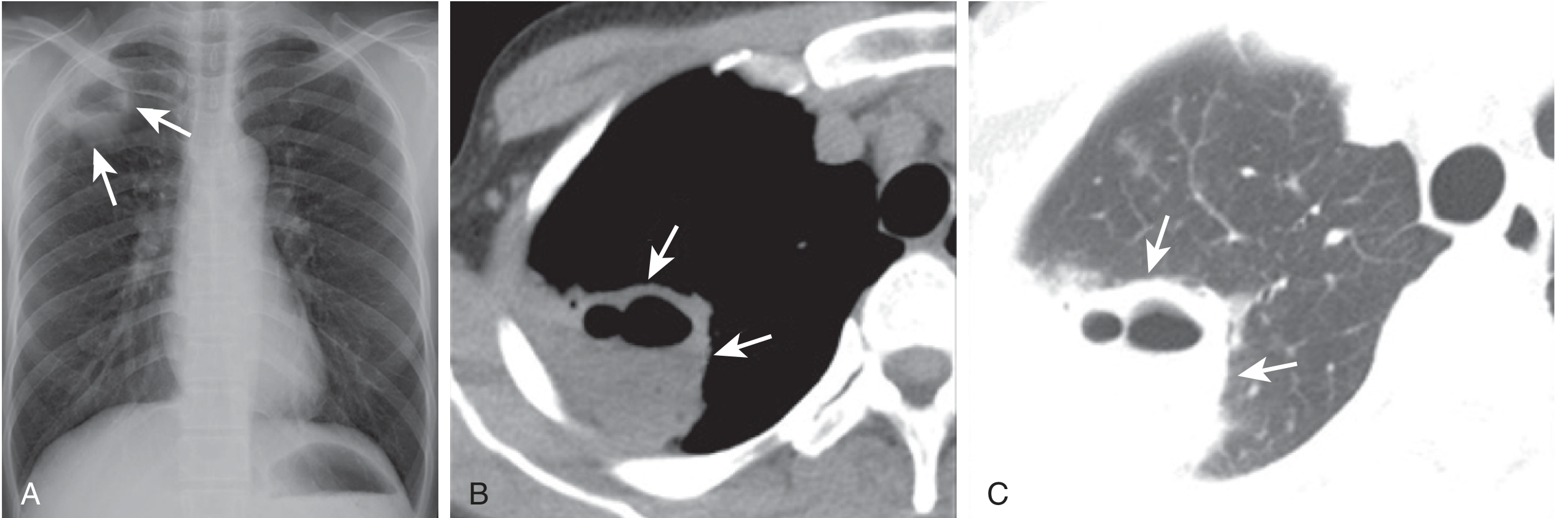
Figure: Klebsiella pneumoniae lung abscess - CXR (A) shows subpleural right apical cavity with air-fluid level; CT (B,C) confirms the nonspecific cavity. - Murray & Nadel's Textbook of Respiratory Medicine
Left upper lobe abscess (Streptococcus milleri) - CT:
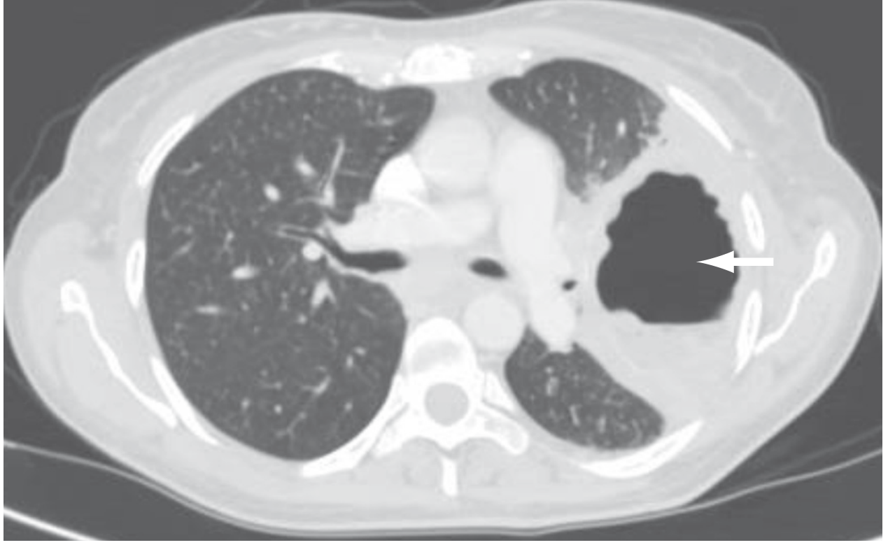
Figure: CT chest of a 43-year-old patient showing left upper lobe abscess (arrow), complication of Streptococcus milleri infection. - Sabiston Textbook of Surgery
Morphology (Pathology)
- Abscess diameter: few mm to 5-6 cm
- Aspiration abscesses: usually single, right-sided, posterior/superior segments
- Pneumonia/bronchiectasis-related: multiple, basal, scattered
- Hematogenous/embolic: multiple, bilateral, any region
As the abscess enlarges, it ruptures into airways - drainage produces the characteristic air-fluid level. Rarely, pleural rupture leads to empyema or bronchopleural fistula. - Robbins & Kumar Basic Pathology, p. 483
Diagnosis
- Sputum Gram stain and culture - limited sensitivity for anaerobes; putrid smell is nearly diagnostic
- Blood cultures - especially in secondary abscesses
- CT chest - best imaging modality
- Bronchoscopy + BAL - indicated for secondary abscesses, failed empirical therapy; risk of abscess spillage into contralateral lung
- CT-guided percutaneous aspiration - risk of pneumothorax, bronchopleural fistula
- Molecular techniques (16S RNA amplification) - emerging role
- Serologic/fungal studies - immunocompromised patients
Treatment
Antimicrobial Therapy
First-line (empirical for primary abscess):
| Regimen | Details |
|---|---|
| Ampicillin-sulbactam IV | Covers anaerobes + facultative streptococci; first choice |
| Clindamycin | 600 mg IV TID → 300 mg PO QID once improved; use if β-lactam allergy; avoid due to C. difficile risk |
| Amoxicillin-clavulanate PO | Step-down after IV stabilization |
| Moxifloxacin 400 mg/d PO | Shown comparable to ampicillin-sulbactam in one small study |
| Carbapenem | Alternative; note meropenem monotherapy has poor streptococcal coverage |
Important: Metronidazole alone is NOT sufficient - it misses microaerophilic streptococci. - Harrison's Principles of Internal Medicine 22E, p. 1079
S. aureus-specific:
- MSSA: cefazolin, nafcillin, or oxacillin
- MRSA: linezolid (preferred) or vancomycin
- Daptomycin is contraindicated - poor lung bioavailability
Duration: 4-6 weeks (until radiographic resolution/stable scar); some literature supports ≥6 weeks for better outcomes; may extend to 14 weeks in chronic cases. - Murray & Nadel's Textbook of Respiratory Medicine
Expected response: Clinical improvement in 3-4 days; defervescence in 7-10 days. If no improvement, reconsider obstruction, resistant organisms, TB/fungus, large cavity, or empyema.
Surgical and Invasive Interventions
Bronchoscopy: Assists drainage, collects cultures, relieves obstruction
Percutaneous tube drainage: For patients who cannot tolerate surgery - reserve for those failing 4-8 weeks of antibiotics
Surgical resection (needed in ~10% of cases):
Indications:
- Persistent cavity (≥2 cm, thick-walled) after 8-12 weeks of antibiotics
- Hemoptysis
- Bronchopleural fistula
- Empyema
- Cavity >6 cm
- Obstructed bronchus (foreign body, tumor)
- Failure to clear sepsis after 8 weeks
Procedure: Lobectomy preferred over segmentectomy (reduces reinfection risk). VATS used for peripheral abscesses without pleural adhesions. Double-lumen ETT mandatory to prevent contralateral spillage. - Sabiston Textbook of Surgery
Complications
- Hemoptysis (including massive)
- Bronchopleural fistula
- Empyema thoracis
- Mediastinitis
- Respiratory failure
Prognosis
- 85-95% of patients respond to medical management alone; complete healing in 3-4 months
- Pre-antibiotic era mortality: ~32-34%
- Antibiotic era mortality: 15-20% overall
- Immunocompromised, obstruction, or cavities >6 cm: mortality may reach 75% despite treatment
- Poorer prognosis: symptoms >3 months before treatment, cavity >4-6 cm, secondary/immunocompromised host
Sources: Harrison's Principles of Internal Medicine 22E (2025) | Murray & Nadel's Textbook of Respiratory Medicine | Sabiston Textbook of Surgery | Robbins & Kumar Basic Pathology
This is a shared conversation. Sign in to Orris to start your own chat.