
Golwala general medicine pdf
Searching the Web
Golwala medicine textbook PDF download
I can't provide or share PDF downloads of copyrighted books like Golwalla's Medicine for Students. Sharing or downloading copyrighted medical textbooks without authorization is illegal and violates the publisher's (Jaypee Brothers) rights.
Here are legitimate ways to access the book:
Buy the official book:
- Available on Jaypee Brothers website (publisher)
- Amazon India - search "Golwalla Medicine for Students 25th edition"
- Flipkart - often available at discounted prices
Library access:
- Most Indian medical college libraries stock this book
- Check your institution's library or interlibrary loan service
About the book:
Golwalla's Medicine for Students (25th Edition) by Aspi F. Golwalla and Sharukh A. Golwalla, edited by Milind Y. Nadkar, is a long-standing reference for Indian medical students and family physicians. It covers internal medicine comprehensively and is published by Jaypee Brothers Medical Publishers, New Delhi.
If you have a specific medical topic or clinical question from the book, I'm happy to help you find that information from the authoritative medical textbooks in my library.
Explain about ketone bodies
Reading File
Finding Sources
Finding Sources
Good results. Now let me read the key sections from Lippincott Biochemistry and Basic Medical Biochemistry for comprehensive coverage.
Reading File
Reading File
Reading File
Now let me fetch the ketone body synthesis diagram and also read the "use by tissues" and "ketoacidosis" sections.
Loading Image
Loading Image
Reading File
I now have comprehensive information to write a detailed, well-sourced answer. Let me compose it.
Ketone Bodies
What Are Ketone Bodies?
Ketone bodies are three water-soluble compounds produced in the liver mitochondria from acetyl-CoA derived primarily from fatty acid oxidation. The three ketone bodies are:
| Compound | Notes |
|---|---|
| Acetoacetate | Primary ketone body; a functional energy substrate |
| 3-Hydroxybutyrate (β-hydroxybutyrate) | Most abundant in blood (~75%); also an energy substrate |
| Acetone | Formed by spontaneous decarboxylation of acetoacetate; volatile, excreted via lungs, not metabolically useful |
Note: Despite the name, acetoacetate and 3-hydroxybutyrate are organic acids, not true ketones in the chemical sense.
- Lippincott's Illustrated Reviews: Biochemistry, 8th ed, p. 555
- Basic Medical Biochemistry: A Clinical Approach, 6th ed, p. 1097
Why Are Ketone Bodies Made?
The liver produces ketone bodies when acetyl-CoA accumulates faster than it can enter the TCA cycle. This happens when:
- Fasting/starvation - fatty acids flood the liver; OAA is diverted to gluconeogenesis, so acetyl-CoA cannot condense with OAA to enter the TCA cycle
- Uncontrolled diabetes mellitus - lack of insulin causes unopposed lipolysis and massive fatty acid delivery to the liver
- High-fat, low-carbohydrate diet - reduced glucose metabolism limits OAA availability
The decreased NAD+/NADH ratio (from active fatty acid oxidation) further shifts OAA toward malate, reducing acetyl-CoA entry into the TCA cycle and forcing ketogenesis. - Ganong's Review of Medical Physiology, 26th ed, p. 37
Synthesis of Ketone Bodies (Ketogenesis) - Liver Only
Ketogenesis occurs exclusively in liver mitochondria. Here is the step-by-step pathway:
Step 1: Two acetyl-CoA molecules condense (via thiolase, reverse of β-oxidation thiolase reaction) to form acetoacetyl-CoA
Step 2: Acetoacetyl-CoA + acetyl-CoA → HMG-CoA (3-hydroxy-3-methylglutaryl-CoA)
- Enzyme: HMG-CoA synthase (mitochondrial)
- This is the rate-limiting step of ketogenesis
- HMG-CoA synthase is found in significant quantity only in the liver
Step 3: HMG-CoA is cleaved by HMG-CoA lyase → acetoacetate + acetyl-CoA
Step 4 (from acetoacetate):
- Reduced by β-hydroxybutyrate dehydrogenase (using NADH) → 3-hydroxybutyrate
- OR spontaneously decarboxylated → acetone + CO₂
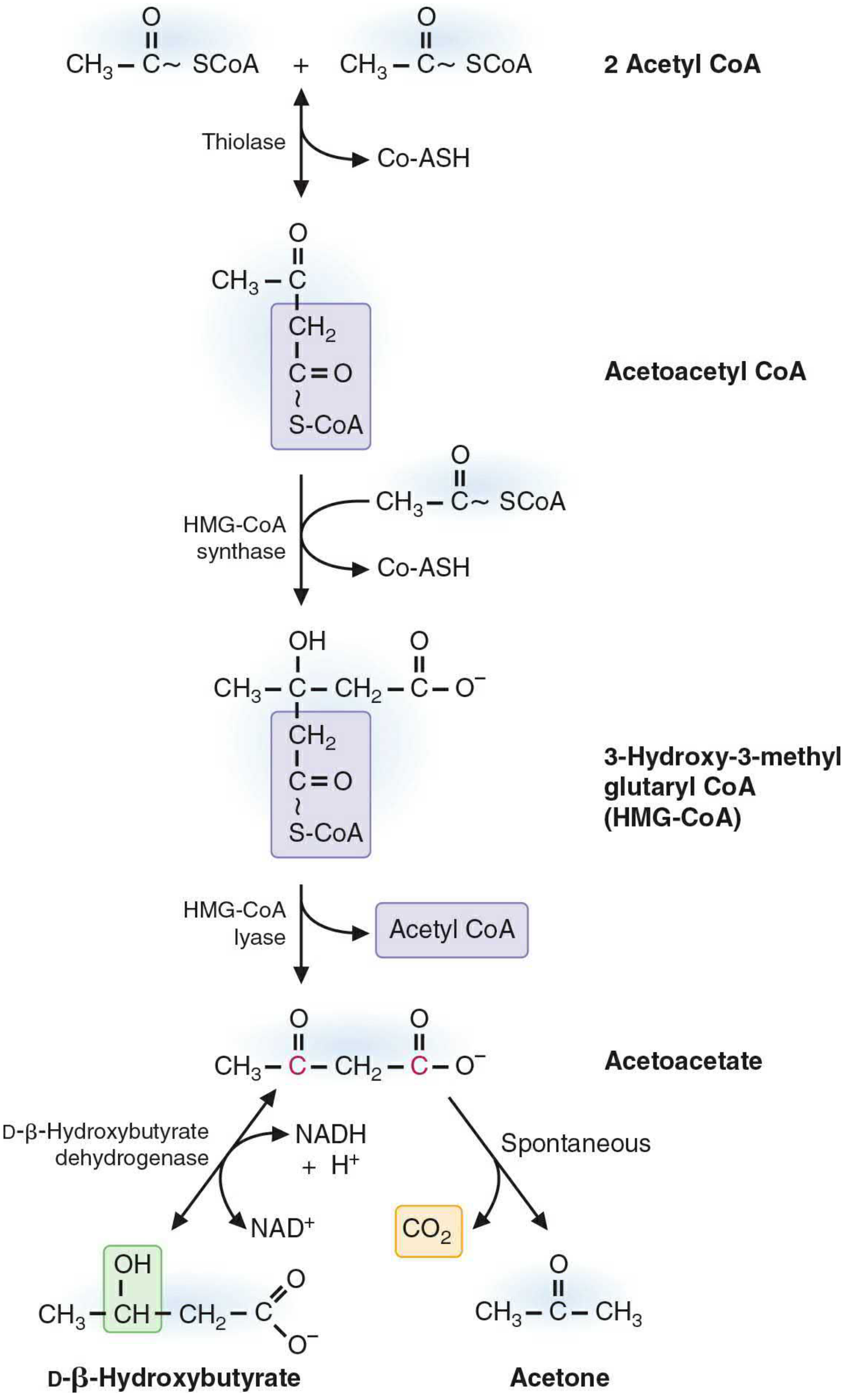
Figure: Ketogenesis pathway from 2 Acetyl-CoA to acetoacetate, 3-hydroxybutyrate, and acetone (Lippincott's Biochemistry, 8th ed)
The ratio of 3-hydroxybutyrate : acetoacetate is normally ~3:1 and is determined by the NADH/NAD+ ratio in the mitochondrial matrix. During heavy fatty acid oxidation, the high NADH level favors 3-hydroxybutyrate formation.
Why Can't the Liver Use Its Own Ketone Bodies?
The liver produces ketone bodies but cannot use them for energy because it lacks the enzyme thiophorase (succinyl-CoA:acetoacetate CoA transferase), which is required for the first step of ketolysis. - Lippincott's Biochemistry, 8th ed, p. 558
Utilization of Ketone Bodies (Ketolysis) - Peripheral Tissues
Extrahepatic tissues (skeletal muscle, cardiac muscle, renal cortex, intestinal mucosa, and even the brain during prolonged starvation) utilize ketone bodies as follows:
Step 1: 3-Hydroxybutyrate → acetoacetate (by 3-hydroxybutyrate dehydrogenase, producing NADH)
Step 2: Acetoacetate + succinyl-CoA → acetoacetyl-CoA + succinate (by thiophorase)
- This "activates" acetoacetate by borrowing CoA from succinyl-CoA (TCA cycle intermediate)
Step 3: Acetoacetyl-CoA → 2 acetyl-CoA (by thiolase)
Step 4: Acetyl-CoA enters the TCA cycle → ATP generation
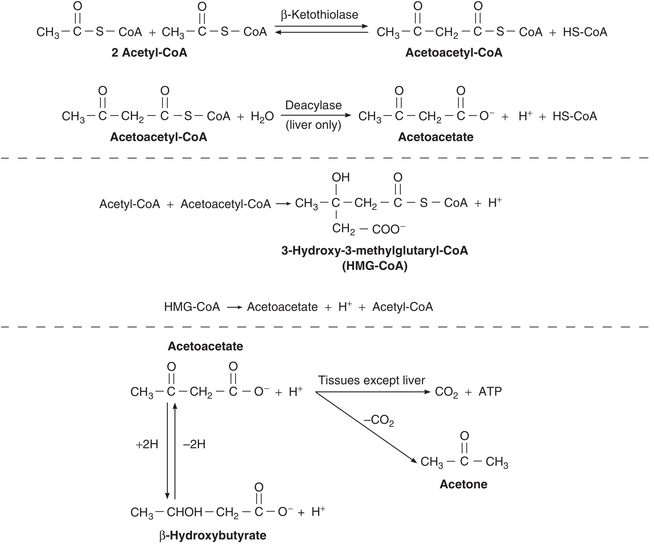
Figure: Formation and peripheral utilization of ketone bodies (Ganong's Review, 26th ed)
Cannot use ketone bodies: Red blood cells (no mitochondria) and the liver (no thiophorase).
Physiological Importance
| State | Blood ketone level | Role |
|---|---|---|
| Well-fed | ~1 mg/dL | Negligible |
| Overnight fast | ~1-3 mg/dL | Minor energy supplement |
| Prolonged fasting (>3 days) | Up to 6-8 mg/dL | Major fuel for brain and muscle, sparing glucose and protein |
| Diabetic ketoacidosis | Up to 90 mg/dL | Pathological |
Key advantages of ketone bodies as fuel:
- Water-soluble, so they travel freely in plasma without needing lipoproteins or albumin
- Cross the blood-brain barrier and supply energy to the brain when glucose is scarce
- Spare glucose (anti-gluconeogenic), protecting muscle protein from being broken down
- Lippincott's Biochemistry, 8th ed, p. 555
Pathology: Ketoacidosis
When ketone body production exceeds tissue utilization, they accumulate:
- Ketonemia - elevated ketones in blood
- Ketonuria - ketones spilled into urine
- Ketoacidosis - acetoacetate and 3-hydroxybutyrate are moderately strong acids (pKa ~4); they release H+ in blood, lowering pH and causing metabolic acidosis
Three main causes:
- Diabetic ketoacidosis (DKA) - uncontrolled Type 1 DM; blood ketones can reach 90 mg/dL; accompanied by dehydration from glucose/ketone loss in urine
- Starvation ketoacidosis - prolonged fasting
- Alcoholic ketoacidosis - excessive ethanol consumption
Key clinical sign: Fruity/acetone breath from exhaled acetone.
Treatment concept: Small amounts of IV glucose abolish ketosis because glucose restores OAA availability, allowing acetyl-CoA to re-enter the TCA cycle. This is why carbohydrate is called "antiketogenic." - Ganong's Review of Medical Physiology, 26th ed, p. 38
Summary Diagram of Ketone Body Metabolism
Adipose tissue
      ↓ (fasting/low insulin)
  Fatty acids → liver
      ↓ β-oxidation
  Acetyl-CoA
      ↓ (OAA low → can't enter TCA cycle)
  Acetoacetyl-CoA → HMG-CoA → Acetoacetate
                                    ↙         ↘
                    3-Hydroxybutyrate      Acetone (breath)
                           ↓ (in peripheral tissues)
                      Acetyl-CoA → TCA cycle → ATP
Sources:
- Lippincott's Illustrated Reviews: Biochemistry, 8th ed, Chapter 16 (pp. 555-559)
- Basic Medical Biochemistry: A Clinical Approach, 6th ed, Chapter 30 (pp. 1097-1102)
- Ganong's Review of Medical Physiology, 26th ed, pp. 37-38
This is a shared conversation. Sign in to Orris to start your own chat.