
All thing about hypertension including emergency
"hypertension"[MeSH Terms] AND "management"[MeSH Terms]
hypertension treatment guidelines
hypertension blood pressure classification stages
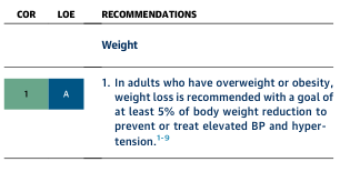
Summary : This figure presents a clinical recommendation regarding weight loss for adults with overweight or obesity, specifically targeting blood pressure and hypertension management. table: Recommendation Overview : • Recommendation is for adults who have overweight or obesity. • Weight loss is recommended with a goal of at least 5% of body weight reduction. • The purpose is to prevent or treat elevated blood pressure (BP) and hypertension. Classification Codes : • COR (Class of Recommendation): 1 • LOE (Level of Evidence): A Reference Notes : • The recommendation is supported by references 1–9. Analysis : • The table provides a high-level, evidence-based clinical guideline emphasizing the importance of modest weight loss (≥5%) in managing or preventing high blood pressure in overweight or obese adults, with a strong recommendation (Class 1, Level A evidence).
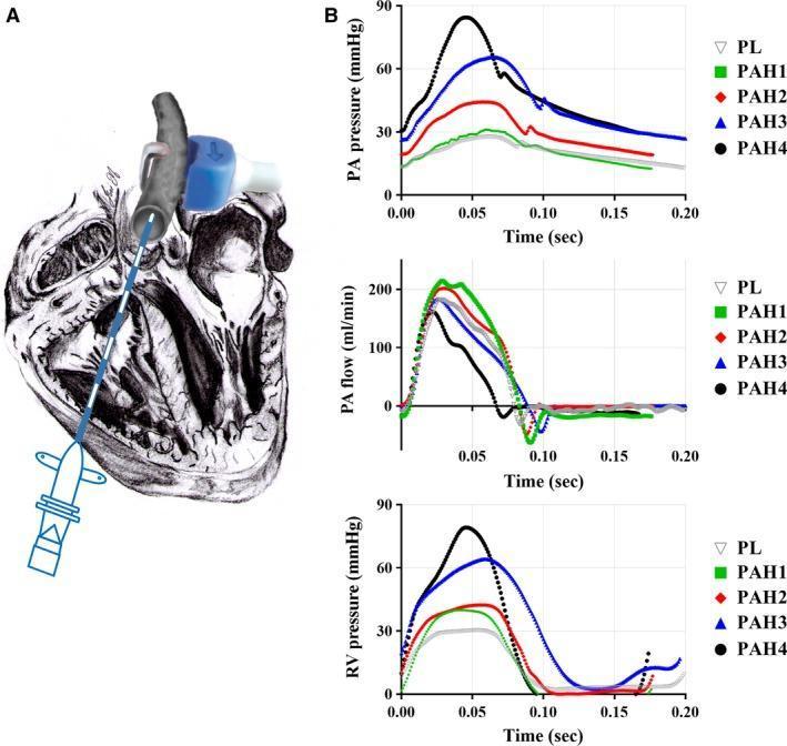
This composite educational graphic details in vivo hemodynamic measurement techniques and results in the context of pulmonary arterial hypertension (PAH). Panel A presents an anatomical diagram of a heart, illustrating the positioning of a dual pressure sensor catheter inserted through the right ventricle into the main pulmonary artery (MPA), complemented by an ultrasonic flow probe wrapped around the MPA. Panel B displays three time-series hemodynamic graphs (0.0 to 0.20 seconds) comparing a placebo (PL) control group with four progressive stages of PAH (PAH1-PAH4). The top graph shows MPA blood pressure (mmHg), demonstrating a progressive increase in peak systolic pressure and steeper systolic slopes as disease severity increases. The middle graph depicts MPA flow (ml/min), showing relatively stable peak flow across stages but altered waveform morphology. The bottom graph shows right ventricular (RV) pressure (mmHg), highlighting a significant rise in RV systolic pressure (RVSP) from ~30 mmHg in PL/PAH1 to nearly 80 mmHg in stage PAH4, indicating increased ventricular afterload and disease progression.
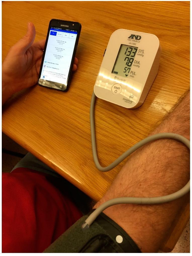
This clinical photograph illustrates a patient-centered remote health monitoring setup for managing hypertension. The central focus is a digital oscillometric blood pressure monitor (A&D Medical UC-651BLE) displaying a systolic pressure of 133 mmHg, a diastolic pressure of 78 mmHg, and a pulse rate of 57 bpm. The device features a Bluetooth connectivity symbol, indicating its role in a wireless telemedicine ecosystem. A gray inflatable cuff is correctly placed on the patient's arm and connected to the base unit via tubing. In the adjacent hand, a smartphone displays a specialized medical application with a history of physiological readings, demonstrating the integration of mobile health (mHealth) technology in chronic disease management. This visual serves to teach the application of 'smart' diagnostic tools in home-based clinical processes, emphasizing data acquisition and real-time patient empowerment through digital tracking.
hypertensive emergency target organ damage management
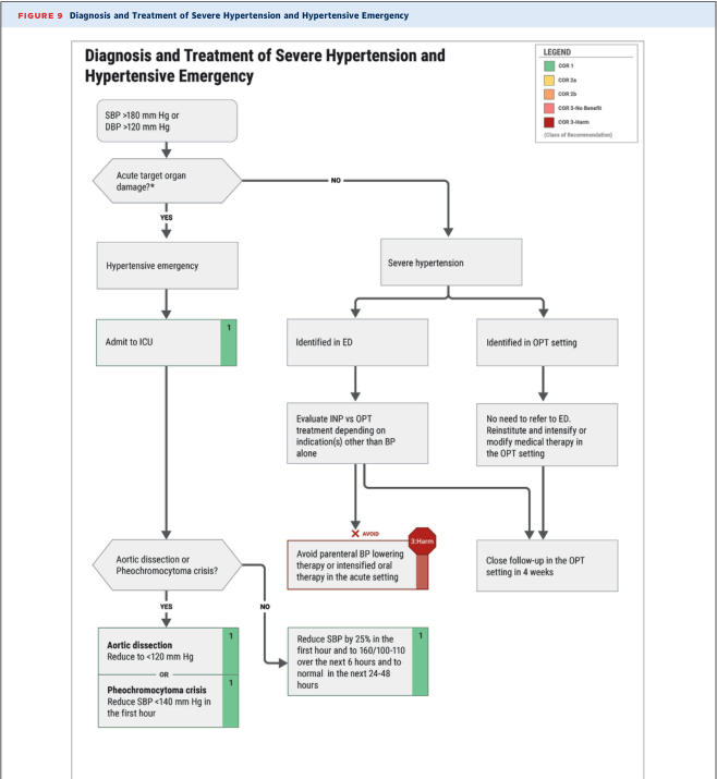
Summary : This flowchart outlines the diagnosis and treatment pathway for severe hypertension and hypertensive emergency, including decision points for acute target organ damage, specific crises (aortic dissection, pheochromocytoma), and recommended interventions based on clinical setting and severity. flowchart: # Nodes : • SBP >180 mm Hg or DBP >120 mm Hg (rectangle) • Acute target organ damage? (diamond) • Hypertensive emergency (rectangle) • Admit to ICU (rectangle, green COR 1) • Aortic dissection or Pheochromocytoma crisis? (diamond) • Aortic dissection: Reduce <120 mm Hg (rectangle, green COR 1) • Pheochromocytoma crisis: Reduce SBP <140 mm Hg in first hour (rectangle, green COR 1) • Severe hypertension (rectangle) • Identified in ED (rectangle) • Evaluate INP vs OPT treatment depending on indication(s) other than BP alone (rectangle) • Avoid parenteral BP lowering therapy or intensified oral therapy in the acute setting (rectangle, red COR 3-Harm) • Reduce SBP by 25% in the first hour and to 160/100-110 over the next 6 hours and to normal in the next 24-48 hours (rectangle, green COR 1) • Identified in OPT setting (rectangle) • No need to refer to ED. Reinstitute and intensify or modify medical therapy in the OPT setting (rectangle) • Close follow-up in the OPT setting in 4 weeks (rectangle) # Connectors : • SBP/DBP node flows to Acute target organ damage? (down arrow) • Acute target organ damage? YES → Hypertensive emergency; NO → Severe hypertension • Hypertensive emergency → Admit to ICU • Admit to ICU → Aortic dissection or Pheochromocytoma crisis? • Aortic dissection or Pheochromocytoma crisis? YES → Aortic dissection or Pheochromocytoma crisis nodes; NO → Reduce SBP by 25% node • Severe hypertension splits to Identified in ED and Identified in OPT setting • Identified in ED → Evaluate INP vs OPT treatment • Evaluate INP vs OPT treatment → Avoid parenteral BP lowering therapy (red X, avoid) • Identified in OPT setting → No need to refer to ED node • No need to refer to ED → Close follow-up in OPT setting in 4 weeks # Layout : • Top-down hierarchical flow, with major split at acute target organ damage. • Left branch for hypertensive emergency, right branch for severe hypertension. • Colour coding for class of recommendation (COR): green (1), yellow (2a), orange (2b), red (3-Harm). • Legend in top right corner for COR colour codes. # Analysis : • The flowchart provides a clear decision-making pathway for clinicians managing severe hypertension, distinguishing between hypertensive emergency (with organ damage) and severe hypertension (without organ damage). • Immediate ICU admission and aggressive BP reduction are recommended for emergencies, with specific targets for aortic dissection and pheochromocytoma crisis. • For severe hypertension, outpatient management and follow-up are emphasised, with a strong warning against inappropriate acute BP lowering in the absence of organ damage. • Colour coding highlights the strength and harm of recommendations, aiding rapid clinical decision-making.
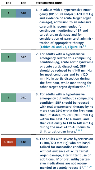
<table><thead><tr><th>COR</th><th>LOE</th><th>RECOMMENDATIONS</th></tr></thead><tbody><tr><td>1</td><td>B-NR</td><td>1. In adults with a hypertensive emergency (BP >180 and/or >120 mm Hg and evidence of acute target organ damage), admission to an intensive care unit is recommended for continuous monitoring of BP and target organ damage and for consideration of parenteral administration of appropriate therapy (Tables 26 and 27, Figure 9).1-3</td></tr><tr><td>1</td><td>C-LD</td><td>2. For adults with a hypertensive emergency related to a compelling condition (eg, acute aortic syndrome or acute aortic dissection), SBP should be reduced to <140 mm Hg for most conditions and to <120 mm Hg in aortic dissection during the first hour, while monitoring for other target organ dysfunction.4-7</td></tr><tr><td>1</td><td>C-LD</td><td>3. For adults with a hypertensive emergency but without a compelling condition, SBP should be reduced with oral or parenteral therapy by no more than 25% within the first hour; then, if stable, to <160/100 mm Hg within the next 2 to 6 hours; and then cautiously to 130 to 140 mm Hg during the next 24 to 48 hours to limit target organ injury.2,8,9</td></tr><tr><td>3: Harm</td><td>B-NR</td><td>4. For adults with severe hypertension (>180/120 mm Hg) who are hospitalized for noncardiac conditions without evidence of acute target organ damage, intermittent use of additional IV or oral antihypertensive medications are not recommended to acutely reduce BP.8,10,11</td></tr></tbody></table>
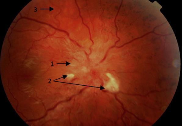
This clinical photograph displays a retinal fundus image highlighting hypertension-mediated end-organ damage (EOD), specifically hypertensive retinopathy. The image is annotated with numerical markers indicating key diagnostic features: (1) points to blurred optic disc margins and papilloedema, signaling severe intracranial pressure or grade IV hypertensive changes; (2) identifies multiple cotton wool spots, which are small, fluffy, whitish lesions representing focal retinal ischemia; and (3) demonstrates generalized arteriolar attenuation and increased vascular tortuosity, where the retinal vessels appear narrowed and more winding than normal. The anatomical focus includes the optic nerve head, the retinal nerve fiber layer, and the retinal vasculature. This material is pedagogically relevant for ophthalmology and internal medicine, illustrating the ocular manifestations of malignant or accelerated hypertension and the importance of fundoscopic examination in assessing systemic cardiovascular risk and subclinical target organ damage.
Hypertension: A Complete Clinical Guide
1. Definition & Classification
| Category | Systolic (mm Hg) | Diastolic (mm Hg) |
|---|---|---|
| Normal | < 120 | < 80 |
| Elevated | 120-129 | < 80 |
| Stage 1 HTN | 130-139 | 80-89 |
| Stage 2 HTN | ≥ 140 | ≥ 90 |
| Hypertensive Urgency | ≥ 180 | ≥ 120 (no TOD) |
| Hypertensive Emergency | ≥ 180 | ≥ 120 + acute target organ damage |
2. Pathophysiology
- Neurohormonal dysregulation: overactivation of the sympathetic nervous system and the renin-angiotensin-aldosterone system (RAAS)
- Vascular modulation: endothelial dysfunction, arterial stiffness
- Sodium/volume: high sodium intake promotes fluid retention
- Genetic factors: heritability ~30%; polygenic; genes for ACE, angiotensinogen, angiotensin II receptor, alpha-adducin involved
- Psychosocial stress, obesity, physical inactivity, alcohol
- Aging: sodium-sensitive BP rise correlates with vascular stiffness
3. Etiology: Primary vs. Secondary
- Onset before age 30 or after 55
- Resistant to ≥3 drugs
- Features suggesting an underlying cause
| Cause | Clinical Clues | Diagnostic Test |
|---|---|---|
| Primary aldosteronism | Hypokalemia, adrenal mass | 24-h urinary aldosterone, plasma aldosterone:renin ratio |
| Pheochromocytoma | Labile/paroxysmal HTN, palpitations, pallor, perspiration | 24-h urinary metanephrine/normetanephrine |
| Renal artery stenosis | Abdominal bruit, refractory HTN, azotemia after ACE inhibitor | Doppler flow study, MRA |
| Cushing's syndrome | Glucose intolerance, purple striae, obesity | Dexamethasone suppression test |
| Chronic kidney disease | Elevated creatinine, proteinuria | eGFR, urine albumin:creatinine |
| Obstructive sleep apnea | Obesity, snoring, daytime somnolence | Sleep study with O2 saturation |
| Thyroid/parathyroid | Weight change, temperature intolerance, hypercalcemia | TSH, serum PTH |
| Coarctation of aorta | Young patient, radiofemoral delay | MRI/CT aorta |
| Oral contraceptives | Clinical history | -- |
4. Risk Factors for End-Organ Damage
- Magnitude of BP elevation
- African American race (higher risk at any BP level)
- Smoking (including secondhand)
- Diabetes mellitus
- Metabolic syndrome (obesity, dyslipidemia)
- Physical inactivity
- Pre-existing end-organ damage at diagnosis
- Family history of cardiovascular disease
- Premenopausal women have relatively lower risk than men
5. Clinical Features & Diagnosis
- Confirmation: ≥2 readings on ≥2 visits; consider ambulatory BP monitoring (ABPM) to exclude white coat hypertension
- Basic labs: CBC, CMP (renal function, electrolytes), fasting glucose, lipid panel, urinalysis (protein, albumin:creatinine ratio)
- ECG: left ventricular hypertrophy
- Fundoscopy: hypertensive retinopathy (arterial narrowing, AV nicking, cotton wool spots, papilledema)
- Echocardiography if LVH suspected
Keith-Wagener-Barker Grading of Hypertensive Retinopathy
| Grade | Finding |
|---|---|
| I | Arteriolar narrowing, increased light reflex |
| II | AV nicking (Gunn sign) |
| III | Cotton wool spots, flame hemorrhages |
| IV | Papilledema (malignant hypertension) |
6. End-Organ Damage (Chronic)
- Heart: Left ventricular hypertrophy → heart failure, coronary artery disease
- Brain: Stroke (ischemic and hemorrhagic), lacunar infarcts, vascular dementia
- Kidneys: Hypertensive nephrosclerosis, CKD, proteinuria
- Eyes: Hypertensive retinopathy, retinal artery occlusion
- Aorta/vessels: Atherosclerosis, aortic aneurysm, PAD
7. Treatment: Lifestyle Modifications (First-line for all)
| Intervention | Expected SBP Reduction |
|---|---|
| Weight loss (≥5% body weight) | 5-20 mmHg per 10 kg lost |
| DASH diet (rich in fruits, vegetables, low-fat dairy) | 8-14 mmHg |
| Sodium restriction (<2.4 g/day) | 2-8 mmHg |
| Physical activity (aerobic, 30 min most days) | 4-9 mmHg |
| Moderation of alcohol | 2-4 mmHg |
| Smoking cessation | Reduces cardiovascular risk broadly |
8. Pharmacological Treatment
First-Line Drug Classes
| Class | Examples | Mechanism | Preferred In |
|---|---|---|---|
| Thiazide/thiazide-like diuretics | Hydrochlorothiazide, chlorthalidone | Reduce plasma volume, then PVR | Most patients (first-line) |
| ACE inhibitors | Enalapril, lisinopril, ramipril | Block ACE → ↓ angiotensin II, ↓ aldosterone | Diabetes, CKD, proteinuria, post-MI, CHF |
| ARBs | Losartan, valsartan, telmisartan | Block AT1 receptor | ACE inhibitor intolerance (cough), DM, CKD |
| Dihydropyridine CCBs | Amlodipine, nifedipine | Block L-type Ca²⁺ channels → vasodilation | Isolated systolic HTN, elderly, angina |
| Beta-blockers | Metoprolol, carvedilol, atenolol | ↓ CO, ↓ renin release | Post-MI, heart failure, arrhythmia |
Compelling Indications (Drug of Choice)
| Condition | Preferred Drug(s) |
|---|---|
| Heart failure with reduced EF | ACE inhibitor/ARB + beta-blocker + loop diuretic + MRA |
| Post-MI | Beta-blocker + ACE inhibitor |
| CKD/diabetic nephropathy | ACE inhibitor or ARB |
| Isolated systolic HTN (elderly) | Thiazide or CCB |
| Angina pectoris | Beta-blocker or CCB |
| Pregnancy | Labetalol, methyldopa, nifedipine (avoid ACE/ARB) |
| Pheochromocytoma | Alpha-blocker (phenoxybenzamine) first, then beta-blocker |
Step-Up Strategy
- Start with one agent at low dose
- Add second agent from different class (ACE/ARB + CCB or thiazide = preferred combination)
- Add third agent (typically thiazide if not already used)
- Consider spironolactone for resistant HTN (see below)
9. Resistant Hypertension
- Poor medication compliance
- Excessive sodium/alcohol intake
- Obesity, sleep apnea, hyperaldosteronism
- Concomitant medications (NSAIDs, sympathomimetics, corticosteroids, oral contraceptives)
- Inadequate drug doses or using drugs with similar mechanisms
- White coat effect
10. Special Populations
| Population | Considerations |
|---|---|
| Elderly | Lower treatment thresholds cautiously; beware orthostatic hypotension; thiazides/CCBs preferred |
| Diabetes | ACE inhibitor or ARB preferred (renoprotective); target BP <130/80 |
| CKD | ACE inhibitor/ARB first-line; monitor K⁺ and creatinine |
| Pregnancy | Labetalol or nifedipine (oral); methyldopa; avoid ACE, ARBs, direct renin inhibitors |
| Stroke (secondary prevention) | ACE inhibitor + thiazide combination shown effective |
| PAD | Beta-blockers are safe and do NOT worsen intermittent claudication |
11. Hypertensive Urgency vs. Emergency
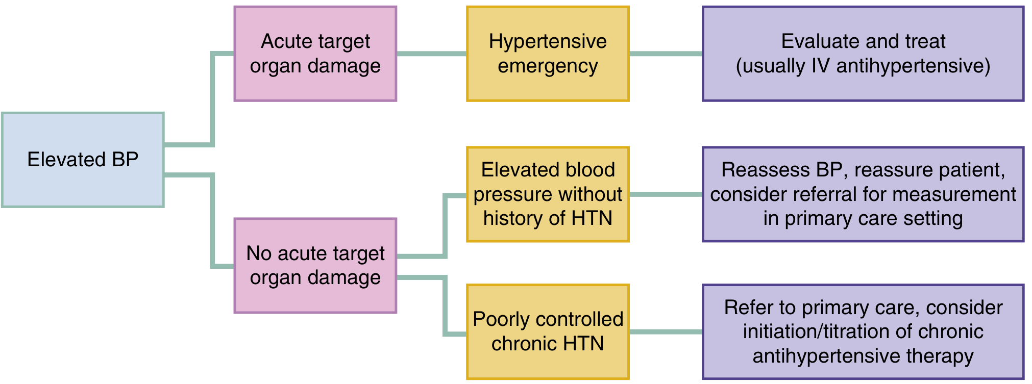
| Feature | Hypertensive Urgency | Hypertensive Emergency |
|---|---|---|
| BP level | ≥180/120 mm Hg | ≥180/120 mm Hg |
| Target organ damage (TOD) | Absent | Present (acute) |
| Symptoms | Headache, anxiety | Chest pain, dyspnea, altered mentation, focal neuro deficits, oliguria |
| Treatment setting | Outpatient; intensify oral therapy | ICU admission; IV antihypertensives |
| Rate of BP reduction | Gradual (24-48 h) | Controlled (see goals below) |
12. Hypertensive Emergency - Detailed
Definition
Organ Systems Involved
| Target Organ | Syndrome | Approximate Incidence |
|---|---|---|
| Heart (cumulative) | Acute heart failure, ACS | 27-49% |
| Acute heart failure | 14-37% | |
| Acute coronary syndrome | 11-12% | |
| Brain (cumulative) | Stroke, encephalopathy | 37-45% |
| Acute ischemic stroke | 6-25% | |
| Spontaneous ICH | 5-23% | |
| Hypertensive encephalopathy | 8-16% | |
| Kidney | AKI | 8-15% |
| Vascular | Aortic dissection | 1-2% |
| Obstetric | Eclampsia | ~2% |
Key Hypertensive Emergency Syndromes
- Mechanism: Failure of cerebral autoregulation → vasogenic cerebral edema, vasospasm, punctate hemorrhages
- Features: Severe headache, vomiting, altered mental status, seizures, papilledema, blurred vision
- Focal neuro deficits may occur on both sides (diffuse cerebral dysfunction, NOT stroke)
- CT/MRI: posterior reversible encephalopathy syndrome (PRES) pattern
- Acute findings: cotton wool spots, focal periarterial transudates, macular/optic disk edema, retinal hemorrhages
- Papilledema confirms Grade IV (malignant) hypertension
Blood Pressure Goals in Emergency
| Scenario | Target |
|---|---|
| General hypertensive emergency | Reduce MAP by ≤25% within 1st hour, then to 160/100-110 over 2-6 hours, then to ~130-140 in 24-48 hours |
| Aortic dissection | SBP < 120 mm Hg within first hour (to reduce shear stress) |
| Pheochromocytoma crisis | SBP < 140 mm Hg in first hour |
| Ischemic stroke | Generally do NOT acutely lower BP unless SBP >220 / DBP >120 (or if thrombolysis planned, keep <185/110) |
| Eclampsia | IV labetalol or hydralazine; magnesium sulfate for seizure prophylaxis |
| Acute CHF | Reduce preload/afterload with IV nitroglycerin or nitroprusside |
IV Agents for Hypertensive Emergency
| Drug | Class | Onset | Dose | Key Use / Notes |
|---|---|---|---|---|
| Labetalol | Mixed α1+β blocker | 2-5 min | IV 20 mg bolus, then 40-80 mg q10 min; or infusion 0.5-2 mg/min | Most versatile; safe in stroke, ACS, aortic dissection; avoid in asthma, severe bradycardia |
| Nicardipine | Dihydropyridine CCB | 5-10 min | 5-15 mg/hr IV infusion | Excellent for stroke, post-op, eclampsia; predictable; preferred over labetalol in some studies |
| Clevidipine | Dihydropyridine CCB | 2-4 min | 1-2 mg/hr titrated up to 32 mg/hr | Rapidly titratable; perioperative HTN |
| Nitroprusside | NO donor (arterial + venous dilator) | Seconds | 0.25-10 mcg/kg/min | Most powerful; risk of cyanide toxicity with prolonged use; avoid in renal failure |
| Nitroglycerin | NO donor (primarily venous) | 2-5 min | 5-100 mcg/min IV | Preferred in ACS, acute CHF; tolerance develops quickly |
| Esmolol | β1-selective blocker | 1-2 min | 250-500 mcg/kg load, then 50-200 mcg/kg/min | Aortic dissection, perioperative; very short-acting |
| Hydralazine | Direct arteriolar vasodilator | 10-20 min | 10-20 mg IV/IM | Eclampsia; unpredictable response; reflex tachycardia |
| Phentolamine | Alpha-blocker | 1-2 min | 5-15 mg IV bolus | Pheochromocytoma, cocaine-induced HTN |
| Fenoldopam | Dopamine D1 agonist | 5-10 min | 0.1-0.3 mcg/kg/min | AKI risk; renal-protective; increases renal blood flow |
| Enalaprilat | IV ACE inhibitor | 15-30 min | 1.25 mg IV q6h | Acute CHF; avoid in bilateral renal artery stenosis |
Diagnostic Workup in Hypertensive Emergency
- All patients: BMP (renal function, electrolytes), CBC, urinalysis with microscopy
- Cardiac symptoms (chest pain/dyspnea): ECG, CXR, troponin, BNP/NT-proBNP
- Neurologic symptoms (focal deficit, AMS): CT brain (rule out ICH, ischemic stroke), then MRI if indicated
- Dissection suspected: CT angiography chest, transesophageal echo, or MRI aorta
- Fundoscopy: for all - acute retinopathy confirms TOD and predicts stroke risk
13. Hypertensive Urgency
- SBP ≥180/120 mm Hg without acute TOD
- Do NOT use IV medications or aggressively lower BP acutely
- ACC/AHA recommendation (Class III: Harm): Do NOT use IV or intensified oral antihypertensives to acutely reduce BP in patients hospitalized for non-cardiac reasons without acute TOD
- Management: Intensify oral regimen, address precipitants (pain, anxiety, medication non-adherence), close follow-up within 1-4 weeks
14. Recent Evidence - 2024 ESC Guidelines (PMID 39210715)
- Introduction of a new category "elevated blood pressure" (SBP 120-139 + DBP <90) warranting lifestyle intervention
- Strong emphasis on combination therapy from the start for most Stage 2 patients
- Continued endorsement of ACE inhibitor/ARB, CCB, and thiazide-like diuretics as first-line pillars
- Ambulatory BP monitoring (ABPM) as gold standard for diagnosis
Summary at a Glance
HYPERTENSION
├── Primary (90%): Multifactorial; treat with lifestyle + drugs
├── Secondary (10%): Find & treat the cause
├── Treatment ladder:
│   Step 1: Lifestyle modification
│   Step 2: Monotherapy (thiazide / ACEI / ARB / CCB)
│   Step 3: Dual combination
│   Step 4: Triple combination
│   Step 5: Resistant HTN → add spironolactone
│
└── EMERGENCY (SBP >180/120 + TOD)
    ├── Admit ICU
    ├── IV antihypertensives (labetalol, nicardipine, nitroprusside...)
    ├── Reduce MAP ≤25% in 1st hour
    ├── Special cases: Dissection (<120 in 1h), Stroke (careful!), Eclampsia (labetalol + MgSO4)
    └── Identify & treat underlying trigger