
CAUSE OF BULLAE IN LUNG FIELD
bullae in lung chest X-ray bullous emphysema
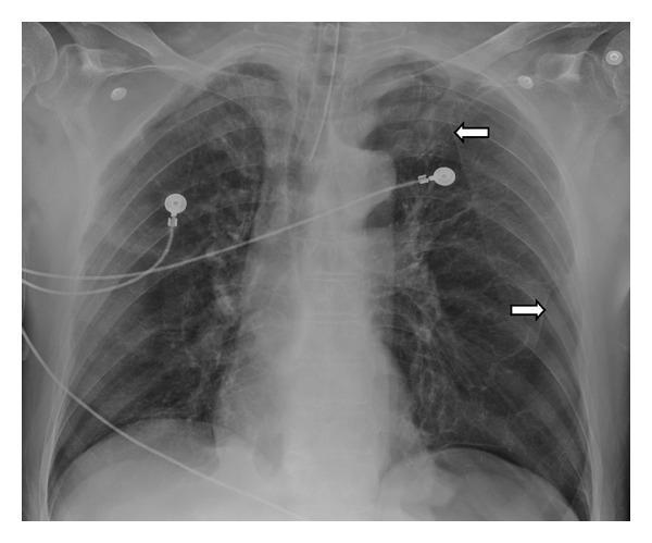
This diagnostic image is an anteroposterior (AP) chest X-ray of a patient in a clinical setting, as evidenced by the presence of an endotracheal tube and ECG leads. The radiograph demonstrates bilateral pulmonary hyperinflation with a notable increase in radiolucency. Specifically, the left lung displays multiple large, focal areas of hyperlucency located at the apex and base. These regions are demarcated by thin, curvilinear, radiopaque borders (indicated by white arrows), which are characteristic of bullae walls. The overall lung volume is increased, with flattening of the diaphragmatic contours. These findings are highly suggestive of Vanishing Lung Syndrome (idiopathic giant bullous emphysema) or advanced bullous emphysema. The image serves as an educational example of how large subpleural bullae can occupy significant portions of the hemithorax, leading to compressive effects on the surrounding lung parenchyma and clinical respiratory compromise.
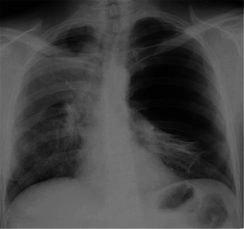
This diagnostic image is an anteroposterior (AP) chest plain film (X-ray) demonstrating advanced bullous emphysema, specifically illustrating features consistent with Vanishing Lung Syndrome. The primary finding is a giant, thin-walled bulla in the right hemithorax, which is significantly hyperextended and occupies approximately two-thirds of the hemithorax volume. This large, space-occupying lesion causes a visible mass effect, resulting in a shift of the mediastinum toward the right and substantial compression of the adjacent right lung parenchyma, which appears poorly expanded. The left lung also demonstrates hyperlucency and signs of bullous disease. Key clinical concepts illustrated include the radiographic presentation of pulmonary bullae, the compressive effects of giant bullae on mediastinal structures, and postoperative complications like secondary pneumothorax. This image serves as an educational example for pulmonology and thoracic surgery, particularly regarding the diagnosis and surgical management of end-stage bullous lung disease and COPD.
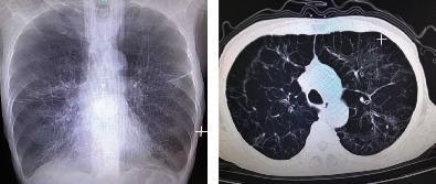
This dual-panel image displays diagnostic thoracic imaging from a patient with Chronic Obstructive Pulmonary Disease (COPD). The left panel is a posterior-anterior (PA) chest X-ray showing classic features of severe emphysema, including hyperinflated lung fields, sparse bronchovascular markings, and a widened chest diameter consistent with a barrel chest deformity. Diaphragmatic flattening and a narrowed mediastinal silhouette are also visible. The right panel is an axial CT scan of the chest (lung window) at the level of the upper lobes, revealing extensive bullous emphysema. Multiple large, thin-walled bullae are visible, particularly in the subpleural regions, along with architectural distortion of the lung parenchyma. These findings demonstrate advanced obstructive lung disease and are clinically significant for explaining symptoms of chronic shortness of breath and impaired gas exchange in elderly patients with a significant smoking history.
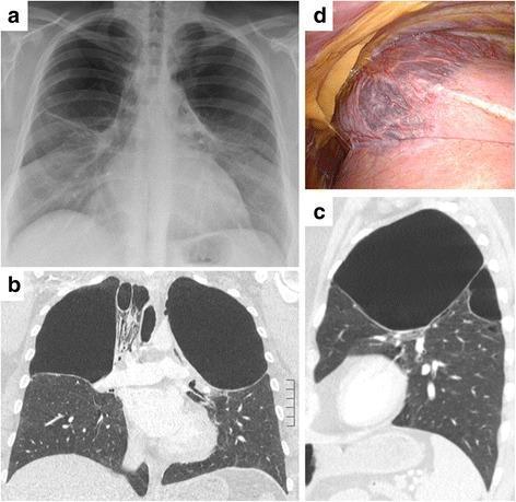
This composite figure illustrates giant apical bullous lung disease through multiple modalities: a posterior-anterior chest X-ray (a), coronal Computed Tomography (CT) scans (b, c), and an intraoperative photograph (d). The radiological images (a-c) demonstrate large, symmetrical, bilateral, and thin-walled bullae located at the pulmonary apices. These air-filled spaces occupy approximately one-third to one-half of the upper thoracic cavity, causing compression of the underlying functional lung parenchyma. The CT scans highlight the sharp demarcation between the hyperlucent bullae and the relatively preserved lower lung lobes. The intraoperative photograph (d) provides a macroscopic view of the right upper lobe, showing a large, distended, dark-colored apical bulla with a thin, translucent wall and visible surface vascularization. The surrounding lung tissue appears relatively healthy in texture. This sequence is representative of severe bullous emphysema, requiring surgical intervention such as thoracoscopic bullectomy to relieve dyspnea and improve respiratory mechanics. Educational focus includes recognizing the radiographic signs of giant bullae versus pneumothorax and understanding their surgical correlation.
Causes of Bullae in the Lung Field
1. Emphysema-related (most common cause)
- Centrilobular/panacinar emphysema from smoking - the classic and most frequent cause. Bullae form when destroyed, confluent alveolar spaces coalesce into larger air-filled cavities.
- Alpha-1 antitrypsin (α1-AT) deficiency - causes panacinar emphysema with a distinctive basal/lower lobe predominance of bullae, unlike the upper-lobe predominance of ordinary smoking-related bullae (Fishman's Pulmonary Diseases and Disorders, p. bullae CT section).
- Giant/vanishing lung syndrome - idiopathic giant bullous emphysema, typically bilateral, upper-lobe, occupying a large portion of the hemithorax and compressing adjacent normal lung (Murray & Nadel's Textbook of Respiratory Medicine).
2. Classification by pathogenesis (Fishman's Pulmonary Diseases)
- Type I bullae - thin-walled, empty interior, narrow neck connecting to lung, usually at the apices and edges of the lingula.
- Type II bullae - arise from subpleural parenchyma with a broad neck of panacinar emphysematous lung tissue.
- Type III bullae - deeper within the lung parenchyma, broad connection to surrounding emphysematous lung.
3. Other/secondary causes
- Connective tissue disease (e.g., Marfan syndrome, Ehlers-Danlos) - apical bullae with pneumothorax risk.
- IV drug use (e.g., talc granulomatosis from injected medications) - can cause bullous change, particularly basilar.
- HIV/Pneumocystis jirovecii pneumonia - upper lobe cystic/bullous change.
- Sarcoidosis, pulmonary fibrosis (honeycombing) - can mimic or coexist with bullous change in advanced fibrotic lung disease.
- Post-infectious/necrotizing pneumonia - pneumatoceles (thin-walled air cysts) can resemble bullae, especially in staphylococcal pneumonia.
- Congenital bullous disease - rare isolated bullous emphysema without generalized COPD.
Clinical significance
- Bullae can enlarge progressively over months to years, sometimes with intervening stability (Grainger & Allison's Diagnostic Radiology).
- Major complications: spontaneous pneumothorax (rupture into pleural space), compression of adjacent functional lung causing dyspnea, infection within a bulla, and rarely hemorrhage.
- Surgical bullectomy is considered when a bulla compresses adjacent functional lung significantly.
COPD xray related viva qs in radiology
COPD Chest X-ray – Viva Questions & Answers
Basic / Definition
- To exclude alternative/coexisting diagnoses (pneumonia, malignancy, heart failure, pneumothorax)
- To assess an acute exacerbation (only ~16% of COPD exacerbations show an abnormal CXR, and only half of those actually change management - Murray & Nadel's)
- To detect complications: bullae, pneumothorax, cor pulmonale
Radiographic Signs
- Hyperinflation of lung fields (best objective sign)
- Flattened hemidiaphragms (loss of normal dome/convex shape) - seen well on lateral view
- Increased retrosternal air space (>2.5-3 cm on lateral view, measured 3 cm below the manubrium)
- Increased AP diameter of the chest ("barrel chest")
- Attenuated, pruned peripheral vascular markings with relatively prominent central pulmonary arteries
- Widened intercostal spaces
- Decreased zone of apposition of diaphragm to rib cage
- Bullae, especially upper-lobe (nearly diagnostic when present, though uncommon)
- Ordinary (centrilobular) emphysema from smoking: upper lobe predominant bullae
- Alpha-1 antitrypsin deficiency (panacinar emphysema): lower lobe/basal predominant bullae - an important discriminator to mention in viva.
Differentiating Chronic Bronchitis vs Emphysema type
- "Pink puffer" (emphysema-predominant): hyperinflation, bullae, flattened diaphragms, paucity of vascular markings
- "Blue bloater" (chronic bronchitis-predominant): may show increased bronchovascular markings, less hyperinflation, signs of cor pulmonale (enlarged central pulmonary arteries, right heart enlargement) This clinical dichotomy is largely outdated but still asked in vivas.
Complications / Related Findings
- Bulla: curvilinear thin wall visible within the lung field, lung markings may be seen crossing over or crowded around it, no mediastinal shift usually unless very large
- Pneumothorax: visceral pleural line paralleling chest wall, absent lung markings peripheral to the line, may show mediastinal shift if tension
- CT resolves ambiguity when uncertain - critical because chest tube placement into a bulla mistaken for pneumothorax is a recognized error.
Correlation/Exam Pearls
Reference Image
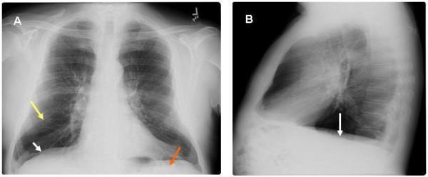
causes of spontaneous pneumothorax
spontaneous pneumothorax chest X-ray
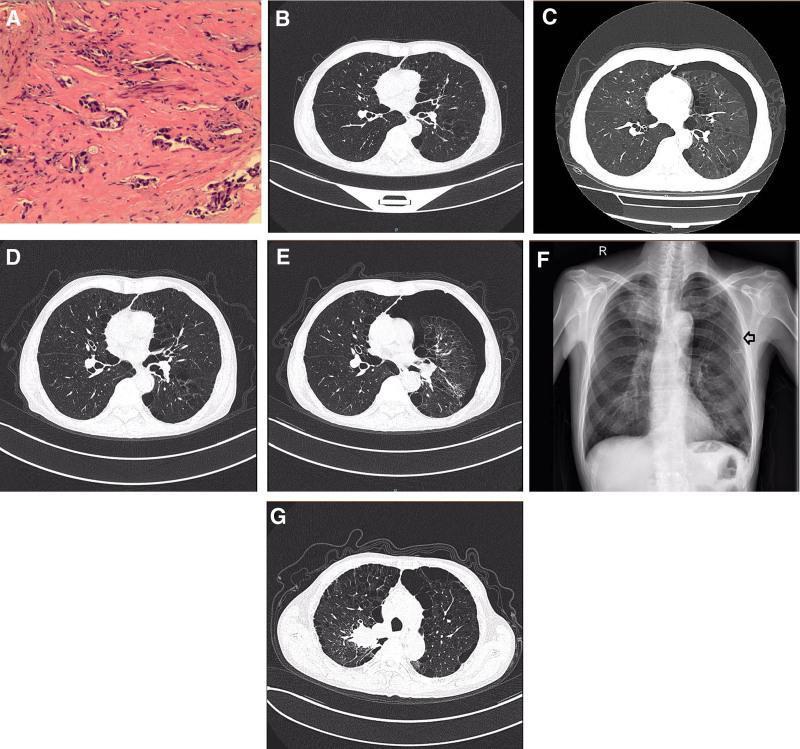
A multi-panel figure illustrating the clinical course of a patient with lung adenocarcinoma and recurring pneumothorax. Panel A displays a histopathological image (H&E stain) showing malignant epithelial cells forming glandular structures, diagnostic of lung adenocarcinoma. Panels B-E and G are axial contrast-enhanced chest CT scans in the lung window. Panel B establishes a baseline with clear lung parenchyma. Panels C and E demonstrate a left-sided pneumothorax, characterized by a visible pleural line and an absence of distal lung markings, indicating partial lung collapse. Panel D shows spontaneous resolution/absorption of the air in the pleural space. Panel F is a posterior-anterior (PA) chest X-ray showing the lung re-expanded following the insertion of a closed thoracic drainage tube (chest tube). Panel G displays a follow-up CT scan with stable lung expansion and no recurrent pneumothorax. The series highlights the management and radiological appearance of secondary spontaneous pneumothorax in the context of advanced pulmonary malignancy and tyrosine kinase inhibitor (anlotinib) therapy.
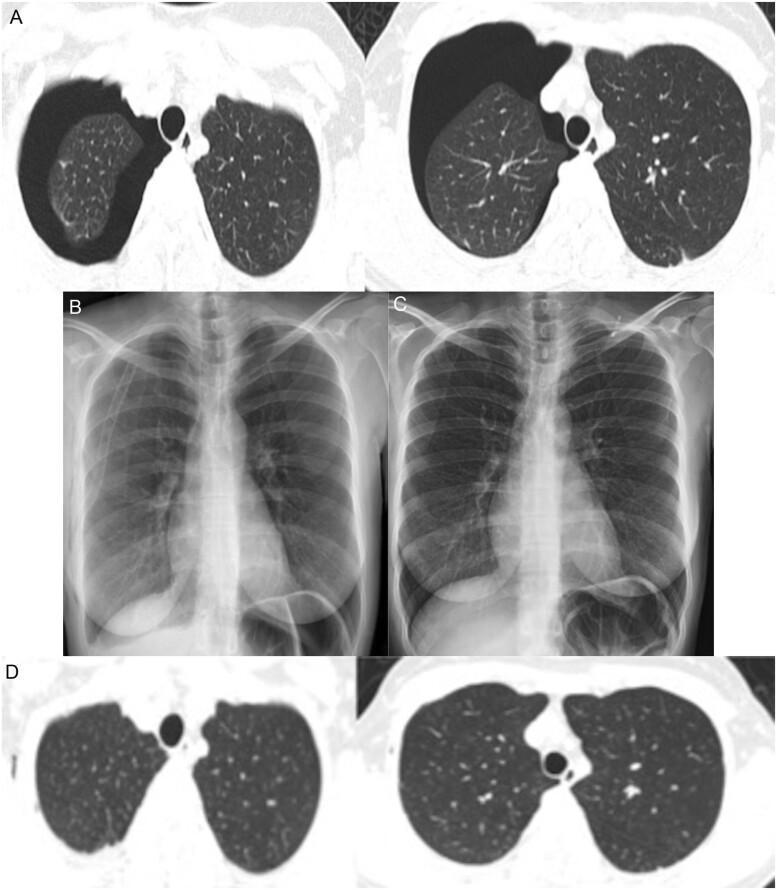
A multi-panel medical image sequence documenting the progression and resolution of a right-sided pneumothorax. Panel A presents axial Thorax Computed Tomography (TCT) lung window slices showing a significant right pneumothorax, characterized by a large, dark, air-filled pleural space (radiolucency) causing collapse of the right lung toward the mediastinum; the left lung remains fully expanded with normal density. Panel B is a Posteroanterior (PA) chest X-ray taken after tube thoracostomy, showing the right lung successfully re-expanded with the chest tube in situ. Panel C displays a follow-up chest X-ray after removal of the chest tube on the fifth day, maintaining full lung expansion and clear costophrenic angles. Panel D shows a follow-up axial TCT image two weeks post-discharge, confirming complete resolution of the pneumothorax with bilateral symmetric lung expansion and normal lung parenchyma. This sequence illustrates the clinical management of a spontaneous pneumothorax in the context of COVID-19 infection, from acute presentation to complete recovery.
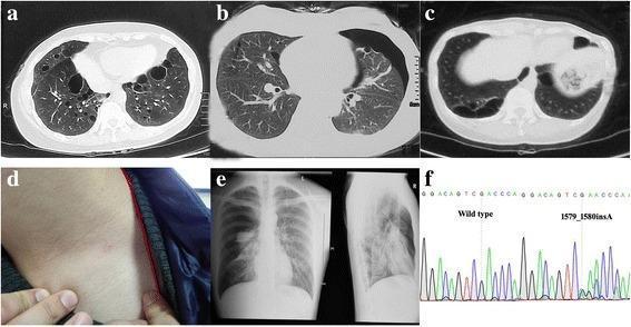
This multi-panel figure presents diagnostic findings consistent with Birt-Hogg-Dubé syndrome in a familial cohort. Panels (a), (b), and (c) are axial chest CT scans demonstrating bilateral, multiple thin-walled pulmonary cysts of varying sizes and distributions, characteristic of the condition. Panel (b) specifically highlights a concurrent pneumothorax. Panel (e) provides a corresponding posterior-anterior chest X-ray showing a collapsed lung with a visible pleural line and peripheral hyperlucency, confirming a pneumothorax. Clinical manifestations are further illustrated in panel (d), showing cutaneous papules (fibrofolliculomas/angiofibromas) on the neck. Panel (f) contains a DNA sequencing chromatogram comparing a wild-type sequence to a frameshift mutation (c.1579_1580insA) in exon 14 of the FLCN gene. The combination of cystic lung disease, spontaneous pneumothorax, cutaneous lesions, and genetic confirmation highlights the multisystemic nature of this autosomal dominant disorder.
Causes of Spontaneous Pneumothorax
1. Primary Spontaneous Pneumothorax (PSP)
- Subpleural blebs/bulla rupture at the lung apex - the leading proposed mechanism, though this remains debated ("Do blebs cause primary spontaneous pneumothorax?" - Fishman's Pulmonary Diseases, referenced controversy)
- Cigarette smoking - strongest modifiable risk factor; increases risk many-fold in a dose-dependent manner
- Tall, thin body habitus - increased apical pleural stress from greater negative pressure gradient at the apex
- Family history/genetic predisposition - familial pneumothorax syndromes
- Connective tissue disorders - Marfan syndrome, Ehlers-Danlos syndrome (apical bleb formation)
2. Secondary Spontaneous Pneumothorax (SSP)
- COPD/emphysema - the most common underlying cause of SSP; ruptured subpleural bullae
- Cystic fibrosis
- Severe acute asthma exacerbation
- Pneumocystis jirovecii pneumonia (PCP) in HIV/immunocompromised patients - classic cause of secondary pneumothorax
- Necrotizing/cavitary pneumonia (staphylococcal, tuberculosis) - rupture of a cavity or pneumatocele
- Lung abscess
- Lymphangioleiomyomatosis (LAM) - important to consider when a woman presents with spontaneous pneumothorax, since PSP is otherwise male-predominant (Murray & Nadel's Textbook of Respiratory Medicine)
- Birt-Hogg-Dube syndrome - multiple thin-walled pulmonary cysts with recurrent pneumothorax, associated skin fibrofolliculomas and FLCN gene mutation
- Pulmonary Langerhans cell histiocytosis
- Idiopathic pulmonary fibrosis / other interstitial lung disease with honeycombing
- Sarcoidosis (advanced fibrocystic stage)
- Primary lung cancer (especially with cavitation, or pleural metastasis) - can present with recurrent pneumothorax
- Metastatic sarcoma to the lung (classically osteosarcoma) causing cavitary nodules that rupture
- Marfan syndrome, Ehlers-Danlos syndrome (also causes PSP-type disease as above)
- Rheumatoid lung disease with necrobiotic nodules
- Thoracic endometriosis - recurrent pneumothorax in women in temporal relation to menstruation, often right-sided with diaphragmatic defects
Clinical Presentation Pearl
Illustrative Image