Give me a photo of typical intercostal nerve
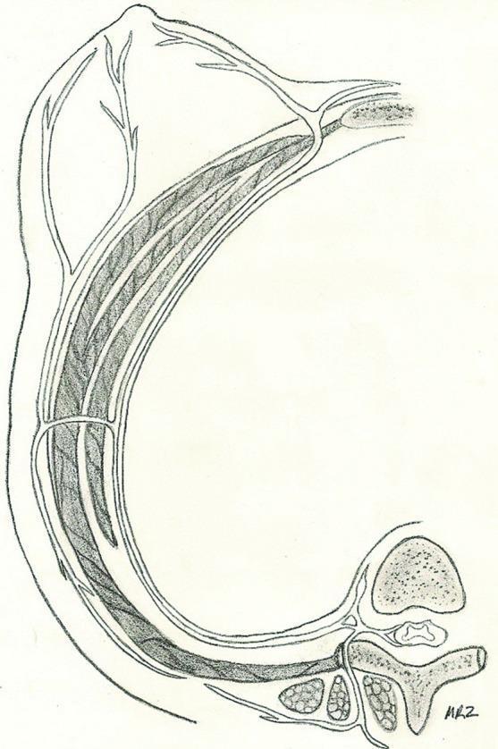
typical intercostal nerve anatomy diagram
intercostal nerves anatomy

Give me a better photo, withdraw and label and clear full
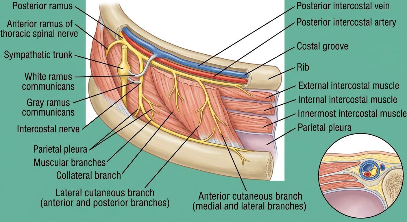
A clear, complete high-resolution educational anatomical illustration of a typical intercostal nerve in one intercostal space, shown in lateral thoracic-wall view and a small cross-section inset. Fully label: posterior ramus, anterior ramus of thoracic spinal nerve, sympathetic trunk, white ramus communicans, gray ramus communicans, intercostal nerve, posterior intercostal vein, posterior intercostal artery, costal groove, rib, external intercostal muscle, internal intercostal muscle, innermost intercostal muscle, parietal pleura, muscular branches, collateral branch, lateral cutaneous branch with anterior and posterior branches, anterior cutaneous branch with medial and lateral branches. Clearly show the neurovascular order in the costal groove as vein-artery-nerve, superior to inferior. Use a clean textbook anatomy style with large legible labels and leader lines, no missing or overlapping labels.
typical intercostal nerve course branches anatomy T3 T6 costal groove VAN
clear labelled diagram course branches typical intercostal nerve
intercostal nerves anatomy